Downloaded 182 times


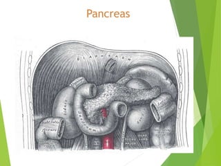
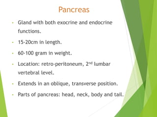

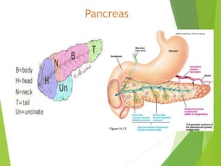
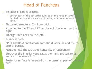


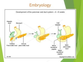


The pancreas is a retroperitoneal gland with both exocrine and endocrine functions. It is 15-20cm in length and divided into the head, neck, body, and tail. The pancreas produces enzymes that are released into the small intestine to aid in digestion and produces hormones like insulin and glucagon that are released into the bloodstream to regulate blood sugar levels. It has both an extensive arterial blood supply and venous drainage that parallels the arteries. The pancreas is innervated by both the sympathetic and parasympathetic nervous systems.


