Downloaded 140 times


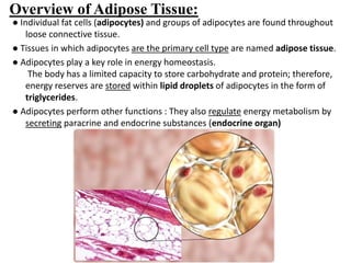

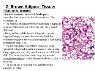
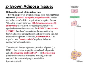
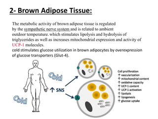
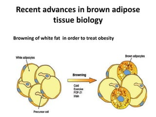
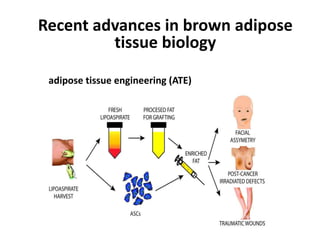
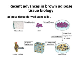
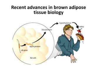


- Adipose tissue contains adipocytes that store triglycerides and regulate energy metabolism through secretion of hormones. There are two types: white and brown adipose tissue. - White adipose tissue is the main site for energy storage. It is found throughout the body. Brown adipose tissue helps generate heat and is found in newborns and certain regions of adults. - White and brown adipocytes differ in lipid droplet size, mitochondrial content, and gene expression factors that regulate their differentiation and function. Recent research focuses on browning of white fat, adipose tissue engineering, and stem cells.


![PERI-PROSTHETIC FRACTURE NAIL-PLATE CONSTRUCT [NPC].pptx](https://cdn.slidesharecdn.com/ss_thumbnails/drarunkumardrmohamedashrafperiprostheticfrasturenail-plateconstructnpc-260209164459-7e9d15a1-thumbnail.jpg?width=640&height=640&fit=bounds)

![ONFH[AVN HIP] -TRIPLE REGIME -A NOVAL SURGICAL CONCEPT .pptx](https://cdn.slidesharecdn.com/ss_thumbnails/onfhavnhip2026koaconcalicutdrgokuldevdrmashraf-260210064517-213ec005-thumbnail.jpg?width=640&height=640&fit=bounds)


