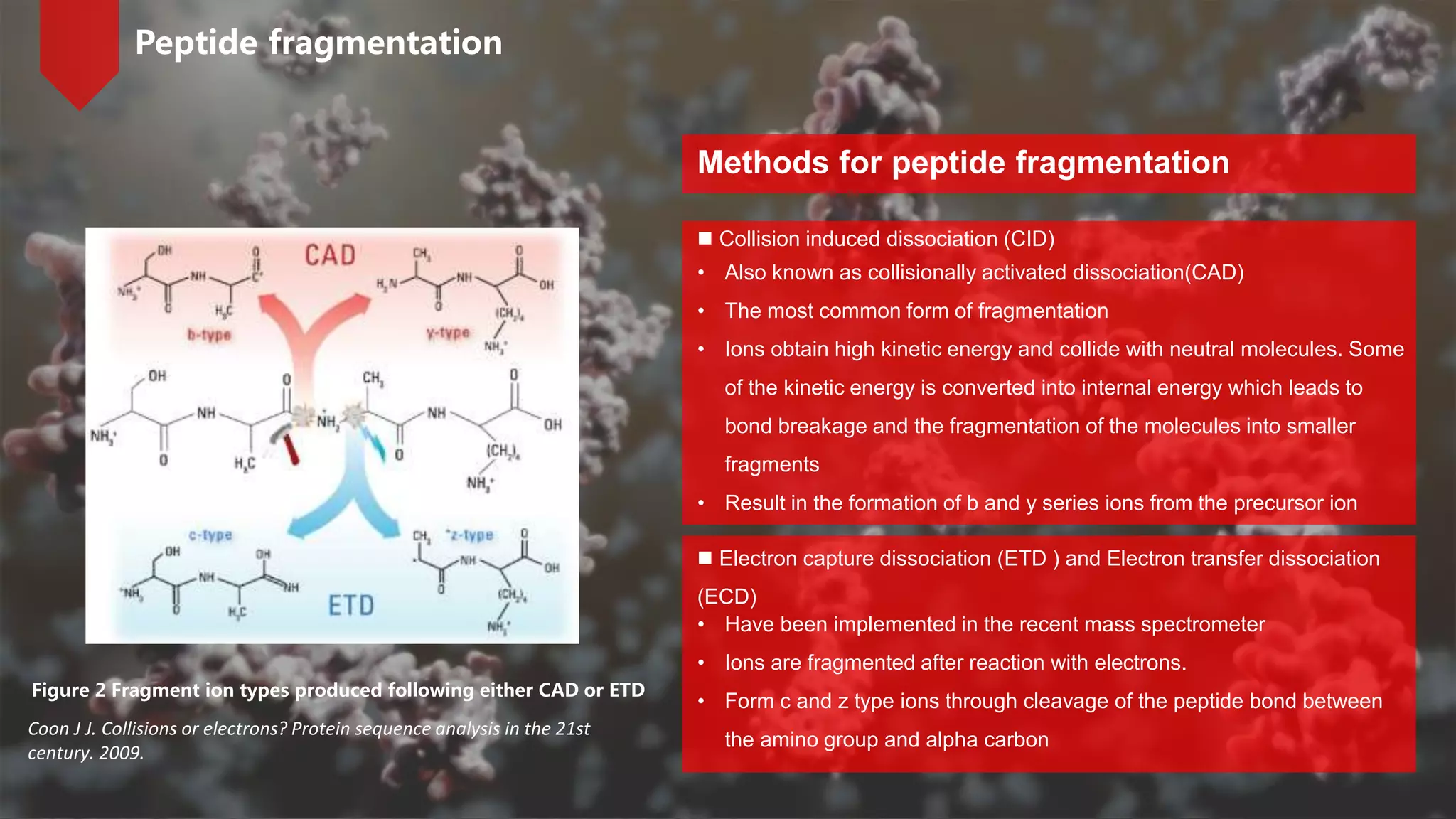
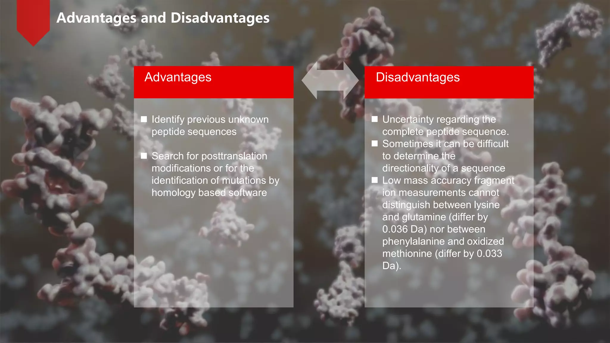
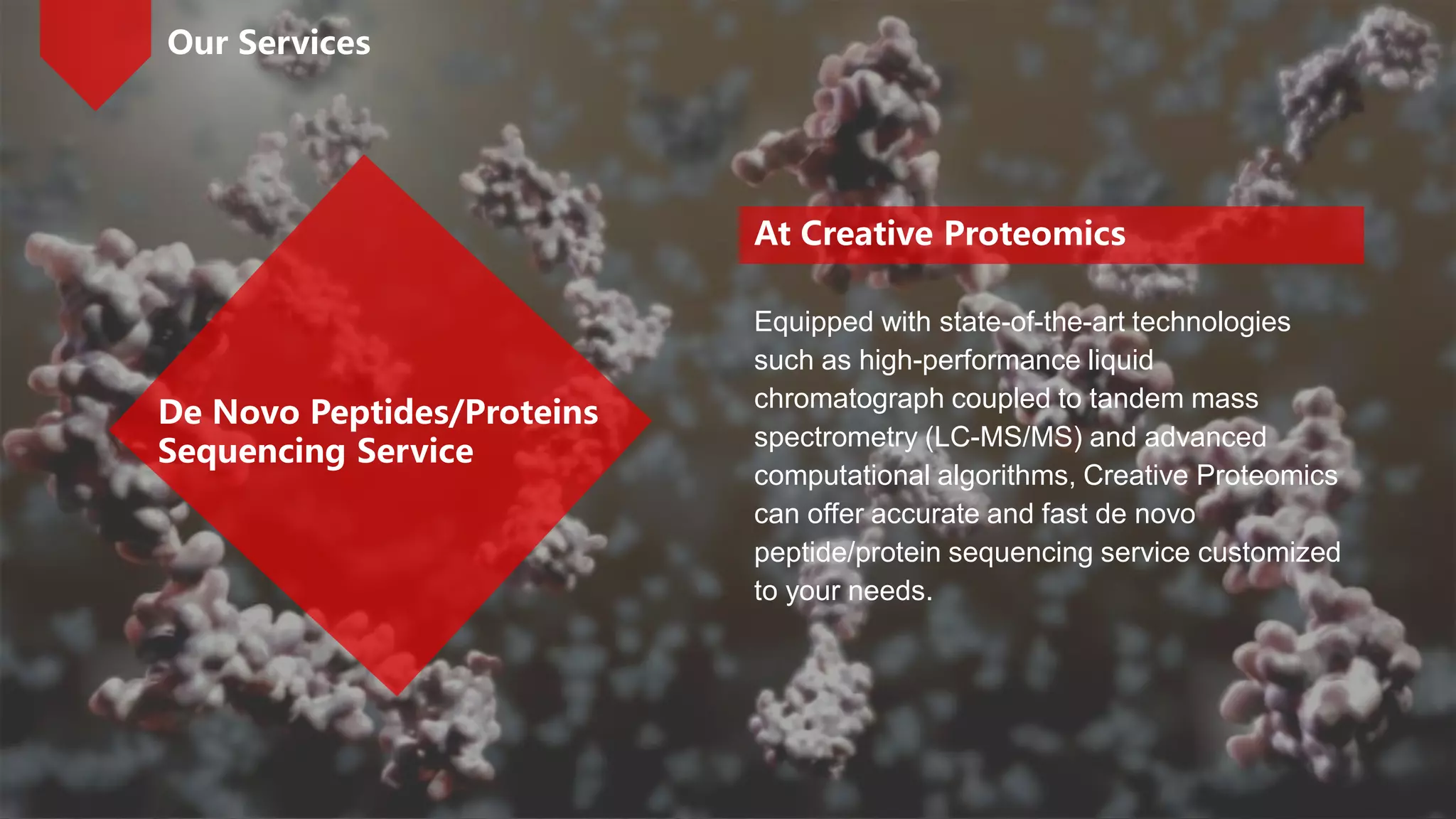
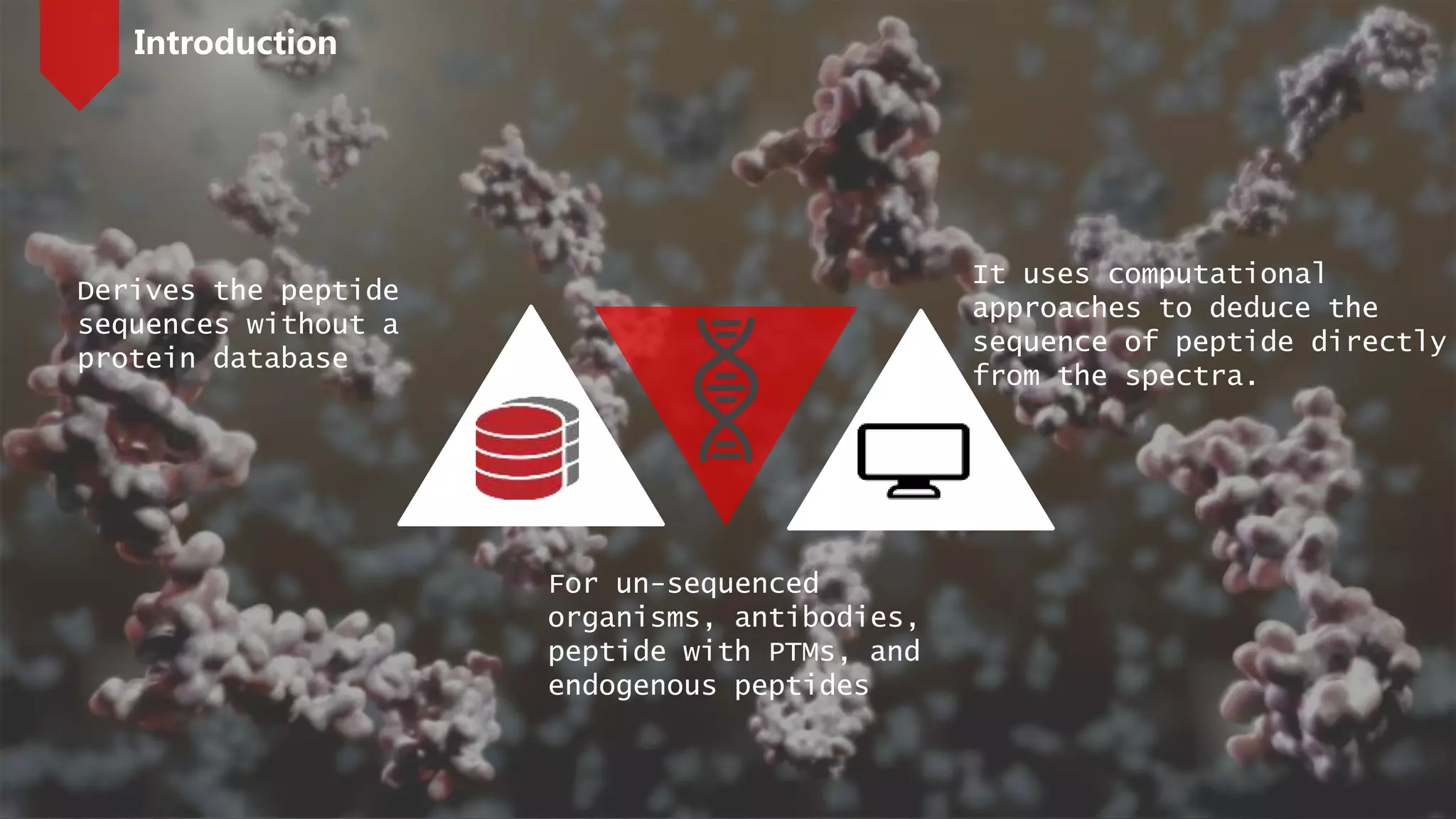
De novo peptide sequencing is performed without prior knowledge of the amino acid sequence. It uses computational approaches to deduce the peptide sequence directly from mass spectrometry spectra. The main principle is to use mass differences between fragment ions like y and b ions to calculate amino acid residues. While it can identify previously unknown sequences, there is uncertainty in the complete sequence and difficulty determining directionality sometimes. Creative Proteomics offers de novo sequencing services using high-performance mass spectrometry and computational algorithms.
This is technique used widely for protein separation from a mixture and is very easy and less costly method. Slides cover all essential points about EMSA and it is quite interesting to know that how it detect and separate different proteins and their mobility shift assay.
This presentation gives you a detailed information about the swiss prot database that comes under UniProtKB. It also covers TrEMBL: a computer annotated supplement to Swiss-Prot.
DNA Protein interaction occur when a protein binds a molecule of DNA, often to regulate the biological function of DNA, usually the expression of a gene. DNA Protein interactions play very vital roles in any living cell. It controls various cellular processes which are very essential for living beings, viz. replication, transcription, recombination, DNA repair etc. There are several types of proteins found in a cell.Direct recognition occurs when the amino acid side chains of a protein interact with specific DNA bases.
Most protein-DNA interactions are mediated by direct physical interaction (hydrogen bonding or hydrophobic interactions) between the protein and the DNA base pairs.
DNA-binding proteins can be identified by many experimental techniques such as chromatin immunoprecipitation on microarrays, X-ray crystallography and nuclear magnetic resonance (NMR).
Primary structure of protein by KK Sahu sirKAUSHAL SAHU
INTRODUCTION
BASIC STRUCTURE OF PROTEIN
TYPES OF PROTEIN
PRIMARY STRUCTURE
IMPORTANCE
SPECIALITY
ROTATION
TRANS ARRANGEMENT
PEPTIDE BOND
CONCLUSIONS
REFERENCES
Cell migration, a key property of live cells, is the process by which cells move from one location to another. There are numerous ways to study cell migrations. Creative Proteomics offers tailored cell migration services and powerful analysis for your research.
https://www.creative-proteomics.com/services/cell-migration-assay.htm
A brief introfuction of label-free protein quantification methodsCreative Proteomics
If you want to know more about our services, please visit https://www.creative-proteomics.com/services/label-free-quantification.htm.
Label-free protein quantification is a mass spectrometry-based method for identifying and quantifying relative changes in two or more biological samples instead of using a stable isotope-containing compound to label proteins.
If you want to know more, please visit https://www.creative-proteomics.com/s...
Stable isotope labeling using amino acids in cell culture (SILAC) is a powerful method based on mass spectrometry that identifies and quantifies relative differential changes in protein abundance. First used in quantitative proteomics in 2002, it provides accurate relative quantification without any chemical derivatization or manipulation.
Mass Spectrometry-Based Proteomics Quantification: iTRAQ Creative Proteomics
For more information, please visit: https://www.creative-proteomics.com/services/itraq-based-proteomics-analysis.htm
iTRAQ (isobaric tag for relative and absolute quantitation), is an isobaric labeling method to determine the amount of proteins from different sources in just one single experiment by mass spectrometry, which was developed by Applied Biosystems Incorporation in 2004.
If you want to know more, please visit https://www.creative-proteomics.com/services/short-chain-fatty-acids-analysis-service.htm. Short chain fatty acids (SCFAs) are defined as fatty acids with two to six carbon atoms. SCFAs have a wide range of metabolic effects. And SCFA profiling has been a major topic in gut bacteria studies.
For more information, you can visit https://www.creative-proteomics.com/services/protein-post-translational-modification-analysis.htm. In this video, we introduce some commonly used methods to detect PPIs and techniques for proteome-scale interactome maps.
Brief Introduction of Protein-Protein Interactions (PPIs)Creative Proteomics
For more information, please visit https://www.creative-proteomics.com/services/protein-protein-interaction-networks.htm. Protein-protein interactions play important roles in various biological processes. PPIs can be classified based on different factors, including composition, affinity, and lifetime.
Peptidomics represents a short version of “peptide proteomics", which means the comprehensive visualization and analysis of small polypeptides, thus covering the mass range between proteomics and metabonomics.
Mass Spectrometry-based Peptidomics for Biomaker DiscoveryCreative Proteomics
Biomarkers are molecules that indicate a physiological state and also the change during a disease process. In human bodies, peptidome biomarkers can be used to forecast disease, diagnose various disorders, guide clinical therapy, and monitor medicine response. The mass spectrometry-based peptidomics for biomarker discovery contains sample preparation, separation, detection and identification, quantitative evaluation, data analysis, as well as biomarkers validation.
Protein phosphorylation, a reversible process, is characterized by adding phosphate donated from ATP and removing phosphate from a phosphorylated protein substrate. For more information, please visit: https://www.creative-proteomics.com/services/phosphorylation.htm
Protein acetylation commonly has two different forms. In humans, almost (80%-90%) proteins become co-translationally acetylated at their Nα-termini of the nascent polypeptide chains. Another type is typically acetylated on lysine residues.
Mass spectrometry (MS) is the suitable method for the analysis of protein modifications because it can provide universal information about protein modifications without a priori knowledge and locating the sites of modification.
If you are interested in our services, please visit: https://www.creative-proteomics.com/services/protein-post-translational-modification-analysis.htm
Brief introduction of post-translational modifications (PTMs)Creative Proteomics
PTMs are chemical alterations to protein structure, typically catalyzed by exceedingly substrate-specific enzymes, which themselves are under strict control by PTMs. They generate a large diversity of gene products because many types of PTMs are covalently attached to amino-acid residues in each protein. For protein post-translational modification analysis at Creative Proteomics, please visit https://www.creative-proteomics.com/services/protein-post-translational-modification-analysis.htm
Glycomics, the study of glycans, is applied to biology and chemistry that focuses on the structure and function of carbohydrates, and on glycoform distributions at the cellular, tissue, organ and organism levels. Mass spectrometry plays an important role in glycomics analysis. If you want to know more, please visit https://www.creative-proteomics.com/services/glycomics-service.htm
Western blot is a commonly used method for protein analysis. It can be used for qualitative and semi-quantitative protein analysis. For the accomplishment of the western blot, there are three elements, separation of proteins by size, transferring proteins to a solid support, and marking proteins by primary and secondary antibodies for visualization.
Two-dimensional gel electrophoresis (2-DE) is considered a powerful tool for proteomics work. 2-DE separates proteins depending on two differ steps: the first one is called isoelectric focusing (IEF) which separates proteins according to isoelectric points (pI); the second step is SDS-polyacrylamide gel electrophoresis (SDS-PAGE) which separates proteins based on the molecular weights.
Our website: www.creative-proteomics.com
Membrane proteins play important roles in various cellular processes, such as cell adhesion, immune response, metabolism and signal transduction. They are popular targets for proteomics research and the common candidates for drug development. Shotgun proteomics methods are available for the identification of membrane proteins.
Proteomics studies play an increasing role in the field of biology. The use of mass spectrometry (MS) in combination with a range of separation methods is the main principal methodology for proteomics. The two principal approaches to identifying and characterizing proteins using MS are the “bottom-up”, which analyze peptides by proteolytic digestion, and “top-down”, which analyze intact proteins.
Peptide mass fingerprinting is a technology to identify proteins. It is a high throughput protein identification technique in which the mass of an unknown protein can be determined. PMF is always performed with MALDI-TOF mass spectrometry
Introduction of mass spectrometer - basic types of ion sourceCreative Proteomics
As we know before, the mass spectrometry consists of three main components, the ion source, the mass analyzer, and the detector. In ion source, a sample is ionized. Today, we are going to introduce several types of ion source, which are usually used in a mass spectrometry.
Multi-source connectivity as the driver of solar wind variability in the heli...Sérgio Sacani
The ambient solar wind that flls the heliosphere originates from multiple
sources in the solar corona and is highly structured. It is often described
as high-speed, relatively homogeneous, plasma streams from coronal
holes and slow-speed, highly variable, streams whose source regions are
under debate. A key goal of ESA/NASA’s Solar Orbiter mission is to identify
solar wind sources and understand what drives the complexity seen in the
heliosphere. By combining magnetic feld modelling and spectroscopic
techniques with high-resolution observations and measurements, we show
that the solar wind variability detected in situ by Solar Orbiter in March
2022 is driven by spatio-temporal changes in the magnetic connectivity to
multiple sources in the solar atmosphere. The magnetic feld footpoints
connected to the spacecraft moved from the boundaries of a coronal hole
to one active region (12961) and then across to another region (12957). This
is refected in the in situ measurements, which show the transition from fast
to highly Alfvénic then to slow solar wind that is disrupted by the arrival of
a coronal mass ejection. Our results describe solar wind variability at 0.5 au
but are applicable to near-Earth observatories.
Nutraceutical market, scope and growth: Herbal drug technologyLokesh Patil
As consumer awareness of health and wellness rises, the nutraceutical market—which includes goods like functional meals, drinks, and dietary supplements that provide health advantages beyond basic nutrition—is growing significantly. As healthcare expenses rise, the population ages, and people want natural and preventative health solutions more and more, this industry is increasing quickly. Further driving market expansion are product formulation innovations and the use of cutting-edge technology for customized nutrition. With its worldwide reach, the nutraceutical industry is expected to keep growing and provide significant chances for research and investment in a number of categories, including vitamins, minerals, probiotics, and herbal supplements.
Earliest Galaxies in the JADES Origins Field: Luminosity Function and Cosmic ...Sérgio Sacani
We characterize the earliest galaxy population in the JADES Origins Field (JOF), the deepest
imaging field observed with JWST. We make use of the ancillary Hubble optical images (5 filters
spanning 0.4−0.9µm) and novel JWST images with 14 filters spanning 0.8−5µm, including 7 mediumband filters, and reaching total exposure times of up to 46 hours per filter. We combine all our data
at > 2.3µm to construct an ultradeep image, reaching as deep as ≈ 31.4 AB mag in the stack and
30.3-31.0 AB mag (5σ, r = 0.1” circular aperture) in individual filters. We measure photometric
redshifts and use robust selection criteria to identify a sample of eight galaxy candidates at redshifts
z = 11.5 − 15. These objects show compact half-light radii of R1/2 ∼ 50 − 200pc, stellar masses of
M⋆ ∼ 107−108M⊙, and star-formation rates of SFR ∼ 0.1−1 M⊙ yr−1
. Our search finds no candidates
at 15 < z < 20, placing upper limits at these redshifts. We develop a forward modeling approach to
infer the properties of the evolving luminosity function without binning in redshift or luminosity that
marginalizes over the photometric redshift uncertainty of our candidate galaxies and incorporates the
impact of non-detections. We find a z = 12 luminosity function in good agreement with prior results,
and that the luminosity function normalization and UV luminosity density decline by a factor of ∼ 2.5
from z = 12 to z = 14. We discuss the possible implications of our results in the context of theoretical
models for evolution of the dark matter halo mass function.
This presentation explores a brief idea about the structural and functional attributes of nucleotides, the structure and function of genetic materials along with the impact of UV rays and pH upon them.
Comparing Evolved Extractive Text Summary Scores of Bidirectional Encoder Rep...University of Maribor
Slides from:
11th International Conference on Electrical, Electronics and Computer Engineering (IcETRAN), Niš, 3-6 June 2024
Track: Artificial Intelligence
https://www.etran.rs/2024/en/home-english/
Deep Behavioral Phenotyping in Systems Neuroscience for Functional Atlasing a...Ana Luísa Pinho
Functional Magnetic Resonance Imaging (fMRI) provides means to characterize brain activations in response to behavior. However, cognitive neuroscience has been limited to group-level effects referring to the performance of specific tasks. To obtain the functional profile of elementary cognitive mechanisms, the combination of brain responses to many tasks is required. Yet, to date, both structural atlases and parcellation-based activations do not fully account for cognitive function and still present several limitations. Further, they do not adapt overall to individual characteristics. In this talk, I will give an account of deep-behavioral phenotyping strategies, namely data-driven methods in large task-fMRI datasets, to optimize functional brain-data collection and improve inference of effects-of-interest related to mental processes. Key to this approach is the employment of fast multi-functional paradigms rich on features that can be well parametrized and, consequently, facilitate the creation of psycho-physiological constructs to be modelled with imaging data. Particular emphasis will be given to music stimuli when studying high-order cognitive mechanisms, due to their ecological nature and quality to enable complex behavior compounded by discrete entities. I will also discuss how deep-behavioral phenotyping and individualized models applied to neuroimaging data can better account for the subject-specific organization of domain-general cognitive systems in the human brain. Finally, the accumulation of functional brain signatures brings the possibility to clarify relationships among tasks and create a univocal link between brain systems and mental functions through: (1) the development of ontologies proposing an organization of cognitive processes; and (2) brain-network taxonomies describing functional specialization. To this end, tools to improve commensurability in cognitive science are necessary, such as public repositories, ontology-based platforms and automated meta-analysis tools. I will thus discuss some brain-atlasing resources currently under development, and their applicability in cognitive as well as clinical neuroscience.
This pdf is about the Schizophrenia.
For more details visit on YouTube; @SELF-EXPLANATORY;
https://www.youtube.com/channel/UCAiarMZDNhe1A3Rnpr_WkzA/videos
Thanks...!
2. De Novo Peptide Sequencing: peptide sequencing performed
without prior knowledge of the amino acid sequence
De novo: Latin for “over again” or
“anew”
De Novo Peptide Sequencing
3. Derives the peptide
sequences without a
protein database
It uses computational
approaches to deduce the
sequence of peptide directly
from the spectra.
For un-sequenced
organisms, antibodies,
peptide with PTMs, and
endogenous peptides
Introduction
4. 3 types of backbone bonds can be broken to
form peptide fragments: alkyl carbonyl (CHR-
CO), peptide amide bond (CO-NH), and amino
alkyl bond (NH-CHR)
6 types of fragmentation ions: the N-terminal
charged fragment ions are classed as a, b or c;
the C-terminal charged ones are classed as x,
y or z
Because the peptide amide bone (CO-NH) is
the most vulnerable, the most common peptide
fragments observed in low energy collisions
are a, b and y ions
Peptide fragmentation
Types of fragmentation ions
Figure 1. Different types of fragmentation ions
Ma B, Johnson R. De novo sequencing and homology searching[J]. Molecular & cellular proteomics, 2012, 11(2): O111. 014902.
5. Collision induced dissociation (CID)
Peptide fragmentation
Methods for peptide fragmentation
Electron capture dissociation (ETD ) and Electron transfer dissociation
(ECD)
• Also known as collisionally activated dissociation(CAD)
• The most common form of fragmentation
• Ions obtain high kinetic energy and collide with neutral molecules. Some
of the kinetic energy is converted into internal energy which leads to
bond breakage and the fragmentation of the molecules into smaller
fragments
• Result in the formation of b and y series ions from the precursor ion
• Have been implemented in the recent mass spectrometer
• Ions are fragmented after reaction with electrons.
• Form c and z type ions through cleavage of the peptide bond between
the amino group and alpha carbon
Figure 2 Fragment ion types produced following either CAD or ETD
Coon J J. Collisions or electrons? Protein sequence analysis in the 21st
century. 2009.
6. The mass can usually uniquely determine the residue. The main principle of de novo sequencing is to use the mass
difference between two fragment ions to calculate the mass of an amino acid residue on the peptide backbone.
Principle
For example, the mass difference between the y7 and y6 ions in the following figure is equal to 101, which is the mass
of residue T.
7. Principle
Thus, if one can identify either the y-ion or b-ion series in the spectrum, the peptide sequence can be
determined. However, the spectrum obtained from the mass spectrometry instrument does not tell the ion
types of the peaks, which need either an expert or a computer algorithm to figure out.
There are couples of software packages used for de novo sequencing, such as PEAKS, Lutefisk,
PepNovo, SHERENGA and so on.
Notes: y and b ion fragments which contain the amino acid residues R, K, Q, and N may appear to lose
ammonia(-17). Y and b ion fragments which contain the amino acid residues S, T, and E may appear to
lose water(-18).
8. Advantages
Identify previous unknown
peptide sequences
Search for posttranslation
modifications or for the
identification of mutations by
homology based software
Disadvantages
Uncertainty regarding the
complete peptide sequence.
Sometimes it can be difficult
to determine the
directionality of a sequence
Low mass accuracy fragment
ion measurements cannot
distinguish between lysine
and glutamine (differ by
0.036 Da) nor between
phenylalanine and oxidized
methionine (differ by 0.033
Da).
Advantages and Disadvantages
9. At Creative Proteomics
Equipped with state-of-the-art technologies
such as high-performance liquid
chromatograph coupled to tandem mass
spectrometry (LC-MS/MS) and advanced
computational algorithms, Creative Proteomics
can offer accurate and fast de novo
peptide/protein sequencing service customized
to your needs.
Our Services
De Novo Peptides/Proteins
Sequencing Service
10. Thank you
Please contact us for more information
Web
Email
www.creative-proteomics.com
info@creative-proteomics.com
Editor's Notes
Hello, welcome to watch Creative Proteomics’Video. As we know, through tandem mass spectrometry is one of the most powerful tool for protein identification. Today, we are going to learn some basic knowledge about de novo peptide sequencing.
De novo is Latin which means “over again” or “anew”. The de novo peptide sequencing is a method for peptide sequencing performed without prior knowledge of the amino acid sequence.
This method can obtain the peptide sequences without a protein database, which can overcome the limitations of database-dependent methods like peptide mass fingerprinting. It can be used for un-sequencd organisms, antibodies, peptide with posttranslational modifications (PTMs), and endogenous peptides. In addition, it uses computational approaches to deduce the sequence of peptide directly from the experimental MS/MS spectra.
In a tandem mass spectrometer, the peptide is fragmented along the peptide backbone and the resulting fragment ions are measured to produce spectrum. There are 3 ways to break bonds to form peptide fragment: alkyl carbonyl (CHR-CO), peptide amide bond (CO-NH), and amino alkyl bond (NH-CHR) Therefore, it can form 6 types of fragmentation ions, including the N-terminal charged fragment ions which are classed as a, b or c, and the C-terminal charged ones which are classed as x, y or z. And because the peptide amide bone (CO-NH) is the most vulnerable, the most common peptide fragments observed in low energy collisions are a, b and y ions
De novo methods use the knowledge of the fragmentation methods employed in the MS, such as CID, or ECD. CID, Collision induced dissociation, also known as collisionally activated dissociation, is the most common form of fragmentation. In this method, The ions can obtain high kinetic energy and collide with neutral molecules. Some of the kinetic energy is converted into internal energy which leads to bond breakage and the fragmentation of the molecules into smaller fragments. This method results in the formation of b and y series ions from the precursor ion. The Electron capture dissociation, ETD and Electron transfer dissociation, ECD, have been implemented in the recent mass spectrometer. In these methods, Ions are fragmented after reaction with electrons. After fragmentation, it Forms c and z type ions through cleavage of the peptide bond between the amino group and alpha carbon.
The mass can usually uniquely determine the residue. The main principle of de novo sequencing is to use the mass difference between two fragment ions to calculate the mass of an amino acid residue on the peptide backbone. For example, the mass difference between the y7 and y6 ions in the following figure is equal to 101, which is the mass of residue T.
Thus, if one can identify either the y-ion or b-ion series in the spectrum, the peptide sequence can be determined. However, the spectrum obtained from the mass spectrometry instrument does not tell the ion types of the peaks, which require either a human expert or a computer algorithm to figure out during the process of de novo sequencing. There are couples of software packages used for de novo sequencing, such as PEAKS, Lutefisk, PepNovo, SHERENGA and so on.But there are some notes you have to mind. y and b ion fragments which contain the amino acid residues R,K,Q, and N may appear to loose ammonia(-17). y and b ion fragments which contain the amino acid residues S, T, and E may appear to lose water(-18).
De novo sequencing can identify previous unknown peptide sequences. In addition, it can search for posttranslational modifications or for identifications of mutations by homology based software. however, de novo sequencing will not be able to derive a complete sequence or will have uncertainty in a portion of the derived sequence. And sometimes it can be difficult to determine the directionality of a sequence. Low mass accuracy fragment ion measurements cannot distinguish between lysine and glutamine which differ by 0.036 Da nor between phenylalanine and oxidized methionine which differ by 0.033 Da.
At Creative Proteomics, we can provide de novo peptides or protein service. Equipped with state-of-the-art technologies such as high-performance liquid chromatograph coupled to tandem mass spectrometry (LC-MS/MS) and advanced computational algorithms, we can offer accurate and fast de novo peptide/protein sequencing service customized to your needs.
Thanks for watching our video. At creative proteomics, we provide the most reliable services for protein identification not only de novo peptide sequencing, but some other technologies. If you have any questions or specific requirements. Please do not hesitate to contact us. We are very glad to cooperate with you.