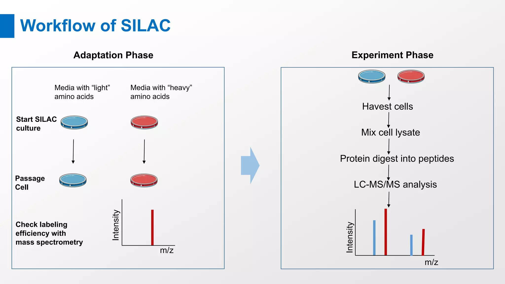
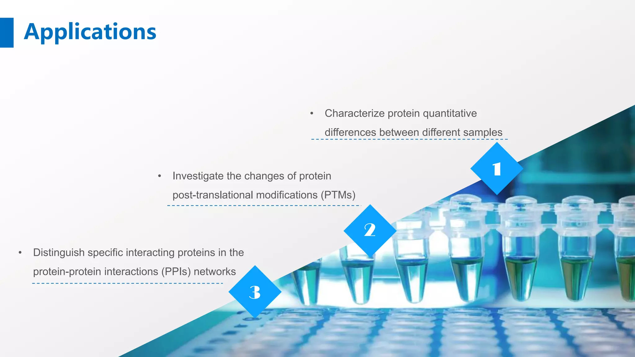
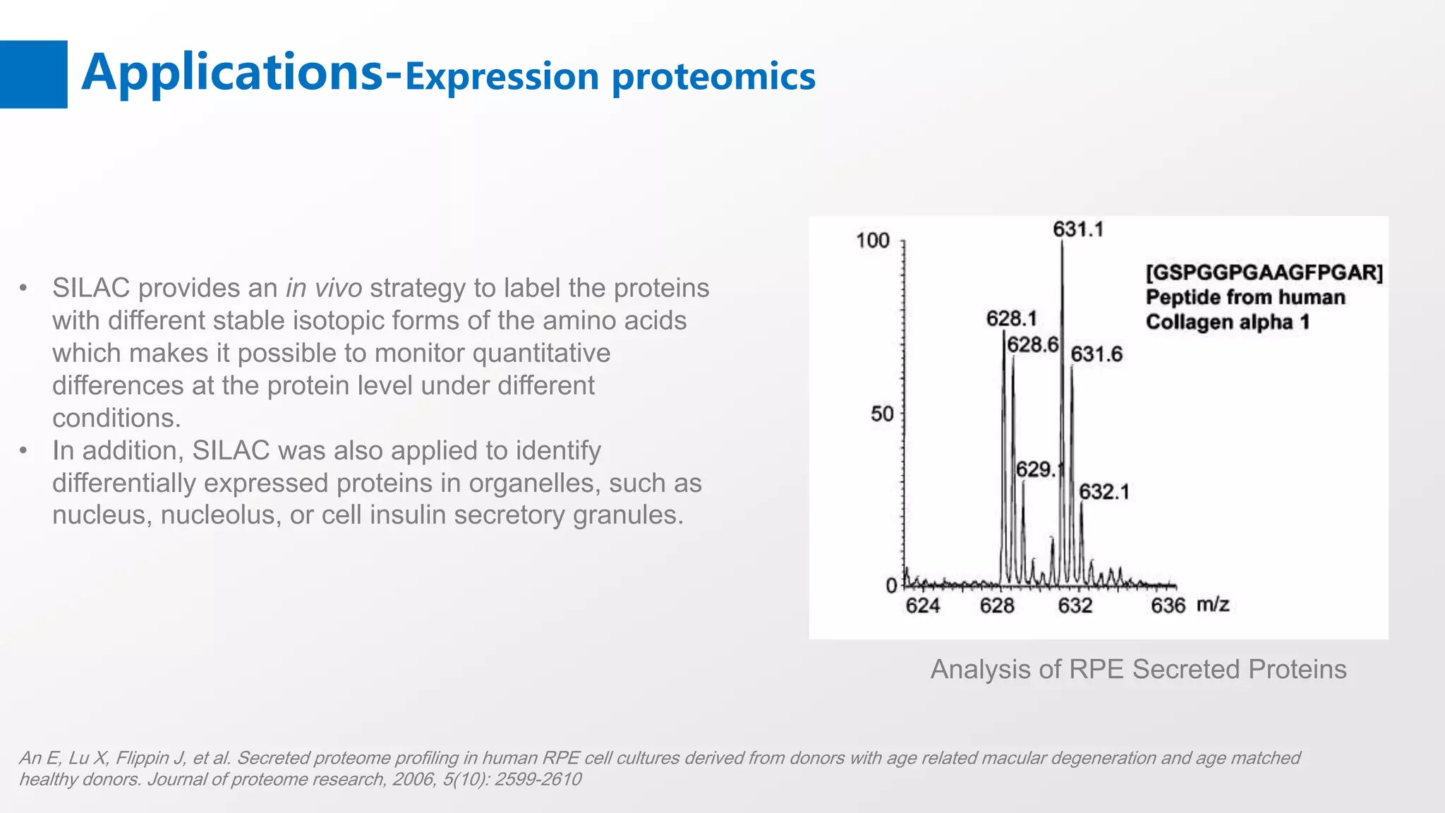
If you want to know more, please visit https://www.creative-proteomics.com/s...
Stable isotope labeling using amino acids in cell culture (SILAC) is a powerful method based on mass spectrometry that identifies and quantifies relative differential changes in protein abundance. First used in quantitative proteomics in 2002, it provides accurate relative quantification without any chemical derivatization or manipulation.
Secondary Structure Prediction of proteins Vijay Hemmadi
Secondary structure prediction has been around for almost a quarter of a century. The early methods suffered from a lack of data. Predictions were performed on single sequences rather than families of homologous sequences, and there were relatively few known 3D structures from which to derive parameters. Probably the most famous early methods are those of Chou & Fasman, Garnier, Osguthorbe & Robson (GOR) and Lim. Although the authors originally claimed quite high accuracies (70-80 %), under careful examination, the methods were shown to be only between 56 and 60% accurate (see Kabsch & Sander, 1984 given below). An early problem in secondary structure prediction had been the inclusion of structures used to derive parameters in the set of structures used to assess the accuracy of the method.
Some good references on the subject:
it will help you to understand how the protein microarrays are made, what are the different types and what all purposes they are used for. its very useful ppt
Peptide mass fingerprinting is a technology to identify proteins. It is a high throughput protein identification technique in which the mass of an unknown protein can be determined. PMF is always performed with MALDI-TOF mass spectrometry
2D-PAGE is a method is used for the separation and identification of proteins in a complex mixture using two separate dimensions that are run perpendicular to one another.
2D-DIGE is an advanced version of classical two-dimensional gel electrophoresis (2D-PAGE).
The protein samples are labeled with fluorescent dyes and then separated by 2D-PAGE.
The three hybrid system of yeast has been described in this ppt. Yeast one Hybrid system, yeast two hybrid system and yeast 3 hybrid system is explained. This explain about the DNA-protein interaction and Protein-DNA-Protein interaction.
protein microarray_k.b institute (m.pharm pharmacology) .pptxNittalVekaria
1: Introduction
Welcome to our presentation on Protein Microarrays.
Discover the revolutionary technology transforming protein analysis and biomolecular research
2: What are Protein Microarrays?
Protein microarrays are high-throughput platforms for studying protein-protein interactions, protein function, and biomarker discovery.
They consist of thousands of immobilized proteins on a solid surface, allowing for simultaneous analysis of multiple proteins.
3Components of Protein Microarrays
Substrate: Glass slides, membranes, or beads.
Proteins: Target proteins immobilized on the substrate.
Detection System: Fluorescent dyes, antibodies, or other probes.
Imaging System: Scanners or cameras for data acquisition.
4: Types of Protein Microarrays
Analytical Microarrays: Used for studying protein-protein interactions, protein expression profiling, and protein function analysis.
Antibody Microarrays: Utilized for detecting and quantifying specific proteins or antibodies in biological samples.
Reverse-Phase Protein Arrays (RPPAs): Designed for high-throughput protein expression profiling and signaling pathway analysis.
5:Applications of Protein Microarrays
Biomarker Discovery: Identification of disease-specific biomarkers for diagnosis, prognosis, and treatment monitoring.
Drug Discovery: High-throughput screening of drug candidates and target validation.
Functional Proteomics: Mapping protein-protein interactions, post-translational modifications, and protein function analysis.
Clinical Diagnostics: Detection of infectious diseases, cancer biomarkers, and autoimmune disorders.
6: Workflow of Protein Microarray Experiment
Protein immobilization: Spotting or printing target proteins onto the microarray substrate.
Sample incubation: Incubating the microarray with biological samples containing proteins of interest.
Detection and analysis: Using fluorescent probes or antibodies to detect bound proteins and quantifying the signals.
Data interpretation: Analyzing and interpreting the results to extract meaningful biological insights.
7: Advantages of Protein Microarrays
-High-throughput analysis of thousands of proteins in parallel.
Small sample volume requirement.
Enables multiplexed assays for comprehensive protein profiling.
Facilitates rapid biomarker discovery and validation.
8: Challenges and Considerations
Standardization of protocols and reagents.
Optimization of protein immobilization and detection methods.
Data analysis and interpretation complexities.
Cost and accessibility of microarray platforms.
9: Future Perspectives
Integration with other omics technologies for holistic biological insights.
Development of miniaturized and portable microarray platforms for point-of-care diagnostics.
Advancements in data analysis algorithms and bioinformatics tools.
Expanding applications in personalized medicine and precision healthcare
10: Conclusion
Protein microarrays offer a powerful and versatile tool for protein analysis and biomarker discover
Cytokines are a class of highly active, multifunctional, soluble small-molecule proteins secreted by activated immune cells and certain stromal cells. Cytokines are widely involved in various biological functions such as immune response, cell migration, and signal transduction through paracrine, autocrine, and endocrine approaches. Cytokine assays can assist in determining the immune function of the body and help in research related to the disease mechanism, diagnosis, and treatment. There are various assays for cytokines, and you can choose the most appropriate assay according to sample size, assay needs, and budget. https://cytokine.creative-proteomics.com/luminex-cytokine-detection-service.htm
Cytokines are a class of highly active, multifunctional, soluble small-molecule proteins secreted by activated immune cells and specific stromal cells. Cytokines are widely involved in various biological functions such as immune response, cell migration, and signal transduction through paracrine, autocrine, and endocrine approaches. Cytokine assays can assist in determining the immune function of the body and help in research related to the disease mechanism, diagnosis, and treatment. There are various assays for cytokines, and you can choose the most appropriate assay according to sample size, assay needs, and budget. https://cytokine.creative-proteomics.com/cytokine-panel-service.htm
Drug Metabolism (DMPK) Assays | MicroConstantsMicroConstants
MicroConstants performs industry-standard assays, custom drug metabolism research, and IND-enabling studies to assess drug-drug interaction potential, metabolic stability, metabolite formation, and protein binding. Whether you are in discovery, lead optimization, or collecting data for regulatory submissions, we will work with you to define the level of research appropriate for your compounds. Results can be presented as a formal report suitable for regulatory submissions, or as an informal report (i.e. raw data tables in Excel).
For more information, you can visit https://www.creative-proteomics.com/services/protein-post-translational-modification-analysis.htm. In this video, we introduce some commonly used methods to detect PPIs and techniques for proteome-scale interactome maps.
Recent analysis method of food allergen rakesh mund
Recent analysis method of food allergen
and case study related to recent method for food allergen analysis
rakesh kumar
Msc scholar IARI PUSA campus new delhi
8385884641 ; rakeshmund94@gmail.com
This presentation gives an insight into the mammalian cell being used as an expression system, it also includes a brief introduction to the strong promoters.
Proteomics: types, protein profiling steps etc.Cherry
Proteome is a set of proteins produced in an organism, system, or biological context or entire set of proteins that is, or can be, expressed by a genome, cell, tissue, or organism at a certain expressed time in a given set of condition. Proteomics is the study of all the proteins produced by a cell.
Cell migration, a key property of live cells, is the process by which cells move from one location to another. There are numerous ways to study cell migrations. Creative Proteomics offers tailored cell migration services and powerful analysis for your research.
https://www.creative-proteomics.com/services/cell-migration-assay.htm
A brief introfuction of label-free protein quantification methodsCreative Proteomics
If you want to know more about our services, please visit https://www.creative-proteomics.com/services/label-free-quantification.htm.
Label-free protein quantification is a mass spectrometry-based method for identifying and quantifying relative changes in two or more biological samples instead of using a stable isotope-containing compound to label proteins.
Mass Spectrometry-Based Proteomics Quantification: iTRAQ Creative Proteomics
For more information, please visit: https://www.creative-proteomics.com/services/itraq-based-proteomics-analysis.htm
iTRAQ (isobaric tag for relative and absolute quantitation), is an isobaric labeling method to determine the amount of proteins from different sources in just one single experiment by mass spectrometry, which was developed by Applied Biosystems Incorporation in 2004.
If you want to know more, please visit https://www.creative-proteomics.com/services/short-chain-fatty-acids-analysis-service.htm. Short chain fatty acids (SCFAs) are defined as fatty acids with two to six carbon atoms. SCFAs have a wide range of metabolic effects. And SCFA profiling has been a major topic in gut bacteria studies.
Brief Introduction of Protein-Protein Interactions (PPIs)Creative Proteomics
For more information, please visit https://www.creative-proteomics.com/services/protein-protein-interaction-networks.htm. Protein-protein interactions play important roles in various biological processes. PPIs can be classified based on different factors, including composition, affinity, and lifetime.
Peptidomics represents a short version of “peptide proteomics", which means the comprehensive visualization and analysis of small polypeptides, thus covering the mass range between proteomics and metabonomics.
Mass Spectrometry-based Peptidomics for Biomaker DiscoveryCreative Proteomics
Biomarkers are molecules that indicate a physiological state and also the change during a disease process. In human bodies, peptidome biomarkers can be used to forecast disease, diagnose various disorders, guide clinical therapy, and monitor medicine response. The mass spectrometry-based peptidomics for biomarker discovery contains sample preparation, separation, detection and identification, quantitative evaluation, data analysis, as well as biomarkers validation.
Protein phosphorylation, a reversible process, is characterized by adding phosphate donated from ATP and removing phosphate from a phosphorylated protein substrate. For more information, please visit: https://www.creative-proteomics.com/services/phosphorylation.htm
Protein acetylation commonly has two different forms. In humans, almost (80%-90%) proteins become co-translationally acetylated at their Nα-termini of the nascent polypeptide chains. Another type is typically acetylated on lysine residues.
Mass spectrometry (MS) is the suitable method for the analysis of protein modifications because it can provide universal information about protein modifications without a priori knowledge and locating the sites of modification.
If you are interested in our services, please visit: https://www.creative-proteomics.com/services/protein-post-translational-modification-analysis.htm
Brief introduction of post-translational modifications (PTMs)Creative Proteomics
PTMs are chemical alterations to protein structure, typically catalyzed by exceedingly substrate-specific enzymes, which themselves are under strict control by PTMs. They generate a large diversity of gene products because many types of PTMs are covalently attached to amino-acid residues in each protein. For protein post-translational modification analysis at Creative Proteomics, please visit https://www.creative-proteomics.com/services/protein-post-translational-modification-analysis.htm
Glycomics, the study of glycans, is applied to biology and chemistry that focuses on the structure and function of carbohydrates, and on glycoform distributions at the cellular, tissue, organ and organism levels. Mass spectrometry plays an important role in glycomics analysis. If you want to know more, please visit https://www.creative-proteomics.com/services/glycomics-service.htm
Western blot is a commonly used method for protein analysis. It can be used for qualitative and semi-quantitative protein analysis. For the accomplishment of the western blot, there are three elements, separation of proteins by size, transferring proteins to a solid support, and marking proteins by primary and secondary antibodies for visualization.
Two-dimensional gel electrophoresis (2-DE) is considered a powerful tool for proteomics work. 2-DE separates proteins depending on two differ steps: the first one is called isoelectric focusing (IEF) which separates proteins according to isoelectric points (pI); the second step is SDS-polyacrylamide gel electrophoresis (SDS-PAGE) which separates proteins based on the molecular weights.
Our website: www.creative-proteomics.com
Membrane proteins play important roles in various cellular processes, such as cell adhesion, immune response, metabolism and signal transduction. They are popular targets for proteomics research and the common candidates for drug development. Shotgun proteomics methods are available for the identification of membrane proteins.
The de novo peptide sequencing is a method for peptide sequencing performed without prior knowledge of the amino acid sequence. It uses computational approaches to deduce the sequence of peptide directly from the experimental MS/MS spectra.This method can obtain the peptide sequences without a protein database. It can be used for un-sequenced organisms, antibodies, peptides with posttranslational modifications, and endogenous peptides.
Proteomics studies play an increasing role in the field of biology. The use of mass spectrometry (MS) in combination with a range of separation methods is the main principal methodology for proteomics. The two principal approaches to identifying and characterizing proteins using MS are the “bottom-up”, which analyze peptides by proteolytic digestion, and “top-down”, which analyze intact proteins.
Introduction of mass spectrometer - basic types of ion sourceCreative Proteomics
As we know before, the mass spectrometry consists of three main components, the ion source, the mass analyzer, and the detector. In ion source, a sample is ionized. Today, we are going to introduce several types of ion source, which are usually used in a mass spectrometry.
Introduction of mass spectrometer - basic types of mass analyzer Creative Proteomics
The mass analyzer is the heart of the mass spectrometer, which takes ionized masses and separates them based on mass to charge ratios. There are several general types of mass analyzers, including magnetic sector, time of flight, quadrupole, ion trap
A brief information about the SCOP protein database used in bioinformatics.
The Structural Classification of Proteins (SCOP) database is a comprehensive and authoritative resource for the structural and evolutionary relationships of proteins. It provides a detailed and curated classification of protein structures, grouping them into families, superfamilies, and folds based on their structural and sequence similarities.
Slide 1: Title Slide
Extrachromosomal Inheritance
Slide 2: Introduction to Extrachromosomal Inheritance
Definition: Extrachromosomal inheritance refers to the transmission of genetic material that is not found within the nucleus.
Key Components: Involves genes located in mitochondria, chloroplasts, and plasmids.
Slide 3: Mitochondrial Inheritance
Mitochondria: Organelles responsible for energy production.
Mitochondrial DNA (mtDNA): Circular DNA molecule found in mitochondria.
Inheritance Pattern: Maternally inherited, meaning it is passed from mothers to all their offspring.
Diseases: Examples include Leber’s hereditary optic neuropathy (LHON) and mitochondrial myopathy.
Slide 4: Chloroplast Inheritance
Chloroplasts: Organelles responsible for photosynthesis in plants.
Chloroplast DNA (cpDNA): Circular DNA molecule found in chloroplasts.
Inheritance Pattern: Often maternally inherited in most plants, but can vary in some species.
Examples: Variegation in plants, where leaf color patterns are determined by chloroplast DNA.
Slide 5: Plasmid Inheritance
Plasmids: Small, circular DNA molecules found in bacteria and some eukaryotes.
Features: Can carry antibiotic resistance genes and can be transferred between cells through processes like conjugation.
Significance: Important in biotechnology for gene cloning and genetic engineering.
Slide 6: Mechanisms of Extrachromosomal Inheritance
Non-Mendelian Patterns: Do not follow Mendel’s laws of inheritance.
Cytoplasmic Segregation: During cell division, organelles like mitochondria and chloroplasts are randomly distributed to daughter cells.
Heteroplasmy: Presence of more than one type of organellar genome within a cell, leading to variation in expression.
Slide 7: Examples of Extrachromosomal Inheritance
Four O’clock Plant (Mirabilis jalapa): Shows variegated leaves due to different cpDNA in leaf cells.
Petite Mutants in Yeast: Result from mutations in mitochondrial DNA affecting respiration.
Slide 8: Importance of Extrachromosomal Inheritance
Evolution: Provides insight into the evolution of eukaryotic cells.
Medicine: Understanding mitochondrial inheritance helps in diagnosing and treating mitochondrial diseases.
Agriculture: Chloroplast inheritance can be used in plant breeding and genetic modification.
Slide 9: Recent Research and Advances
Gene Editing: Techniques like CRISPR-Cas9 are being used to edit mitochondrial and chloroplast DNA.
Therapies: Development of mitochondrial replacement therapy (MRT) for preventing mitochondrial diseases.
Slide 10: Conclusion
Summary: Extrachromosomal inheritance involves the transmission of genetic material outside the nucleus and plays a crucial role in genetics, medicine, and biotechnology.
Future Directions: Continued research and technological advancements hold promise for new treatments and applications.
Slide 11: Questions and Discussion
Invite Audience: Open the floor for any questions or further discussion on the topic.
Introduction:
RNA interference (RNAi) or Post-Transcriptional Gene Silencing (PTGS) is an important biological process for modulating eukaryotic gene expression.
It is highly conserved process of posttranscriptional gene silencing by which double stranded RNA (dsRNA) causes sequence-specific degradation of mRNA sequences.
dsRNA-induced gene silencing (RNAi) is reported in a wide range of eukaryotes ranging from worms, insects, mammals and plants.
This process mediates resistance to both endogenous parasitic and exogenous pathogenic nucleic acids, and regulates the expression of protein-coding genes.
What are small ncRNAs?
micro RNA (miRNA)
short interfering RNA (siRNA)
Properties of small non-coding RNA:
Involved in silencing mRNA transcripts.
Called “small” because they are usually only about 21-24 nucleotides long.
Synthesized by first cutting up longer precursor sequences (like the 61nt one that Lee discovered).
Silence an mRNA by base pairing with some sequence on the mRNA.
Discovery of siRNA?
The first small RNA:
In 1993 Rosalind Lee (Victor Ambros lab) was studying a non- coding gene in C. elegans, lin-4, that was involved in silencing of another gene, lin-14, at the appropriate time in the
development of the worm C. elegans.
Two small transcripts of lin-4 (22nt and 61nt) were found to be complementary to a sequence in the 3' UTR of lin-14.
Because lin-4 encoded no protein, she deduced that it must be these transcripts that are causing the silencing by RNA-RNA interactions.
Types of RNAi ( non coding RNA)
MiRNA
Length (23-25 nt)
Trans acting
Binds with target MRNA in mismatch
Translation inhibition
Si RNA
Length 21 nt.
Cis acting
Bind with target Mrna in perfect complementary sequence
Piwi-RNA
Length ; 25 to 36 nt.
Expressed in Germ Cells
Regulates trnasposomes activity
MECHANISM OF RNAI:
First the double-stranded RNA teams up with a protein complex named Dicer, which cuts the long RNA into short pieces.
Then another protein complex called RISC (RNA-induced silencing complex) discards one of the two RNA strands.
The RISC-docked, single-stranded RNA then pairs with the homologous mRNA and destroys it.
THE RISC COMPLEX:
RISC is large(>500kD) RNA multi- protein Binding complex which triggers MRNA degradation in response to MRNA
Unwinding of double stranded Si RNA by ATP independent Helicase
Active component of RISC is Ago proteins( ENDONUCLEASE) which cleave target MRNA.
DICER: endonuclease (RNase Family III)
Argonaute: Central Component of the RNA-Induced Silencing Complex (RISC)
One strand of the dsRNA produced by Dicer is retained in the RISC complex in association with Argonaute
ARGONAUTE PROTEIN :
1.PAZ(PIWI/Argonaute/ Zwille)- Recognition of target MRNA
2.PIWI (p-element induced wimpy Testis)- breaks Phosphodiester bond of mRNA.)RNAse H activity.
MiRNA:
The Double-stranded RNAs are naturally produced in eukaryotic cells during development, and they have a key role in regulating gene expression .
Seminar of U.V. Spectroscopy by SAMIR PANDASAMIR PANDA
Spectroscopy is a branch of science dealing the study of interaction of electromagnetic radiation with matter.
Ultraviolet-visible spectroscopy refers to absorption spectroscopy or reflect spectroscopy in the UV-VIS spectral region.
Ultraviolet-visible spectroscopy is an analytical method that can measure the amount of light received by the analyte.
Earliest Galaxies in the JADES Origins Field: Luminosity Function and Cosmic ...Sérgio Sacani
We characterize the earliest galaxy population in the JADES Origins Field (JOF), the deepest
imaging field observed with JWST. We make use of the ancillary Hubble optical images (5 filters
spanning 0.4−0.9µm) and novel JWST images with 14 filters spanning 0.8−5µm, including 7 mediumband filters, and reaching total exposure times of up to 46 hours per filter. We combine all our data
at > 2.3µm to construct an ultradeep image, reaching as deep as ≈ 31.4 AB mag in the stack and
30.3-31.0 AB mag (5σ, r = 0.1” circular aperture) in individual filters. We measure photometric
redshifts and use robust selection criteria to identify a sample of eight galaxy candidates at redshifts
z = 11.5 − 15. These objects show compact half-light radii of R1/2 ∼ 50 − 200pc, stellar masses of
M⋆ ∼ 107−108M⊙, and star-formation rates of SFR ∼ 0.1−1 M⊙ yr−1
. Our search finds no candidates
at 15 < z < 20, placing upper limits at these redshifts. We develop a forward modeling approach to
infer the properties of the evolving luminosity function without binning in redshift or luminosity that
marginalizes over the photometric redshift uncertainty of our candidate galaxies and incorporates the
impact of non-detections. We find a z = 12 luminosity function in good agreement with prior results,
and that the luminosity function normalization and UV luminosity density decline by a factor of ∼ 2.5
from z = 12 to z = 14. We discuss the possible implications of our results in the context of theoretical
models for evolution of the dark matter halo mass function.
This presentation explores a brief idea about the structural and functional attributes of nucleotides, the structure and function of genetic materials along with the impact of UV rays and pH upon them.
Observation of Io’s Resurfacing via Plume Deposition Using Ground-based Adapt...Sérgio Sacani
Since volcanic activity was first discovered on Io from Voyager images in 1979, changes
on Io’s surface have been monitored from both spacecraft and ground-based telescopes.
Here, we present the highest spatial resolution images of Io ever obtained from a groundbased telescope. These images, acquired by the SHARK-VIS instrument on the Large
Binocular Telescope, show evidence of a major resurfacing event on Io’s trailing hemisphere. When compared to the most recent spacecraft images, the SHARK-VIS images
show that a plume deposit from a powerful eruption at Pillan Patera has covered part
of the long-lived Pele plume deposit. Although this type of resurfacing event may be common on Io, few have been detected due to the rarity of spacecraft visits and the previously low spatial resolution available from Earth-based telescopes. The SHARK-VIS instrument ushers in a new era of high resolution imaging of Io’s surface using adaptive
optics at visible wavelengths.
Cancer cell metabolism: special Reference to Lactate PathwayAADYARAJPANDEY1
Normal Cell Metabolism:
Cellular respiration describes the series of steps that cells use to break down sugar and other chemicals to get the energy we need to function.
Energy is stored in the bonds of glucose and when glucose is broken down, much of that energy is released.
Cell utilize energy in the form of ATP.
The first step of respiration is called glycolysis. In a series of steps, glycolysis breaks glucose into two smaller molecules - a chemical called pyruvate. A small amount of ATP is formed during this process.
Most healthy cells continue the breakdown in a second process, called the Kreb's cycle. The Kreb's cycle allows cells to “burn” the pyruvates made in glycolysis to get more ATP.
The last step in the breakdown of glucose is called oxidative phosphorylation (Ox-Phos).
It takes place in specialized cell structures called mitochondria. This process produces a large amount of ATP. Importantly, cells need oxygen to complete oxidative phosphorylation.
If a cell completes only glycolysis, only 2 molecules of ATP are made per glucose. However, if the cell completes the entire respiration process (glycolysis - Kreb's - oxidative phosphorylation), about 36 molecules of ATP are created, giving it much more energy to use.
IN CANCER CELL:
Unlike healthy cells that "burn" the entire molecule of sugar to capture a large amount of energy as ATP, cancer cells are wasteful.
Cancer cells only partially break down sugar molecules. They overuse the first step of respiration, glycolysis. They frequently do not complete the second step, oxidative phosphorylation.
This results in only 2 molecules of ATP per each glucose molecule instead of the 36 or so ATPs healthy cells gain. As a result, cancer cells need to use a lot more sugar molecules to get enough energy to survive.
introduction to WARBERG PHENOMENA:
WARBURG EFFECT Usually, cancer cells are highly glycolytic (glucose addiction) and take up more glucose than do normal cells from outside.
Otto Heinrich Warburg (; 8 October 1883 – 1 August 1970) In 1931 was awarded the Nobel Prize in Physiology for his "discovery of the nature and mode of action of the respiratory enzyme.
WARNBURG EFFECT : cancer cells under aerobic (well-oxygenated) conditions to metabolize glucose to lactate (aerobic glycolysis) is known as the Warburg effect. Warburg made the observation that tumor slices consume glucose and secrete lactate at a higher rate than normal tissues.
Richard's entangled aventures in wonderlandRichard Gill
Since the loophole-free Bell experiments of 2020 and the Nobel prizes in physics of 2022, critics of Bell's work have retreated to the fortress of super-determinism. Now, super-determinism is a derogatory word - it just means "determinism". Palmer, Hance and Hossenfelder argue that quantum mechanics and determinism are not incompatible, using a sophisticated mathematical construction based on a subtle thinning of allowed states and measurements in quantum mechanics, such that what is left appears to make Bell's argument fail, without altering the empirical predictions of quantum mechanics. I think however that it is a smoke screen, and the slogan "lost in math" comes to my mind. I will discuss some other recent disproofs of Bell's theorem using the language of causality based on causal graphs. Causal thinking is also central to law and justice. I will mention surprising connections to my work on serial killer nurse cases, in particular the Dutch case of Lucia de Berk and the current UK case of Lucy Letby.
(May 29th, 2024) Advancements in Intravital Microscopy- Insights for Preclini...Scintica Instrumentation
Intravital microscopy (IVM) is a powerful tool utilized to study cellular behavior over time and space in vivo. Much of our understanding of cell biology has been accomplished using various in vitro and ex vivo methods; however, these studies do not necessarily reflect the natural dynamics of biological processes. Unlike traditional cell culture or fixed tissue imaging, IVM allows for the ultra-fast high-resolution imaging of cellular processes over time and space and were studied in its natural environment. Real-time visualization of biological processes in the context of an intact organism helps maintain physiological relevance and provide insights into the progression of disease, response to treatments or developmental processes.
In this webinar we give an overview of advanced applications of the IVM system in preclinical research. IVIM technology is a provider of all-in-one intravital microscopy systems and solutions optimized for in vivo imaging of live animal models at sub-micron resolution. The system’s unique features and user-friendly software enables researchers to probe fast dynamic biological processes such as immune cell tracking, cell-cell interaction as well as vascularization and tumor metastasis with exceptional detail. This webinar will also give an overview of IVM being utilized in drug development, offering a view into the intricate interaction between drugs/nanoparticles and tissues in vivo and allows for the evaluation of therapeutic intervention in a variety of tissues and organs. This interdisciplinary collaboration continues to drive the advancements of novel therapeutic strategies.
Body fluids_tonicity_dehydration_hypovolemia_hypervolemia.pptx
Brief Introduction of SILAC
1. Brief Introduction of
Stable Isotope Labeling
Using Amino Acids in Cell
Culture (SILAC)
Creative Proteomics Presentation
2. Introduction
• Stable isotope labeling using amino acids in cell
culture (SILAC) is a powerful method based on
mass spectrometry that identifies and quantifies
relative differential changes in protein abundance.
SILAC Based on MS Quantitative Proteomics
• First used in quantitative proteomics in 2002, it
provides accurate relative quantification without any
chemical derivatization or manipulation.
3. The Principle of SILAC
The principle of SILAC is based on metabolically incorporating stable isotope labeled amino
acids into the entire proteome.
“Light” “Heavy”
m/z m/z
m/z
Intensity
Intensity
Intensity
• The “light” medium that contains amino
acids with the natural isotope and the
“heavy” medium that contains stable
isotope labeled amino acids.
• After a sufficient number of cell divisions,
all the proteins from the cells cultured in
"heavy" medium contain amino acids in
the heavy state.
• Analyzed with LC-MS/MS, the
quantification of SILAC is based on
testing the ratio of introduced isotope-
labeled peptides to unlabeled peptides.
• The signal intensities from light and
heavy samples allow for quantitative
comparison of their relative abundances
in the mixture.
4. Workflow of SILAC
Adaptation Phase
Havest cells
Mix cell lysate
Protein digest into peptides
LC-MS/MS analysis
m/z
Intensity
Media with “light”
amino acids
Media with “heavy”
amino acids
m/z
IntensityStart SILAC
culture
Passage
Cell
Check labeling
efficiency with
mass spectrometry
Experiment Phase
5. Applications
1
2
3
• Characterize protein quantitative
differences between different samples
• Investigate the changes of protein
post-translational modifications (PTMs)
• Distinguish specific interacting proteins in the
protein-protein interactions (PPIs) networks
6. Applications-Expression proteomics
• SILAC provides an in vivo strategy to label the proteins
with different stable isotopic forms of the amino acids
which makes it possible to monitor quantitative
differences at the protein level under different
conditions.
• In addition, SILAC was also applied to identify
differentially expressed proteins in organelles, such as
nucleus, nucleolus, or cell insulin secretory granules.
An E, Lu X, Flippin J, et al. Secreted proteome profiling in human RPE cell cultures derived from donors with age related macular degeneration and age matched
healthy donors. Journal of proteome research, 2006, 5(10): 2599-2610
Analysis of RPE Secreted Proteins
7. Applications-Dynamic changes of PTMs
• For PTMomics analysis, SILAC labeling peptides are
subject to a fractionation and an enrichment step to
improve identification of PTM-peptides.
• Combined with MS technologies, SILAC allows for the
global and dynamic analysis of PTMs, including
phosphorylation, acetylation, glycosylation,
ubiquitination, and methylation.
• For example, researchers used SILAC and
immobilized metal affinity chromatography (IMAC) for
phosphopeptide enrichment to quantify
phosphorylation changes of proteins in G-protein-
coupled receptor signaling pathways in response to
pheromone signals in yeast.
Gruhler, A., Olsen, J. V., Mohammed, S., Mortensen, P. et al., Quantitative phosphoproteomics applied to the yeast
pheromone signaling pathway. Mol. Cell Proteomics 2005, 4, 310–327.
8. Applications-PPIs study
• When studying PPIs, protein complexes are
immuno-precipitated from the mixture of SILAC-
labeled cell lysates. Combined with SILAC,
specifically interacting proteins can be efficiently
distinguished from nonspecific background proteins.
• The abundance of specific interaction partners
purified from the bait sample is significantly higher
than the one from the control sample, resulting in
quantified ratios much higher than 1. In contrast,
the abundance of nonspecific background proteins
should be comparable from both the bait and the
control sample, such that their ratio is close to 1.
• SILAC-based quantitative proteomics can be used
to identify the specifically interacting proteins in
investigating exogenous PPIs, endogenous PPIs, or
inducible PPIs.
Chen X, Wei S, Ji Y, et al. Quantitative proteomics using SILAC: principles, applications, and developments. Proteomics, 2015, 15(18): 3175-3192
9. SILAC
SILAC is a simple and powerful method for quantitative analysis of proteins, characterized by quantitative accuracy
and reproducibility. Differentially treated samples can be combined at the level of intact cells or proteins, namely at
the very first step of the experimental workflow, and can be processed together to minimize experimental error or
bias. But it is only appropriate for cell samples, which requires a long time due to cell culture.
10. Our Sevices
At Creative Proteomics, we are confident to provide professional
and reliable SILAC services and other proteomics quantification
services, including:
• iTRAQ-based proteomics analysis service
• TMT-based proteomics analysis service
• Absolute quantification (AQUA) service
• Label-free quantification service
• Semi-quantitative proteomics analysis service
11. Thanks for Your Attention
Email: info@creative-proteomics.com
Web: www.creative-proteomics.com/services/itraq-based-
proteomics-analysis.htm