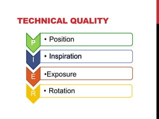
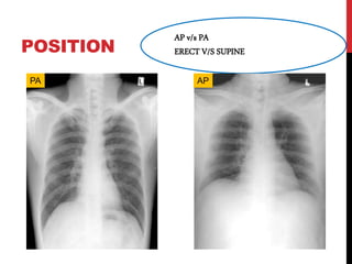
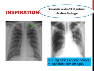
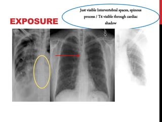
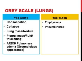
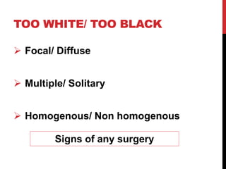
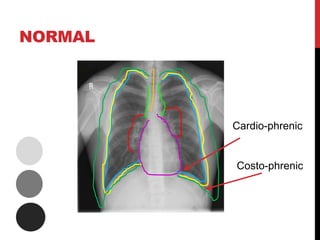
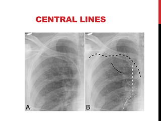
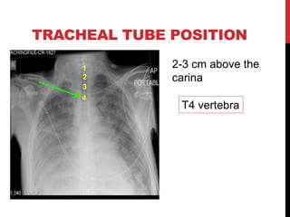
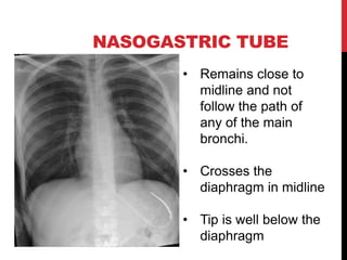
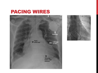
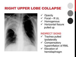
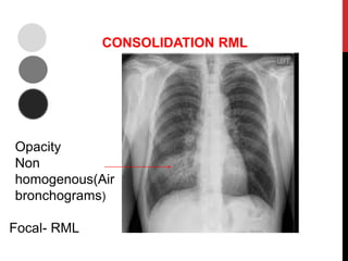
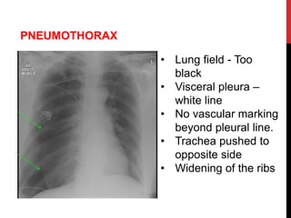
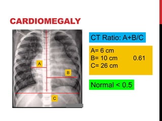
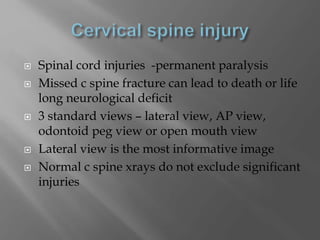
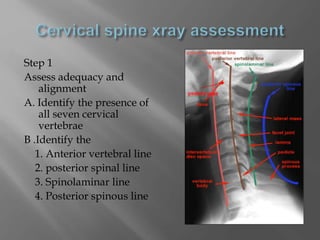
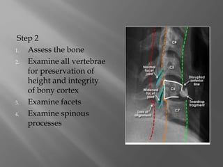
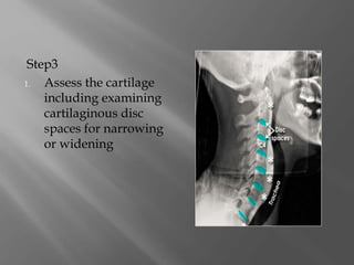
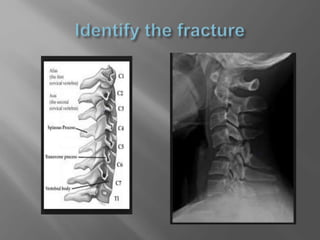
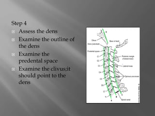
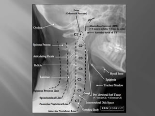
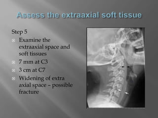
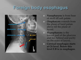
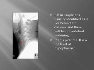
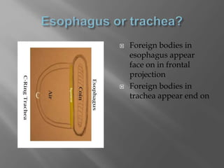
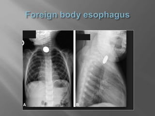
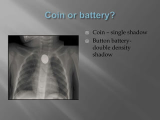
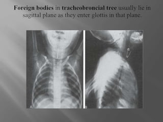
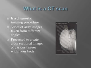
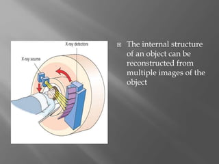
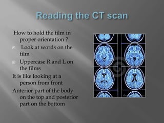
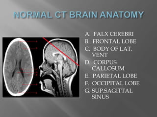
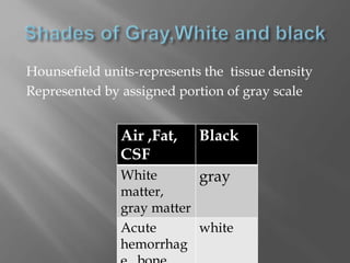
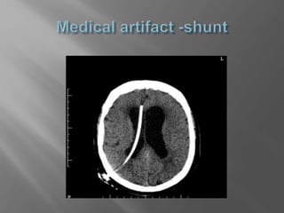
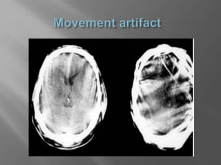
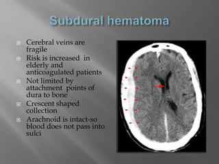
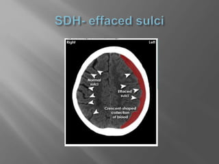
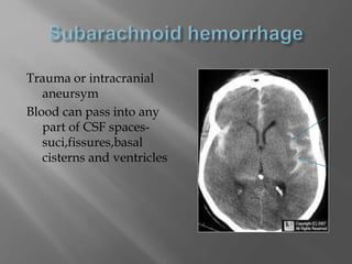
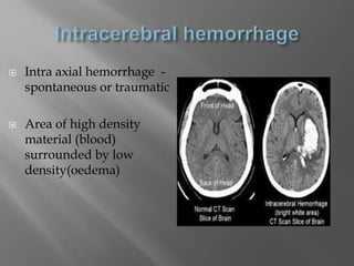
CHEST X RAY
CONTENTS
1. Identifying lung zones
2. Stepwise approach to describe/read a
CXR.
3. Coming to a diagnosis
4. Hardware/ Equipment
CONCEPT OF ZONES
Anterior ribs Zones
2 - 4 UPPER zone
4 - 6 MIDDLE zone
BELOW 6 LOWER zone
Loss of normal silhouettes of structures
aid in identifying lobes involved.
E.g. Right heart border= right middle lobe
STEPWISE APPROACH
NAME, AGE, SEX & IP NO.
DATE AND TIME OF STUDY
Do this before putting it on screen..
Ramu or Gopal
Sequential improvement
or worsening
TECHNICAL QUALITY
P • Position
I
E •Exposure
R • Rotation
POSITION
AP v/s PA
ERECT V/S SUPINE
PA AP
INSPIRATION
5-6 ant ribs in MCL/ 8-10 posterior
ribs above diaphragm
 Lung bases appear denser
 Apparent cardiomegaly
EXPOSURE
Just visible Intervertebral spaces, spinous
process / T4 visible through cardiac
shadow
ROTATION
Medial ends of clavicle equidistant
from spinous process
 Distort Mediastinal image
 Lung lesions hidden behind mediastinum
 Lung on rotated side appear denser
CENTRE PERIPHERY
 Trachea & Bronchi
 Heart & Mediastinum
 Hila
 Lungs
 Pleura & angles
 Chest wall
- Diaphragm
- Soft tissue
- Bony
AT EACH STEP LOOK AT..
• Grey scale
• Too white/Too black?
G
• Position
• Normal/ shifted?
P
• Size/Shape
• Normal/ altered
S
GREY SCALE
Bones- Denser – opaque
Tissue – Air – Grey
Air – Lucent - Darker
GREY SCALE (LUNGS)
TOO WHITE
• Consolidation
• Collapse
• Lung mass/Nodule
• Pleural mass/fluid/
thickening
• ARDS/ Pulmonary
edema (Ground glass
appearance)
TOO BLACK
• Emphysema
• Pneumothorax
TOO WHITE/ TOO BLACK
 Focal/ Diffuse
 Multiple/ Solitary
 Homogenous/ Non homogenous
Signs of any surgery
NORMAL
Cardio-phrenic
Costo-phrenic
CENTRAL LINES
TRACHEAL TUBE POSITION
2-3 cm above the
carina
T4 vertebra
1
3
2
4
NASOGASTRIC TUBE
• Remains close to
midline and not
follow the path of
any of the main
bronchi.
• Crosses the
diaphragm in midline
• Tip is well below the
diaphragm
PACING WIRES
RIGHT UPPER LOBE COLLAPSE
 Opacity
 Focal – R UL
 Homogenous
 Horizontal fissure
pulled up
INDIRECT SIGNS
 Trachea pulled
ipsilaterally
 Compensatory
hyperinflation of RML
 Elevation of
hemidiaphragm
CONSOLIDATION RML
Opacity
Non
homogenous(Air
bronchograms)
Focal- RML
PNEUMOTHORAX
• Lung field - Too
black
• Visceral pleura –
white line
• No vascular marking
beyond pleural line.
• Trachea pushed to
opposite side
• Widening of the ribs
CARDIOMEGALY
A
B
C
A= 6 cm
B= 10 cm 0.61
C= 26 cm
CT Ratio: A+B/C
Normal < 0.5
BATWING APPEARANCE
CONGESTIVE HEART FAILIURE
PNEUMONECTOMY
Hemithorax opacified
Ipsilateral
mediastinal shiftContralateral lung
hyper-inflated
Absent left main
bronchus
DR CHARULATHA R MD
Assistant professor MGMCRI
 Cervical spine injury in x ray
 Level of foreign body in chest x ray
 Ct brain in trauma
 CERVICAL SPINE INJURY IN XRAY
 Spinal cord injuries -permanent paralysis
 Missed c spine fracture can lead to death or life
long neurological deficit
 3 standard views – lateral view, AP view,
odontoid peg view or open mouth view
 Lateral view is the most informative image
 Normal c spine xrays do not exclude significant
injuries
Step 1
Assess adequacy and
alignment
A. Identify the presence of
all seven cervical
vertebrae
B .Identify the
1. Anterior vertebral line
2. posterior spinal line
3. Spinolaminar line
4. Posterior spinous line
Step 2
1. Assess the bone
2. Examine all vertebrae
for preservation of
height and integrity
of bony cortex
3. Examine facets
4. Examine spinous
processes
Step3
1. Assess the cartilage
including examining
cartilaginous disc
spaces for narrowing
or widening
Step 4
 Assess the dens
 Examine the outline of
the dens
 Examine the
predental space
 Examine the clivus;it
should point to the
dens
Step 5
 Examine the
extraaxial space and
soft tissues
 7 mm at C3
 3 cm at C7
 Widening of extra
axial space – possible
fracture
 FOREIGN BODY ESOPHAGUS
 Nasopharynx is from base
of skull till soft palate.
 Oropharynx extends from
the plane of hard palate
above till the plane of
hyoid.
 Hypopharynx is the
lowest part of the pharynx
and lies behind and partly
on the sides of larynx.
 Cervical esophagus starts
at C6 level. Below this
level F.B is in esophagus.
 F.B in esophagus
usually identified as it
lies behind air
column, and there
will be prevertebral
widening.
 In this picture F.B is a
the level of
hypopharynx.
 Foreign bodies in
esophagus appear
face on in frontal
projection
 Foreign bodies in
trachea appear end on
 Coin – single shadow
 Button battery-
double density
shadow
 CT brain in trauma
 Is a diagnostic
imaging procedure
 Series of Xray images
taken from different
angles
 Processed to create
cross sectional images
of various tissues
within our body
 The internal structure
of an object can be
reconstructed from
multiple images of the
object
How to hold the film in
proper orientation ?
 Look at words on the
film
 Uppercase R and L on
the films
It is like looking at a
person from front
Anterior part of the body
on the top and posterior
part on the bottom
A. FALX CEREBRI
B. FRONTAL LOBE
C. BODY OF LAT.
VENT
D. CORPUS
CALLOSUM
E. PARIETAL LOBE
F. OCCIPITAL LOBE
G. SUP.SAGITTAL
SINUS
Hounsefield units-represents the tissue density
Represented by assigned portion of gray scale
Air ,Fat,
CSF
Black
White
matter,
gray matter
gray
Acute
hemorrhag
white
 Check patient and image information
 Check date and time
 Check image quality
 Scalp and skull bones
 Brain volume
 Ischemia and Hemorrhage
 Mass effect
 Look at old images and reports
 Check for movement artifacts and medical
artifacts
 Do not view only a single slice in isolation
 If you suspect brain stem pathology ,consider
MRI
 Sutures
 found in typical anatomical
locations
 Jagged in appearance and corticated
 Fracture
 passes across both inner and outer
table of the skull in a straight line
 Edges of fractured skull bones are
not corticated
 Extradural hematoma
 Subdural hematoma
 Subarachnoid hemorrhage
 Post traumatic event
 Injury to an intracranial
artery –middle
meningeal artery
 Leakage of injured
artery –collection of
blood which strips the
dura mater away from
inner table of skull
 Lens shaped collection –
dura is strongly
adherent to the skull in
the region of sutures
 Cerebral veins are
fragile
 Risk is increased in
elderly and
anticoagulated patients
 Not limited by
attachment points of
dura to bone
 Crescent shaped
collection
 Arachnoid is intact-so
blood does not pass into
sulci
Trauma or intracranial
aneursym
Blood can pass into any
part of CSF spaces-
suci,fissures,basal
cisterns and ventricles
 Intra axial hemorrhage -
spontaneous or traumatic
 Area of high density
material (blood)
surrounded by low
density(oedema)
 Assess brain volume by
assessing volume of
CSF spaces
 Cerebral oedema-
can cause the brain to
swell – generalised
reduction of CSF
volume and loss of
differentiation between
grey and white matter
 Can be caused by
intracranial
masses,hemorrhage and
oedema
 Effacement of sulci,
partial or complete
effacement of adjacent
ventricles
 Displacement of
midline structures
 Effacement of
contralateral ventricles
and sulci
Cxr

Cxr

  • 1.
  • 2.
    CONTENTS 1. Identifying lungzones 2. Stepwise approach to describe/read a CXR. 3. Coming to a diagnosis 4. Hardware/ Equipment
  • 3.
    CONCEPT OF ZONES Anteriorribs Zones 2 - 4 UPPER zone 4 - 6 MIDDLE zone BELOW 6 LOWER zone Loss of normal silhouettes of structures aid in identifying lobes involved. E.g. Right heart border= right middle lobe
  • 4.
  • 5.
    NAME, AGE, SEX& IP NO. DATE AND TIME OF STUDY Do this before putting it on screen.. Ramu or Gopal Sequential improvement or worsening
  • 6.
    TECHNICAL QUALITY P •Position I E •Exposure R • Rotation
  • 7.
    POSITION AP v/s PA ERECTV/S SUPINE PA AP
  • 8.
    INSPIRATION 5-6 ant ribsin MCL/ 8-10 posterior ribs above diaphragm  Lung bases appear denser  Apparent cardiomegaly
  • 9.
    EXPOSURE Just visible Intervertebralspaces, spinous process / T4 visible through cardiac shadow
  • 10.
    ROTATION Medial ends ofclavicle equidistant from spinous process  Distort Mediastinal image  Lung lesions hidden behind mediastinum  Lung on rotated side appear denser
  • 11.
    CENTRE PERIPHERY  Trachea& Bronchi  Heart & Mediastinum  Hila  Lungs  Pleura & angles  Chest wall - Diaphragm - Soft tissue - Bony
  • 12.
    AT EACH STEPLOOK AT.. • Grey scale • Too white/Too black? G • Position • Normal/ shifted? P • Size/Shape • Normal/ altered S
  • 13.
    GREY SCALE Bones- Denser– opaque Tissue – Air – Grey Air – Lucent - Darker
  • 14.
    GREY SCALE (LUNGS) TOOWHITE • Consolidation • Collapse • Lung mass/Nodule • Pleural mass/fluid/ thickening • ARDS/ Pulmonary edema (Ground glass appearance) TOO BLACK • Emphysema • Pneumothorax
  • 15.
    TOO WHITE/ TOOBLACK  Focal/ Diffuse  Multiple/ Solitary  Homogenous/ Non homogenous Signs of any surgery
  • 16.
  • 17.
  • 18.
    TRACHEAL TUBE POSITION 2-3cm above the carina T4 vertebra 1 3 2 4
  • 19.
    NASOGASTRIC TUBE • Remainsclose to midline and not follow the path of any of the main bronchi. • Crosses the diaphragm in midline • Tip is well below the diaphragm
  • 20.
  • 22.
    RIGHT UPPER LOBECOLLAPSE  Opacity  Focal – R UL  Homogenous  Horizontal fissure pulled up INDIRECT SIGNS  Trachea pulled ipsilaterally  Compensatory hyperinflation of RML  Elevation of hemidiaphragm
  • 23.
  • 24.
    PNEUMOTHORAX • Lung field- Too black • Visceral pleura – white line • No vascular marking beyond pleural line. • Trachea pushed to opposite side • Widening of the ribs
  • 25.
    CARDIOMEGALY A B C A= 6 cm B=10 cm 0.61 C= 26 cm CT Ratio: A+B/C Normal < 0.5
  • 26.
  • 27.
  • 28.
  • 30.
    DR CHARULATHA RMD Assistant professor MGMCRI
  • 31.
     Cervical spineinjury in x ray  Level of foreign body in chest x ray  Ct brain in trauma
  • 32.
     CERVICAL SPINEINJURY IN XRAY
  • 33.
     Spinal cordinjuries -permanent paralysis  Missed c spine fracture can lead to death or life long neurological deficit  3 standard views – lateral view, AP view, odontoid peg view or open mouth view  Lateral view is the most informative image  Normal c spine xrays do not exclude significant injuries
  • 34.
    Step 1 Assess adequacyand alignment A. Identify the presence of all seven cervical vertebrae B .Identify the 1. Anterior vertebral line 2. posterior spinal line 3. Spinolaminar line 4. Posterior spinous line
  • 35.
    Step 2 1. Assessthe bone 2. Examine all vertebrae for preservation of height and integrity of bony cortex 3. Examine facets 4. Examine spinous processes
  • 36.
    Step3 1. Assess thecartilage including examining cartilaginous disc spaces for narrowing or widening
  • 38.
    Step 4  Assessthe dens  Examine the outline of the dens  Examine the predental space  Examine the clivus;it should point to the dens
  • 40.
    Step 5  Examinethe extraaxial space and soft tissues  7 mm at C3  3 cm at C7  Widening of extra axial space – possible fracture
  • 41.
  • 42.
     Nasopharynx isfrom base of skull till soft palate.  Oropharynx extends from the plane of hard palate above till the plane of hyoid.  Hypopharynx is the lowest part of the pharynx and lies behind and partly on the sides of larynx.  Cervical esophagus starts at C6 level. Below this level F.B is in esophagus.
  • 43.
     F.B inesophagus usually identified as it lies behind air column, and there will be prevertebral widening.  In this picture F.B is a the level of hypopharynx.
  • 44.
     Foreign bodiesin esophagus appear face on in frontal projection  Foreign bodies in trachea appear end on
  • 46.
     Coin –single shadow  Button battery- double density shadow
  • 48.
     CT brainin trauma
  • 49.
     Is adiagnostic imaging procedure  Series of Xray images taken from different angles  Processed to create cross sectional images of various tissues within our body
  • 50.
     The internalstructure of an object can be reconstructed from multiple images of the object
  • 51.
    How to holdthe film in proper orientation ?  Look at words on the film  Uppercase R and L on the films It is like looking at a person from front Anterior part of the body on the top and posterior part on the bottom
  • 52.
    A. FALX CEREBRI B.FRONTAL LOBE C. BODY OF LAT. VENT D. CORPUS CALLOSUM E. PARIETAL LOBE F. OCCIPITAL LOBE G. SUP.SAGITTAL SINUS
  • 53.
    Hounsefield units-represents thetissue density Represented by assigned portion of gray scale Air ,Fat, CSF Black White matter, gray matter gray Acute hemorrhag white
  • 54.
     Check patientand image information  Check date and time  Check image quality  Scalp and skull bones  Brain volume  Ischemia and Hemorrhage  Mass effect
  • 55.
     Look atold images and reports  Check for movement artifacts and medical artifacts  Do not view only a single slice in isolation  If you suspect brain stem pathology ,consider MRI
  • 58.
     Sutures  foundin typical anatomical locations  Jagged in appearance and corticated  Fracture  passes across both inner and outer table of the skull in a straight line  Edges of fractured skull bones are not corticated
  • 59.
     Extradural hematoma Subdural hematoma  Subarachnoid hemorrhage
  • 60.
     Post traumaticevent  Injury to an intracranial artery –middle meningeal artery  Leakage of injured artery –collection of blood which strips the dura mater away from inner table of skull  Lens shaped collection – dura is strongly adherent to the skull in the region of sutures
  • 62.
     Cerebral veinsare fragile  Risk is increased in elderly and anticoagulated patients  Not limited by attachment points of dura to bone  Crescent shaped collection  Arachnoid is intact-so blood does not pass into sulci
  • 64.
    Trauma or intracranial aneursym Bloodcan pass into any part of CSF spaces- suci,fissures,basal cisterns and ventricles
  • 65.
     Intra axialhemorrhage - spontaneous or traumatic  Area of high density material (blood) surrounded by low density(oedema)
  • 67.
     Assess brainvolume by assessing volume of CSF spaces  Cerebral oedema- can cause the brain to swell – generalised reduction of CSF volume and loss of differentiation between grey and white matter
  • 68.
     Can becaused by intracranial masses,hemorrhage and oedema  Effacement of sulci, partial or complete effacement of adjacent ventricles  Displacement of midline structures  Effacement of contralateral ventricles and sulci

Editor's Notes

  • #11 Clavicle appears away from midline on the side to which the film is rotatted
  • #27 Perihilar edema - cardiogenic