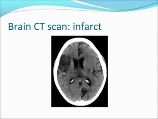
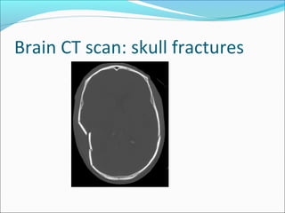
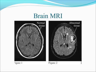
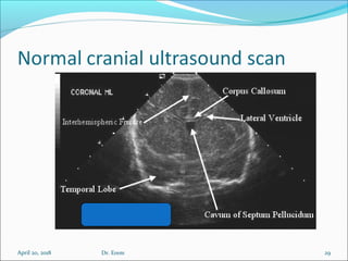
CHS 3111: CENTRAL NERVOUS
SYSTEM (CNS) PATHOLOGY
Dr. Erem Geoffrey
Lecturer, Radiology Department
School of Medicine
Learning objectives
To learn the different terminologies used to describe
CNS radiological findings
To describe the different imaging modalities and
their indications, in investigation of the CNS
To describe the radiological anatomy of the CNS at
the different imaging modalities
Learning objectives
To describe the common pathology of the CNS as
demonstrated on radiological imaging
To identify emergencies of the CNS that are
demonstrable on radiological imaging
Common terminologies used to
describe CNS radiological findings
Plain X-ray
Opacity: opaque/dense area on an X-ray- appears
white
Sclerosis: Thickening & increase in bone density-
appears white
Radiolucency: Non opaque area on an X-ray- appears
as dark/ black area
Terminologies cont.
CT scan
Hypodense: Area with a lower density in comparison to another
area-appears dark/black
Hyperdense: Area with a higher density in comparison to
another area-appears bright/white
Isodense: Area with same density in comparison to another
area-similar appearance
Terminologies cont.
Ultrasound scan
Echogenicity: Intensity of echoes reflected by tissues
or structures from inside the body
Hyperechoic: An area with higher intensity echoes in
comparison to another area-appears brighter
Hypoechoic: An area with lower intensity echoes in
comparison to another area-appears darker/blacker
Anechoic: An area with no echoes-appears black
Isoechoic: An area with same/similar echo intensity in
comparison to another area-similar appearance
Terminologies cont.
MRI
Hyperintense: areas which appear birght on MRI
Hypointense: areas which appear black
Radiological imaging modalities for investigation
of CNS & their indications
Plain X-ray: Skull & spine X-rays
Indications:
Infections & inflammatory diseases
Trauma
Neoplasm
Foreign body
Imaging modalities & indications cont.
Ultrasound
Indications:
 Evaluation of intracranial diseases & anomalies in children with
open anterior fontanelle: e.g. hydrocephalus, intraventricular
haemorrhage, subdural effusion, various congenital anomalies
such as Dandy-Walker cyst
 Evaluation for spinal dysraphism in neonates & young infants
April 20, 2018 10Dr. Erem
Imaging modalities & indications cont.
CT scan/ MRI
Indications
Infections & inflammatory diseases
Congenital anomalies of CNS
Trauma
Neoplasm
April 20, 2018 12Dr. Erem
April 20, 2018 13Dr. Erem
Imaging modalities & indications cont.
Radionuclide imaging
Indications:
Functional studies e.g brain perfusion
Angiography
Indications:
Pts with stroke/cerebral infarcts
Pts with transient ischaemic disease
Preoperative evaluation e.g. in pts with certain
tumours
Normal Skull X-rays (SXR)
Lateral view AP view
April 20, 2018 16
Sella turcica
Anterior clinoid process
Posterior clinoid process
Tuberculum sellae
Dorsum sellae
Dr. Erem
Normal CT anatomy of brain
23: Interhemispheric fissure
24: Head of caudate nucleus
25: Lentiform nucleus
(globus pallidus & putamen)
26: Thalamus
27: Atrium of lateral
ventricles
28: Anterior limb of internal
capsule
29: corpus callosum (body)
30: Posterior limb of internal
capsule
Anatomy
April 20, 2018 18Dr. Erem
Anatomy….
April 20, 2018 19Dr. Erem
Normal CT anatomy of brain: level of
basal ganglia
April 20, 2018 21Dr. Erem
April 20, 2018 22Dr. Erem
Brain CT scan: posterior fossa tumour
Brain CT scan: brain abscess
Brain CT scan: right subdural
haemorrhage
Brain CT scan: infarct
Brain CT scan: skull fractures
Brain MRI
April 20, 2018 29Dr. Erem
April 20, 2018 30Dr. Erem
April 20, 2018 31Dr. Erem
April 20, 2018 32Dr. Erem
April 20, 2018 33Dr. Erem
Conventional myelogram CT myelogram
April 20, 2018 35Dr. Erem
Emergencies of CNS that demonstrable are
on radiological imaging
Trauma:
Spinal injury with cord compression
Intracranial haemorrhages
Cardiovascular diseases
Stroke especially haemorrhagic
Increased intracranial pressure
Tumours
Infection with brain oedema

Chs 3111 cns pathology radiology

  • 1.
    CHS 3111: CENTRALNERVOUS SYSTEM (CNS) PATHOLOGY Dr. Erem Geoffrey Lecturer, Radiology Department School of Medicine
  • 2.
    Learning objectives To learnthe different terminologies used to describe CNS radiological findings To describe the different imaging modalities and their indications, in investigation of the CNS To describe the radiological anatomy of the CNS at the different imaging modalities
  • 3.
    Learning objectives To describethe common pathology of the CNS as demonstrated on radiological imaging To identify emergencies of the CNS that are demonstrable on radiological imaging
  • 4.
    Common terminologies usedto describe CNS radiological findings Plain X-ray Opacity: opaque/dense area on an X-ray- appears white Sclerosis: Thickening & increase in bone density- appears white Radiolucency: Non opaque area on an X-ray- appears as dark/ black area
  • 5.
    Terminologies cont. CT scan Hypodense:Area with a lower density in comparison to another area-appears dark/black Hyperdense: Area with a higher density in comparison to another area-appears bright/white Isodense: Area with same density in comparison to another area-similar appearance
  • 6.
    Terminologies cont. Ultrasound scan Echogenicity:Intensity of echoes reflected by tissues or structures from inside the body Hyperechoic: An area with higher intensity echoes in comparison to another area-appears brighter Hypoechoic: An area with lower intensity echoes in comparison to another area-appears darker/blacker Anechoic: An area with no echoes-appears black Isoechoic: An area with same/similar echo intensity in comparison to another area-similar appearance
  • 7.
    Terminologies cont. MRI Hyperintense: areaswhich appear birght on MRI Hypointense: areas which appear black
  • 8.
    Radiological imaging modalitiesfor investigation of CNS & their indications Plain X-ray: Skull & spine X-rays Indications: Infections & inflammatory diseases Trauma Neoplasm Foreign body
  • 9.
    Imaging modalities &indications cont. Ultrasound Indications:  Evaluation of intracranial diseases & anomalies in children with open anterior fontanelle: e.g. hydrocephalus, intraventricular haemorrhage, subdural effusion, various congenital anomalies such as Dandy-Walker cyst  Evaluation for spinal dysraphism in neonates & young infants
  • 10.
    April 20, 201810Dr. Erem
  • 11.
    Imaging modalities &indications cont. CT scan/ MRI Indications Infections & inflammatory diseases Congenital anomalies of CNS Trauma Neoplasm
  • 12.
    April 20, 201812Dr. Erem
  • 13.
    April 20, 201813Dr. Erem
  • 14.
    Imaging modalities &indications cont. Radionuclide imaging Indications: Functional studies e.g brain perfusion Angiography Indications: Pts with stroke/cerebral infarcts Pts with transient ischaemic disease Preoperative evaluation e.g. in pts with certain tumours
  • 15.
    Normal Skull X-rays(SXR) Lateral view AP view
  • 16.
    April 20, 201816 Sella turcica Anterior clinoid process Posterior clinoid process Tuberculum sellae Dorsum sellae Dr. Erem
  • 17.
    Normal CT anatomyof brain 23: Interhemispheric fissure 24: Head of caudate nucleus 25: Lentiform nucleus (globus pallidus & putamen) 26: Thalamus 27: Atrium of lateral ventricles 28: Anterior limb of internal capsule 29: corpus callosum (body) 30: Posterior limb of internal capsule
  • 18.
  • 19.
  • 20.
    Normal CT anatomyof brain: level of basal ganglia
  • 21.
    April 20, 201821Dr. Erem
  • 22.
    April 20, 201822Dr. Erem
  • 23.
    Brain CT scan:posterior fossa tumour
  • 24.
    Brain CT scan:brain abscess
  • 25.
    Brain CT scan:right subdural haemorrhage
  • 26.
  • 27.
    Brain CT scan:skull fractures
  • 28.
  • 29.
    April 20, 201829Dr. Erem
  • 30.
    April 20, 201830Dr. Erem
  • 31.
    April 20, 201831Dr. Erem
  • 32.
    April 20, 201832Dr. Erem
  • 33.
    April 20, 201833Dr. Erem
  • 34.
  • 35.
    April 20, 201835Dr. Erem
  • 36.
    Emergencies of CNSthat demonstrable are on radiological imaging Trauma: Spinal injury with cord compression Intracranial haemorrhages Cardiovascular diseases Stroke especially haemorrhagic Increased intracranial pressure Tumours Infection with brain oedema