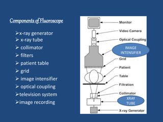
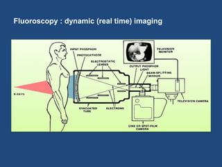
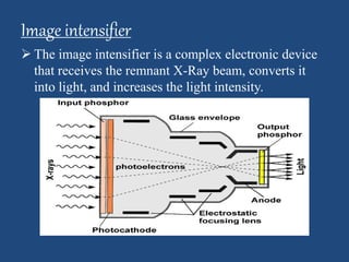
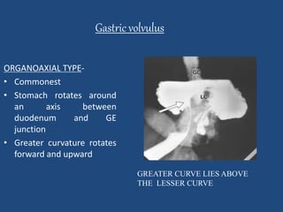
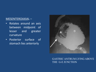
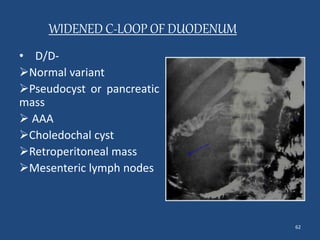
FLOUROSCOPIC 
PROCEDURES 
DR NANI LAMPUNG 
JR1 DEPT OF RADIODIAGNOSIS 
JNMCH AMU ALIGARH
TO BE DISCUSSED 
 FLOUROSCOPY 
 CONTRAST AGENTS 
 PROCEDURES 
o BARIUM SWALLOW 
o BA MEAL 
o BA MEAL FOLLOW THROUGH 
o ENTEROCLYSIS 
o BARIUM ENEMA 
o DISTAL LOOPOGRAM 
o MCU, RGU AND HSG 
 AND FINDINGS
FLOUROSCOPY 
Imaging technique that uses x-rays to 
obtain real time images
HISTORY 
• Thomas Edison 1896 
• Screen (zinc-cadmium 
sulfide) placed over 
patient’s body 
• Red goggles-30 minutes 
before exam 
• 1950s image 
intensifiers developed
Fluoroscopic Equipment 
General purpose fluoroscopic system
Mobile fluoroscopic 
system for routine 
procedures during 
surgery 
Fluoroscopic Equipment (cont)
Components of Fluoroscope 
x-ray generator 
 x-ray tube 
 collimator 
 filters 
 patient table 
 grid 
 image intensifier 
 optical coupling 
television system 
image recording 
IMAGE 
INTENSIFIER 
XRAY 
TUBE
Fluoroscopy : dynamic (real time) imaging
Image intensifier 
 The image intensifier is a complex electronic device 
that receives the remnant X-Ray beam, converts it 
into light, and increases the light intensity.
 The principal advantage- increased image 
brightness. 
Image monitoring: 
Two methods -used to electronically convert the 
visible image on the output phosphor of the image 
intensifier into an electronic signal. 
1. Thermionic television camera tube 
2. The solid state charge-coupled device (CCD).
Camera tube: 
• The TV camera consists of cylindrical housing, approx 15 mm 
in diameter by 25 cm in length, that contains the heart of the 
camera, TV camera tube. 
• It also contains electromagnetic coils (used to properly steer 
the electron beam inside the tube) 
• Vidicon and its modified version, the Plumbicon, are used 
most often.
• Two methods are commonly used to couple the 
television camera tube to the image-intensifier 
tube. 
– Fiber optics. 
– Lens system.
• The video signal is amplified and is transmitted by cable to 
the television monitor, where it is transformed back into a 
visible image.
Fluoroscopy Uses 
• Barium studies 
• Catheter Insertion 
• Blood Flow Studies 
• Orthopedic Surgery
OTHERS 
• Injections into the knees (Viscosupplementation injections) 
• Locating foreign bodies 
• Percutaneous Vertebroplasty (Treating compressed fractures of 
the spine) 
• Injections into joints or spine 
• Image-guided anesthetic injections
CONTRAST used 
1. BARIUM SULPHATE 
SUSPENSION 
• Inert 
• Insoluble in water 
• Good mucosal coating 
• No major sequalae if aspirated 
• Cost effective 
DISADVANTAGES 
• Mediastinitis 
• Peritonitis 
• may collect in GIT
2. WATER SOLUBLE CONTRAST 
• Gastrograffin, Gastromiro 
• USE- suspected perforation 
DISADVANTAGES 
• Poor mucosal coating 
• Allergic reactions 
• Chemical pneumonitis
3. GASES 
• Co2 
• Air 
• room air for Ba Enema
TYPES OF BARIUM STUDIES 
1. SINGLE CONTRAST- single contrast medium 
2. DOUBLE CONTRAST- two contrast media 
• Ba sulphate suspension (+ve contrast ) 
• Air (-ve contrast)
SINGLE CONTRAST 
• Elderly & uncooperative pts 
• Low concentration 
• Poor mucosal coating 
• Less sensitive for polyps, 
erosions, linear ulcerations, 
superficial gastric ca, subtle 
mucosal abnormalities 
DOUBLE CONTRAST 
• younger 
• higher conc. 
• good mucosal coating 
• more sensitive polyps & 
small mucosal lesions
DOUBLE CONTRAST SINGLE CONTRAST
PREPARATION FOR GI BARIUM STUDIES 
• Maintain Low residual diet 
• Laxatives 
• Increase fluid intake 
• NPO from midnight 
• Follow up care –colour change/constipation 
• 2 times cleansing enema for barium enema studies
PREPARATION FOR GI BARIUM STUDIES 
• Take h/o related to medications & any allergy to 
contrast agents 
• Ask patient to remove jewellery, eyes glasses or any 
metallic objects / clothings 
• Explain the procedure to patient & take consent
BARIUM SWALLOW 
• Radiographic evaluation of pharynx and entire 
oesophagus upto gastroesophageal junction
INDICATIONS 
• DYSPHAGIA,ODYNOPHAGIA 
• HEARTBURN,RETROSTERNAL PAIN 
• HIATUS HERNIA,STRICTURE FORMATION 
• ESOPHAGEAL CA 
• MOTILITY D/O-ACHALASIA,DIFFUSE OESOPHAGEAL SPASMS 
• THORACIC MASS LESIONS 
• MITRAL VALVE DISEASE 
• OESOPHAGEAL VARICES 
• ASSESSMENT OF ABNORMALITY OF 
A. PHARYNGOESOPHAGEAL J/N INCLUDING ZENKERS DIVERTICULUM 
B. CRICOID WEB 
C. CRICOPHARYNGEAL ACHALASIA
CONTRAINDICATIONS 
• Suspected leakage into the mediastinum, 
pleural or peritoneal cavities 
• Tracheo-esophageal fistula
TECHNIQUE 
• PHARYNX 
• Mouthful contrast bolus with high density (250%w/v) 
• Pt is asked to swallow once & stop swallowing 
thereafter( to get optimum mucosal coating) 
• frontal and lateral view x-ray taken
SP-soft palate 
V-valeculla 
P-pyriform sinus 
LATERAL VIEW AP VIEW
ESOPHAGUS (SINGLE CONTRAST) 
• Multiple mouthful 80% w/v barium suspension is given 
• Take the flouroscopic unit to pt neck command to 
swallow 
• Observe the Ba passing down the esophagus upto GE J/N 
USEFUL IN – 
Ext compression, Displacement, Motility d/o 
• Prone swallow to assess esophageal contraction 
(esophageal web, hiatal hernia & esophageal varices)
DOUBLE CONTRAST 
• Effervescent powder is added to the Ba mixture 
250% w/v 
• Inj buscopan i.v may be given to keep esophagus 
distended for a longer time 
• FILMS 
1. Control film 
2. Spot films- AP/Lateral views of upper, mid & lower 
oesophagus
32 
AP VIEW LATERAL VIEW
COMPLICATIONS 
• Aspiration 
• Leakage of Ba from perforation l/t mediastinitis
SPECIFIC FINDINGS
OESOPHAGEAL WEB 
• SHELF-LIKE INFOLDING OF 
MUCOSA(1-2 MM THICK) 
• PARTIALLY OBSTRUCTING 
CERVICAL ESOPHAGEAL 
WEB 
• JET PHENOMENON 
• ASSOCIATION-EB,BP,PVS
ZENKERS DIVERTICULUM 
• ZENKER’S 
DIVERTICULUM • Pulsion false diverticulum 
• Killian’s dehiscence 
• Defect in cricopharyngeus 
muscle
KILLIAN JAMIESON DIVERTICULUM 
• Pulsion diverticulum 
• Lateral anatomic weak site 
• Below cricopharyngeus
DIFFUSE ESOPHAGEAL SPASM 
• Irregular areas of 
narrowing & 
dilatations 
• Cork screw/shish 
kebab/rosary bead 
appearance
ACHALSIA CARDIA 
*With short segment 
stricture. 
* A “bird-peak " like 
tapering of the 
esophagus at the GE 
junction. OR 
• DEGENERATION OF 
NEURONS OF 
AURBACH’S PLEXUS 
• Dilatation of body 
• Short segment stricture 
• BIRD BEAK like tapering 
at GE J/N
ESOPHAGEAL VARICES 
• Mild dilatation of the 
esophagus 
with multiple 
persistent filling 
defects in the lower 
third of the 
esophagus and/or 
longitudinal furrows. 
• Mild dilatation 
with multiple 
persistent filling 
defects
TRACHEOESOPHAGEAL FISTULA 
• Congenital/Acquired 
• Ideal contrast non ionic 
water soluble media 
• In case fistula not 
identified laterally, put in 
prone. 
• If fistula seen, stop 
procedure as barium 
aspiration result in 
inflammation and 
granuloma
Diaphragmatic hernia 
Herniation of abdominal viscera into thoracic 
cavity through- 
DIAPHRAGMATIC HIATUS:- 
 sliding hernia 
 rolling hiatus hernia 
CONGENITAL DEFECT:- 
 Bochdalek hernia 
 Morgagni hernia
SLIDING HIATUS HERNIA 
Develop due to stretching or 
tear of phrenico-esophageal 
ligament 
stomach protrudes >2cm 
above the hiatus 
>3 folds seen across the 
hiatus 
diameter of hiatus >3cm
Rolling hiatus hernia 
Fundus herniates 
alongside lower esophagus 
while cardia remains below 
the diaphragm 
 T/T– 
Nissen’s fundoplication
Esophageal carcinoma 
NARROWING OF LUMEN WITH MUCOSAL 
IRREGULARITY AND SHOULDERING
BARIUM MEAL 
• Radiological study of esophagus, stomach, duodenum 
till the Gastroduodenal J/N
INDICATIONS 
• Peptic ulceration 
• Malignancies of GE junction, stomach, duodenum 
• Motility d/o 
• Hiatus hernia 
• Gastric or duodenal obstruction 
• Children- GER, pyloric obstruction, malrotation
CONTRAINDICATIONS 
• Complete large bowel obstruction 
• Suspected perforation (unless water soluble 
contrast medium is used)
TECHNIQUE 
DOUBLE CONTRAST 
kilovolt range-70 – 120 kv 
• Inj buscopan(20mg) /glucagon(0.1-0.2 mg) iv-relax 
stomach & suspend peristalsis 
• Effervescent agent is given 
• Standing in RAO position- 120ml of high 
density barium(250%w/v) double contrast 
views of lower esophagus is obtained
• FOR STOMACH-Pt 
placed in recumbent position & roll the 
patient from side to side ( encourages mucosal 
coating & adequate distension) 
• FOR DUODENUM-When 
Ba enters duodenum pt is turned to RAO 
position to fill duodenum with gas and then DC 
films are taken
DOUBLE CONTRAST STUDY 
ADVANTAGES 
• Highly accurate detecting abnormalities 
following gastric sx, 
• Bile reflex gastritis, marginal ulcerations 
• Recurrent Ca
TYPICAL FILMING SEQUENCES 
POSITIONING 
• ERECT RAO 
AP 
• SUPINE-RAO 
AP 
LAO 
RL 
• PRONE (PAD ↓ ANTRUM)-AP 
• SUPINE-RAO 
DEMONSTRATES 
 ESOPHAGUS {PRONE LPO(SC)} 
 FUNDUS 
 BODY+ANTRUM WITH LESSER CURVE 
 BODY + ANTRUM 
 BODY WITH LESSER CURVE 
 FUNDUS 
 DUODENAL LOOP 
 DUODENAL CAP
SINGLE CONTRAST STUDY 
• Ba mixture given in erect position wherever possible 
• Compression films are taken to obtain good mucosal detail 
• kilovolt range-120 to 150 kv 
ADVANTAGES- 
• pylorospasm, fistulae, enlarged gastric rugae best seen 
• filling defect due to large mass easily identifiable 
DISADVANTAGES- 
• lack of sensitivity for small polyps, ulcers, superficial Ca
MODIFICATIONS 
• FRAIL IMMOBILE ELDERLY PT- SC study with 100% w/v barium 
• PARTIAL GASTRECTOMY OR GASTRIC DRAINAGE 
PROCEDURE(PYLOROPLASTY/GASTROENTROSTOMY)- 
early flooding 
- start examination in prone swallow using high density barium 
-when Ba reaches duodenum or gastroentrostomy pt is quickly 
turned supine and dc films are taken 
• HIATUS HERNIA- patient put in prone with abdominal 
compression applied by pillow or pad
Normal mucosal pattern 
Areae gastricae Rugal folds
56 
Duodenal cap 
• Symmetric triangular 
structure formed by 
first part of duodenum 
• Shows a fine velvety 
surface pattern due to 
presence of villi
Peptic ulcer 
An ulcer is a focal area of mucosal disruption 
On barium meal examination- an ulcer 
dependent wall – round or ovoid collection of 
barium filling the ulcer crater 
nondependent wall – circular or hemispheric ring 
due to barium coating the rim of unfilled ulcer
BENIGN GASTRIC ULCER MALIGNANT GASTRIC ULCER 
• Smooth round/ovoid 
ulcer 
• Hampton’s 
hump/smooth ulcer 
collar 
• Smooth, straight 
radiating folds 
• Projects outside luminal 
contour 
• Irregular shaped, 
abnormally surfaced 
ulcer 
• Mucosal nodularity at 
edge 
• Lobulated, enlarged or 
club shaped folds 
• Lies within the outline 
of stomach
BENIGN ULCER MALIGNANT ULCER
Gastric volvulus 
ORGANOAXIAL TYPE- 
• Commonest 
• Stomach rotates around 
an axis between 
duodenum and GE 
junction 
• Greater curvature rotates 
forward and upward 
GREATER CURVE LIES ABOVE 
THE LESSER CURVE
MESENTEROAXIAL – 
• Rotates around an axis 
between midpoint of 
lesser and greater 
curvature 
• Posterior surface of 
stomach lies anteriorly 
GASTRIC ANTRUM LYING ABOVE 
THE G-E JUNCTION
62 
WIDENED C-LOOP OF DUODENUM 
• D/D- 
Normal variant 
Pseudocyst or pancreatic 
mass 
 AAA 
Choledochal cyst 
Retroperitoneal mass 
Mesenteric lymph nodes
BARIUM MEAL FOLLOW THROUGH 
• Demonstrate the whole of small bowel from 
duodenal flexure to ileocaecal junction
INDICATIONS 
• MALABSORPTIONS 
• INTESTINAL TB 
• MALROTATION IN INFANTS/NEONATES- TO KNOW LEVEL & 
CAUSE OF OBSTRUCTION
TECHNIQUE 
• Following drinking contrast(50-100%w/v) 
patient lies in prone or on right side until ba 
has left the stomach 
• AIM- to produce continous column of Ba in 
small bowel 
• spot films may be made of any loops or 
segments of bowels
WELL PERFORMED BARIUM MEAL 
• Entire BL should be included in each 
radiograph 
• Stomach & duodenum should be documented 
in initial films 
• Time marker should be clearly visible 
• Ileocaecal j/n should be well visualised
Feathery app. of 
jejunum 
Smooth contour 
of ileum
Enteroclysis/small bowel enema 
• Radiographic procedure in which contrast 
medium is injected directly beyond duodenum 
• ADVANTAGE- Eliminates the obscuration of 
individual segment of small bowel by 
complete filling of tract with Ba
TECHNIQUE 
• Under flouroscopic control the tube with stiff 
guidewire is advanced upto duodenojejunal flexure 
• Contrast (200 ml of 110% w/v) mixed with 350 ml of 
H2O infused ↓ gravity control followed by 500 to 
1000 ml of fresh water is given @ 100 ml/min 
• When Ba is injected the radiologist follows the 
barium column carefully with fluoroscopy & spot 
radiographs are taken at appropriate times
ADVANTAGES 
Better visualization 
of mucosal lesions 
Distension can be 
controlled by rate of 
infusion 
Shorter duration 
DISADVANTAGES 
More exposure 
Patient discomfort
Ileocecal tuberculosis 
EARLY PHASE- 
Incompetence and 
thickening of illeoceacal 
valve with narrowing of 
terminal ileum 
ADVANCED STAGE – 
Narrowing of ileum 
with shrunken, 
retracted( out of illiac 
fossa) caecum
BARIUM ENEMA 
• LARGE BOWEL EXAMINATION
INDICATIONS 
1. BLEEDING PR 
2. COLITIS 
3. SUSPECTED LARGE BOWEL OBSTRUCTION 
4. COLONIC POLYPS & COLORECTAL CA 
5. FAILED COLONOSCOPY 
6. FOLLOW UP SCREENING FOR POST OP COLORECTAL CA 
7. NON SPECIFIC ABDOMINAL PAIN
CONTRAINDICATIONS 
• TOXIC MEGACOLON 
• PSEUDOMEMBRANOUS COLITIS 
• SUSPECTED PERFORATION 
• WEEK BEFORE OR AFTER RECTAL BIOPSY 
• POSSIBBILITY OF CA IN ULCERATIVE COLITIS 
• PREGNANCY
TECHNIQUE 
• Perform P/R examination 
• Insert enema tip with patient in lt lateral position 
with knees flexed 
• Check for any blocks 
• Look intermittently under flouroscopy to check for 
progression of Ba column 
• Gas insufflation done intermitently 
• change position of patient and table accordingly
SPOT FILMS 
• Rectum & sigmoid colon-RAO in lying position 
• Splenic flexure-LAO(erect) 
• Hepatic flexure-RAO(erect) 
• Caecum-supine lying on rt side with head down tilt 
and compression 
• Over head film- lying position
COMPLICATIONS 
• BOWEL PERFORATION 
• INJURY TO RECTAL MUCOSA OR ANAL CANAL DUE TO ENEMA 
TIP 
• VENOUS INTRAVASATION 
• Ba IMPACTION,WATER INTOXICATION 
• ALLERGIC RXNS & CARDIAC ARRHYTMIAS 
• TRANSIENT BACTERAEMIA
Colonic polyps 
Sessile polyps- BOWLER’S HAT sign 
(dome of hat points towards the lumen) 
Colonic diverticula-dome 
points away from lumen
80 
Pedunculated polyp - Mexican hat sign 
(outer ring- head of polyp 
inner ring- stalk seen through the head)
Ulcerative colitis 
ACUTE STAGE- 
• Fine mucosal granularity(d/t 
edema and hyperemia) 
• ULCERATIONS - 
Collar button (flask shaped ulcer) 
Double tracking (longitudinal ulcer 
in submucosa)
• Pseudopolyps-raised 
area of inflammed 
tissue 
CHRONIC STAGE- 
Loss of haustrations 
shortening & narrowing 
of colon 
( lead pipe colon)
Crohn’s disease 
• Mucosal granularity with 
aphthous ulcer 
• Cobblestone app.(deep 
longitudinal and 
trasverse ulcer with 
adjacent mucosal edema) 
• Skip lesions with rectal 
sparing
84 
STRING SIGN- 
• narrowing of terminal 
ileum due to edema, 
spasm and fibrosis
diverticulosis 
• Small outpouchings 
from colonic wall 
which are filled by 
barium
86 
Sigmoid volvulus 
Barium fills to point of 
obstruction and twist of sigmoid 
colon 
Xray abdomen-Massively 
distended sigmoid colon – 
COFFEE BEAN sign
87 
Colonic cancer 
Annular, irregular and 
ulcerating lesion 
appear as 
circumferential 
irregular filling defect 
with narrowing of 
lumen(apple core sign)
INSTANT BARIUM ENEMA 
• DEVELOPED BY YOUNG (1963) 
INDICATIONS 
• Identify level of suspected obstruction 
• Show d/s extent & severity of mucosal lesions during an a/c 
episode of uc 
CONTRAINDICATIONS 
• Toxic megacolon, suspected perforation 
• Recent rectal biopsy 
ADVANTAGE- DONE IN UNPREPARED BOWEL 
GUIDE TREATMENT
DISTAL LOOPOGRAM 
• MAIN INDICATION - 
to know the patency of 
distal bowel prior to 
reclosure of 
ileostomy/colostomy 
• Mainly single contrast study
MICTURATING CYSTOGRAPHY (MCU) 
INDICATIONS 
1. Outflow obstruction 
2. Stress incontinence 
3. Vesicoureteric reflux (children) 
CONTRAINDICATIONS 
Acute infections bladder / urethra
PATIENT PREPARATION 
• Micturates immediately before examination 
• In cathetherised patient, clamp release to 
decompress the bladder 
CONTRAST MEDIUM-sodium 
iodide conc 12% w/v
TECHNIQUE 
• Cathether introduction-by drop 
infusion/manually through huggusons syringe 
• Full bladder AP radiograph in erect 
• Pt positioned in oblique position 
• Next radiograph taken moment urine is seen on 
external meatus 
why in oblique ? 
• assess urethra in its entire length free of bony 
superimposition 
• then post micturition film
POSTERIOR URETHRAL VALVE
CHRISTMASS TREE SIGN
RETROGRADE URETHROGRAPHY 
INDICATIONS 
1. Stricture 
2. Diverticula 
3. False passages in urethra 
CONTRAST MEDIUM 
60% Urograffin or NaI
TECHNIQUE 
• Preliminary film of bladder base & urethra 
• Film in oblique position- bladder neck &urethra 
• Brodney clamp introduction 
• Radiographs during injection/distension of urethra 
• Procedure repeated in other oblique positions & 
appropriate films are taken
STRICTURES
HYSTROSALPHINGOGRAM 
INDICATION- Infertility 
• Establish tubal patency 
• Delineate- contour & cavity of uterus 
• BEST PERIOD- 7-10 days of menstruation 
• Jointly performed by gynaecologist & 
radiologist
TECHNIQUE 
• Preliminary film- Supine 
• CONTRAST AGENT 
60% urograffin or diaginol viscous 
• cannulation 
• contrast loaded syringe attached to cannula ( airtight) 
• slowly injected – fill uterus & tubes until free peritoneal 
spillage 
• under couch exposure made to assess uterus & tubes 
• another exposure after intruments removal 
• occasionally 45 mins film (pooling d/t adhesions)
HYDROSALPHINGES
CT FLUOROSCOPY 
• Recently developed acquisition mode that allows faster image 
reconstruction,near-continous image update,& image 
viewing during a CT guided procedures 
• kV 120, tube current settings in CTF 
10mA paeds,10-40 mA chest,40-50 mA abdominal 
• In typical CTF system cross sectional images are reconstructed 
at reduced spatial resolution & updated continually at a rate 
of several frames per second by using high speed array 
processor
TWO OPERATIONAL MODES- 
• continuous(real- time) 
• intermittent(quick- check) 
ADVANTAGES 
• convenient room table controL 
• potential for decrease in pt radiation & 
• increased procedure efficiency 
HOWEVER GREATER POTENTIAL FOR RADIATION INJURY TO 
PATIENT & HEALTH PERSONNEL IF IMPROPERLY DONE
APPLICATIONS 
• Core biopsies 
• RF Ablation 
• Fluid collection aspirations 
• Local drug inj, lumbar nerve root blocks, Precise needle 
placement 
• vertebroplasty , arthrography, etc
THANK YOU…

Flouroscopic procedures

  • 1.
    FLOUROSCOPIC PROCEDURES DRNANI LAMPUNG JR1 DEPT OF RADIODIAGNOSIS JNMCH AMU ALIGARH
  • 2.
    TO BE DISCUSSED  FLOUROSCOPY  CONTRAST AGENTS  PROCEDURES o BARIUM SWALLOW o BA MEAL o BA MEAL FOLLOW THROUGH o ENTEROCLYSIS o BARIUM ENEMA o DISTAL LOOPOGRAM o MCU, RGU AND HSG  AND FINDINGS
  • 3.
    FLOUROSCOPY Imaging techniquethat uses x-rays to obtain real time images
  • 4.
    HISTORY • ThomasEdison 1896 • Screen (zinc-cadmium sulfide) placed over patient’s body • Red goggles-30 minutes before exam • 1950s image intensifiers developed
  • 5.
    Fluoroscopic Equipment Generalpurpose fluoroscopic system
  • 6.
    Mobile fluoroscopic systemfor routine procedures during surgery Fluoroscopic Equipment (cont)
  • 7.
    Components of Fluoroscope x-ray generator  x-ray tube  collimator  filters  patient table  grid  image intensifier  optical coupling television system image recording IMAGE INTENSIFIER XRAY TUBE
  • 8.
    Fluoroscopy : dynamic(real time) imaging
  • 9.
    Image intensifier The image intensifier is a complex electronic device that receives the remnant X-Ray beam, converts it into light, and increases the light intensity.
  • 10.
     The principaladvantage- increased image brightness. Image monitoring: Two methods -used to electronically convert the visible image on the output phosphor of the image intensifier into an electronic signal. 1. Thermionic television camera tube 2. The solid state charge-coupled device (CCD).
  • 11.
    Camera tube: •The TV camera consists of cylindrical housing, approx 15 mm in diameter by 25 cm in length, that contains the heart of the camera, TV camera tube. • It also contains electromagnetic coils (used to properly steer the electron beam inside the tube) • Vidicon and its modified version, the Plumbicon, are used most often.
  • 13.
    • Two methodsare commonly used to couple the television camera tube to the image-intensifier tube. – Fiber optics. – Lens system.
  • 14.
    • The videosignal is amplified and is transmitted by cable to the television monitor, where it is transformed back into a visible image.
  • 15.
    Fluoroscopy Uses •Barium studies • Catheter Insertion • Blood Flow Studies • Orthopedic Surgery
  • 16.
    OTHERS • Injectionsinto the knees (Viscosupplementation injections) • Locating foreign bodies • Percutaneous Vertebroplasty (Treating compressed fractures of the spine) • Injections into joints or spine • Image-guided anesthetic injections
  • 17.
    CONTRAST used 1.BARIUM SULPHATE SUSPENSION • Inert • Insoluble in water • Good mucosal coating • No major sequalae if aspirated • Cost effective DISADVANTAGES • Mediastinitis • Peritonitis • may collect in GIT
  • 18.
    2. WATER SOLUBLECONTRAST • Gastrograffin, Gastromiro • USE- suspected perforation DISADVANTAGES • Poor mucosal coating • Allergic reactions • Chemical pneumonitis
  • 19.
    3. GASES •Co2 • Air • room air for Ba Enema
  • 20.
    TYPES OF BARIUMSTUDIES 1. SINGLE CONTRAST- single contrast medium 2. DOUBLE CONTRAST- two contrast media • Ba sulphate suspension (+ve contrast ) • Air (-ve contrast)
  • 21.
    SINGLE CONTRAST •Elderly & uncooperative pts • Low concentration • Poor mucosal coating • Less sensitive for polyps, erosions, linear ulcerations, superficial gastric ca, subtle mucosal abnormalities DOUBLE CONTRAST • younger • higher conc. • good mucosal coating • more sensitive polyps & small mucosal lesions
  • 22.
  • 23.
    PREPARATION FOR GIBARIUM STUDIES • Maintain Low residual diet • Laxatives • Increase fluid intake • NPO from midnight • Follow up care –colour change/constipation • 2 times cleansing enema for barium enema studies
  • 24.
    PREPARATION FOR GIBARIUM STUDIES • Take h/o related to medications & any allergy to contrast agents • Ask patient to remove jewellery, eyes glasses or any metallic objects / clothings • Explain the procedure to patient & take consent
  • 25.
    BARIUM SWALLOW •Radiographic evaluation of pharynx and entire oesophagus upto gastroesophageal junction
  • 26.
    INDICATIONS • DYSPHAGIA,ODYNOPHAGIA • HEARTBURN,RETROSTERNAL PAIN • HIATUS HERNIA,STRICTURE FORMATION • ESOPHAGEAL CA • MOTILITY D/O-ACHALASIA,DIFFUSE OESOPHAGEAL SPASMS • THORACIC MASS LESIONS • MITRAL VALVE DISEASE • OESOPHAGEAL VARICES • ASSESSMENT OF ABNORMALITY OF A. PHARYNGOESOPHAGEAL J/N INCLUDING ZENKERS DIVERTICULUM B. CRICOID WEB C. CRICOPHARYNGEAL ACHALASIA
  • 27.
    CONTRAINDICATIONS • Suspectedleakage into the mediastinum, pleural or peritoneal cavities • Tracheo-esophageal fistula
  • 28.
    TECHNIQUE • PHARYNX • Mouthful contrast bolus with high density (250%w/v) • Pt is asked to swallow once & stop swallowing thereafter( to get optimum mucosal coating) • frontal and lateral view x-ray taken
  • 29.
    SP-soft palate V-valeculla P-pyriform sinus LATERAL VIEW AP VIEW
  • 30.
    ESOPHAGUS (SINGLE CONTRAST) • Multiple mouthful 80% w/v barium suspension is given • Take the flouroscopic unit to pt neck command to swallow • Observe the Ba passing down the esophagus upto GE J/N USEFUL IN – Ext compression, Displacement, Motility d/o • Prone swallow to assess esophageal contraction (esophageal web, hiatal hernia & esophageal varices)
  • 31.
    DOUBLE CONTRAST •Effervescent powder is added to the Ba mixture 250% w/v • Inj buscopan i.v may be given to keep esophagus distended for a longer time • FILMS 1. Control film 2. Spot films- AP/Lateral views of upper, mid & lower oesophagus
  • 32.
    32 AP VIEWLATERAL VIEW
  • 33.
    COMPLICATIONS • Aspiration • Leakage of Ba from perforation l/t mediastinitis
  • 34.
  • 35.
    OESOPHAGEAL WEB •SHELF-LIKE INFOLDING OF MUCOSA(1-2 MM THICK) • PARTIALLY OBSTRUCTING CERVICAL ESOPHAGEAL WEB • JET PHENOMENON • ASSOCIATION-EB,BP,PVS
  • 36.
    ZENKERS DIVERTICULUM •ZENKER’S DIVERTICULUM • Pulsion false diverticulum • Killian’s dehiscence • Defect in cricopharyngeus muscle
  • 37.
    KILLIAN JAMIESON DIVERTICULUM • Pulsion diverticulum • Lateral anatomic weak site • Below cricopharyngeus
  • 38.
    DIFFUSE ESOPHAGEAL SPASM • Irregular areas of narrowing & dilatations • Cork screw/shish kebab/rosary bead appearance
  • 39.
    ACHALSIA CARDIA *Withshort segment stricture. * A “bird-peak " like tapering of the esophagus at the GE junction. OR • DEGENERATION OF NEURONS OF AURBACH’S PLEXUS • Dilatation of body • Short segment stricture • BIRD BEAK like tapering at GE J/N
  • 40.
    ESOPHAGEAL VARICES •Mild dilatation of the esophagus with multiple persistent filling defects in the lower third of the esophagus and/or longitudinal furrows. • Mild dilatation with multiple persistent filling defects
  • 41.
    TRACHEOESOPHAGEAL FISTULA •Congenital/Acquired • Ideal contrast non ionic water soluble media • In case fistula not identified laterally, put in prone. • If fistula seen, stop procedure as barium aspiration result in inflammation and granuloma
  • 42.
    Diaphragmatic hernia Herniationof abdominal viscera into thoracic cavity through- DIAPHRAGMATIC HIATUS:-  sliding hernia  rolling hiatus hernia CONGENITAL DEFECT:-  Bochdalek hernia  Morgagni hernia
  • 43.
    SLIDING HIATUS HERNIA Develop due to stretching or tear of phrenico-esophageal ligament stomach protrudes >2cm above the hiatus >3 folds seen across the hiatus diameter of hiatus >3cm
  • 44.
    Rolling hiatus hernia Fundus herniates alongside lower esophagus while cardia remains below the diaphragm  T/T– Nissen’s fundoplication
  • 45.
    Esophageal carcinoma NARROWINGOF LUMEN WITH MUCOSAL IRREGULARITY AND SHOULDERING
  • 46.
    BARIUM MEAL •Radiological study of esophagus, stomach, duodenum till the Gastroduodenal J/N
  • 47.
    INDICATIONS • Pepticulceration • Malignancies of GE junction, stomach, duodenum • Motility d/o • Hiatus hernia • Gastric or duodenal obstruction • Children- GER, pyloric obstruction, malrotation
  • 48.
    CONTRAINDICATIONS • Completelarge bowel obstruction • Suspected perforation (unless water soluble contrast medium is used)
  • 49.
    TECHNIQUE DOUBLE CONTRAST kilovolt range-70 – 120 kv • Inj buscopan(20mg) /glucagon(0.1-0.2 mg) iv-relax stomach & suspend peristalsis • Effervescent agent is given • Standing in RAO position- 120ml of high density barium(250%w/v) double contrast views of lower esophagus is obtained
  • 50.
    • FOR STOMACH-Pt placed in recumbent position & roll the patient from side to side ( encourages mucosal coating & adequate distension) • FOR DUODENUM-When Ba enters duodenum pt is turned to RAO position to fill duodenum with gas and then DC films are taken
  • 51.
    DOUBLE CONTRAST STUDY ADVANTAGES • Highly accurate detecting abnormalities following gastric sx, • Bile reflex gastritis, marginal ulcerations • Recurrent Ca
  • 52.
    TYPICAL FILMING SEQUENCES POSITIONING • ERECT RAO AP • SUPINE-RAO AP LAO RL • PRONE (PAD ↓ ANTRUM)-AP • SUPINE-RAO DEMONSTRATES  ESOPHAGUS {PRONE LPO(SC)}  FUNDUS  BODY+ANTRUM WITH LESSER CURVE  BODY + ANTRUM  BODY WITH LESSER CURVE  FUNDUS  DUODENAL LOOP  DUODENAL CAP
  • 53.
    SINGLE CONTRAST STUDY • Ba mixture given in erect position wherever possible • Compression films are taken to obtain good mucosal detail • kilovolt range-120 to 150 kv ADVANTAGES- • pylorospasm, fistulae, enlarged gastric rugae best seen • filling defect due to large mass easily identifiable DISADVANTAGES- • lack of sensitivity for small polyps, ulcers, superficial Ca
  • 54.
    MODIFICATIONS • FRAILIMMOBILE ELDERLY PT- SC study with 100% w/v barium • PARTIAL GASTRECTOMY OR GASTRIC DRAINAGE PROCEDURE(PYLOROPLASTY/GASTROENTROSTOMY)- early flooding - start examination in prone swallow using high density barium -when Ba reaches duodenum or gastroentrostomy pt is quickly turned supine and dc films are taken • HIATUS HERNIA- patient put in prone with abdominal compression applied by pillow or pad
  • 55.
    Normal mucosal pattern Areae gastricae Rugal folds
  • 56.
    56 Duodenal cap • Symmetric triangular structure formed by first part of duodenum • Shows a fine velvety surface pattern due to presence of villi
  • 57.
    Peptic ulcer Anulcer is a focal area of mucosal disruption On barium meal examination- an ulcer dependent wall – round or ovoid collection of barium filling the ulcer crater nondependent wall – circular or hemispheric ring due to barium coating the rim of unfilled ulcer
  • 58.
    BENIGN GASTRIC ULCERMALIGNANT GASTRIC ULCER • Smooth round/ovoid ulcer • Hampton’s hump/smooth ulcer collar • Smooth, straight radiating folds • Projects outside luminal contour • Irregular shaped, abnormally surfaced ulcer • Mucosal nodularity at edge • Lobulated, enlarged or club shaped folds • Lies within the outline of stomach
  • 59.
  • 60.
    Gastric volvulus ORGANOAXIALTYPE- • Commonest • Stomach rotates around an axis between duodenum and GE junction • Greater curvature rotates forward and upward GREATER CURVE LIES ABOVE THE LESSER CURVE
  • 61.
    MESENTEROAXIAL – •Rotates around an axis between midpoint of lesser and greater curvature • Posterior surface of stomach lies anteriorly GASTRIC ANTRUM LYING ABOVE THE G-E JUNCTION
  • 62.
    62 WIDENED C-LOOPOF DUODENUM • D/D- Normal variant Pseudocyst or pancreatic mass  AAA Choledochal cyst Retroperitoneal mass Mesenteric lymph nodes
  • 63.
    BARIUM MEAL FOLLOWTHROUGH • Demonstrate the whole of small bowel from duodenal flexure to ileocaecal junction
  • 64.
    INDICATIONS • MALABSORPTIONS • INTESTINAL TB • MALROTATION IN INFANTS/NEONATES- TO KNOW LEVEL & CAUSE OF OBSTRUCTION
  • 65.
    TECHNIQUE • Followingdrinking contrast(50-100%w/v) patient lies in prone or on right side until ba has left the stomach • AIM- to produce continous column of Ba in small bowel • spot films may be made of any loops or segments of bowels
  • 66.
    WELL PERFORMED BARIUMMEAL • Entire BL should be included in each radiograph • Stomach & duodenum should be documented in initial films • Time marker should be clearly visible • Ileocaecal j/n should be well visualised
  • 67.
    Feathery app. of jejunum Smooth contour of ileum
  • 68.
    Enteroclysis/small bowel enema • Radiographic procedure in which contrast medium is injected directly beyond duodenum • ADVANTAGE- Eliminates the obscuration of individual segment of small bowel by complete filling of tract with Ba
  • 69.
    TECHNIQUE • Underflouroscopic control the tube with stiff guidewire is advanced upto duodenojejunal flexure • Contrast (200 ml of 110% w/v) mixed with 350 ml of H2O infused ↓ gravity control followed by 500 to 1000 ml of fresh water is given @ 100 ml/min • When Ba is injected the radiologist follows the barium column carefully with fluoroscopy & spot radiographs are taken at appropriate times
  • 70.
    ADVANTAGES Better visualization of mucosal lesions Distension can be controlled by rate of infusion Shorter duration DISADVANTAGES More exposure Patient discomfort
  • 71.
    Ileocecal tuberculosis EARLYPHASE- Incompetence and thickening of illeoceacal valve with narrowing of terminal ileum ADVANCED STAGE – Narrowing of ileum with shrunken, retracted( out of illiac fossa) caecum
  • 72.
    BARIUM ENEMA •LARGE BOWEL EXAMINATION
  • 73.
    INDICATIONS 1. BLEEDINGPR 2. COLITIS 3. SUSPECTED LARGE BOWEL OBSTRUCTION 4. COLONIC POLYPS & COLORECTAL CA 5. FAILED COLONOSCOPY 6. FOLLOW UP SCREENING FOR POST OP COLORECTAL CA 7. NON SPECIFIC ABDOMINAL PAIN
  • 74.
    CONTRAINDICATIONS • TOXICMEGACOLON • PSEUDOMEMBRANOUS COLITIS • SUSPECTED PERFORATION • WEEK BEFORE OR AFTER RECTAL BIOPSY • POSSIBBILITY OF CA IN ULCERATIVE COLITIS • PREGNANCY
  • 75.
    TECHNIQUE • PerformP/R examination • Insert enema tip with patient in lt lateral position with knees flexed • Check for any blocks • Look intermittently under flouroscopy to check for progression of Ba column • Gas insufflation done intermitently • change position of patient and table accordingly
  • 76.
    SPOT FILMS •Rectum & sigmoid colon-RAO in lying position • Splenic flexure-LAO(erect) • Hepatic flexure-RAO(erect) • Caecum-supine lying on rt side with head down tilt and compression • Over head film- lying position
  • 77.
    COMPLICATIONS • BOWELPERFORATION • INJURY TO RECTAL MUCOSA OR ANAL CANAL DUE TO ENEMA TIP • VENOUS INTRAVASATION • Ba IMPACTION,WATER INTOXICATION • ALLERGIC RXNS & CARDIAC ARRHYTMIAS • TRANSIENT BACTERAEMIA
  • 79.
    Colonic polyps Sessilepolyps- BOWLER’S HAT sign (dome of hat points towards the lumen) Colonic diverticula-dome points away from lumen
  • 80.
    80 Pedunculated polyp- Mexican hat sign (outer ring- head of polyp inner ring- stalk seen through the head)
  • 81.
    Ulcerative colitis ACUTESTAGE- • Fine mucosal granularity(d/t edema and hyperemia) • ULCERATIONS - Collar button (flask shaped ulcer) Double tracking (longitudinal ulcer in submucosa)
  • 82.
    • Pseudopolyps-raised areaof inflammed tissue CHRONIC STAGE- Loss of haustrations shortening & narrowing of colon ( lead pipe colon)
  • 83.
    Crohn’s disease •Mucosal granularity with aphthous ulcer • Cobblestone app.(deep longitudinal and trasverse ulcer with adjacent mucosal edema) • Skip lesions with rectal sparing
  • 84.
    84 STRING SIGN- • narrowing of terminal ileum due to edema, spasm and fibrosis
  • 85.
    diverticulosis • Smalloutpouchings from colonic wall which are filled by barium
  • 86.
    86 Sigmoid volvulus Barium fills to point of obstruction and twist of sigmoid colon Xray abdomen-Massively distended sigmoid colon – COFFEE BEAN sign
  • 87.
    87 Colonic cancer Annular, irregular and ulcerating lesion appear as circumferential irregular filling defect with narrowing of lumen(apple core sign)
  • 88.
    INSTANT BARIUM ENEMA • DEVELOPED BY YOUNG (1963) INDICATIONS • Identify level of suspected obstruction • Show d/s extent & severity of mucosal lesions during an a/c episode of uc CONTRAINDICATIONS • Toxic megacolon, suspected perforation • Recent rectal biopsy ADVANTAGE- DONE IN UNPREPARED BOWEL GUIDE TREATMENT
  • 89.
    DISTAL LOOPOGRAM •MAIN INDICATION - to know the patency of distal bowel prior to reclosure of ileostomy/colostomy • Mainly single contrast study
  • 90.
    MICTURATING CYSTOGRAPHY (MCU) INDICATIONS 1. Outflow obstruction 2. Stress incontinence 3. Vesicoureteric reflux (children) CONTRAINDICATIONS Acute infections bladder / urethra
  • 91.
    PATIENT PREPARATION •Micturates immediately before examination • In cathetherised patient, clamp release to decompress the bladder CONTRAST MEDIUM-sodium iodide conc 12% w/v
  • 92.
    TECHNIQUE • Cathetherintroduction-by drop infusion/manually through huggusons syringe • Full bladder AP radiograph in erect • Pt positioned in oblique position • Next radiograph taken moment urine is seen on external meatus why in oblique ? • assess urethra in its entire length free of bony superimposition • then post micturition film
  • 93.
  • 94.
  • 95.
    RETROGRADE URETHROGRAPHY INDICATIONS 1. Stricture 2. Diverticula 3. False passages in urethra CONTRAST MEDIUM 60% Urograffin or NaI
  • 96.
    TECHNIQUE • Preliminaryfilm of bladder base & urethra • Film in oblique position- bladder neck &urethra • Brodney clamp introduction • Radiographs during injection/distension of urethra • Procedure repeated in other oblique positions & appropriate films are taken
  • 97.
  • 98.
    HYSTROSALPHINGOGRAM INDICATION- Infertility • Establish tubal patency • Delineate- contour & cavity of uterus • BEST PERIOD- 7-10 days of menstruation • Jointly performed by gynaecologist & radiologist
  • 99.
    TECHNIQUE • Preliminaryfilm- Supine • CONTRAST AGENT 60% urograffin or diaginol viscous • cannulation • contrast loaded syringe attached to cannula ( airtight) • slowly injected – fill uterus & tubes until free peritoneal spillage • under couch exposure made to assess uterus & tubes • another exposure after intruments removal • occasionally 45 mins film (pooling d/t adhesions)
  • 100.
  • 101.
    CT FLUOROSCOPY •Recently developed acquisition mode that allows faster image reconstruction,near-continous image update,& image viewing during a CT guided procedures • kV 120, tube current settings in CTF 10mA paeds,10-40 mA chest,40-50 mA abdominal • In typical CTF system cross sectional images are reconstructed at reduced spatial resolution & updated continually at a rate of several frames per second by using high speed array processor
  • 102.
    TWO OPERATIONAL MODES- • continuous(real- time) • intermittent(quick- check) ADVANTAGES • convenient room table controL • potential for decrease in pt radiation & • increased procedure efficiency HOWEVER GREATER POTENTIAL FOR RADIATION INJURY TO PATIENT & HEALTH PERSONNEL IF IMPROPERLY DONE
  • 103.
    APPLICATIONS • Corebiopsies • RF Ablation • Fluid collection aspirations • Local drug inj, lumbar nerve root blocks, Precise needle placement • vertebroplasty , arthrography, etc
  • 104.

Editor's Notes

  • #30 Double contrast
  • #56 flat polygonal-shaped tufts of mucosa separated by narrow grooves fiiled with barium Longitudinal folds present in fundus and body
  • #62 Displacement of gastric antrum above the gi junction(arrow)
  • #63 abd aorta aneurysm
  • #80 Barium coated nodules projecting into the lumen
  • #84 Barium collects in the center surrounded by radiolucent halo
  • #86 Some of them showing formation of air-fluid level
  • #87 Twisting of sigmoid loop around mesenteric axis