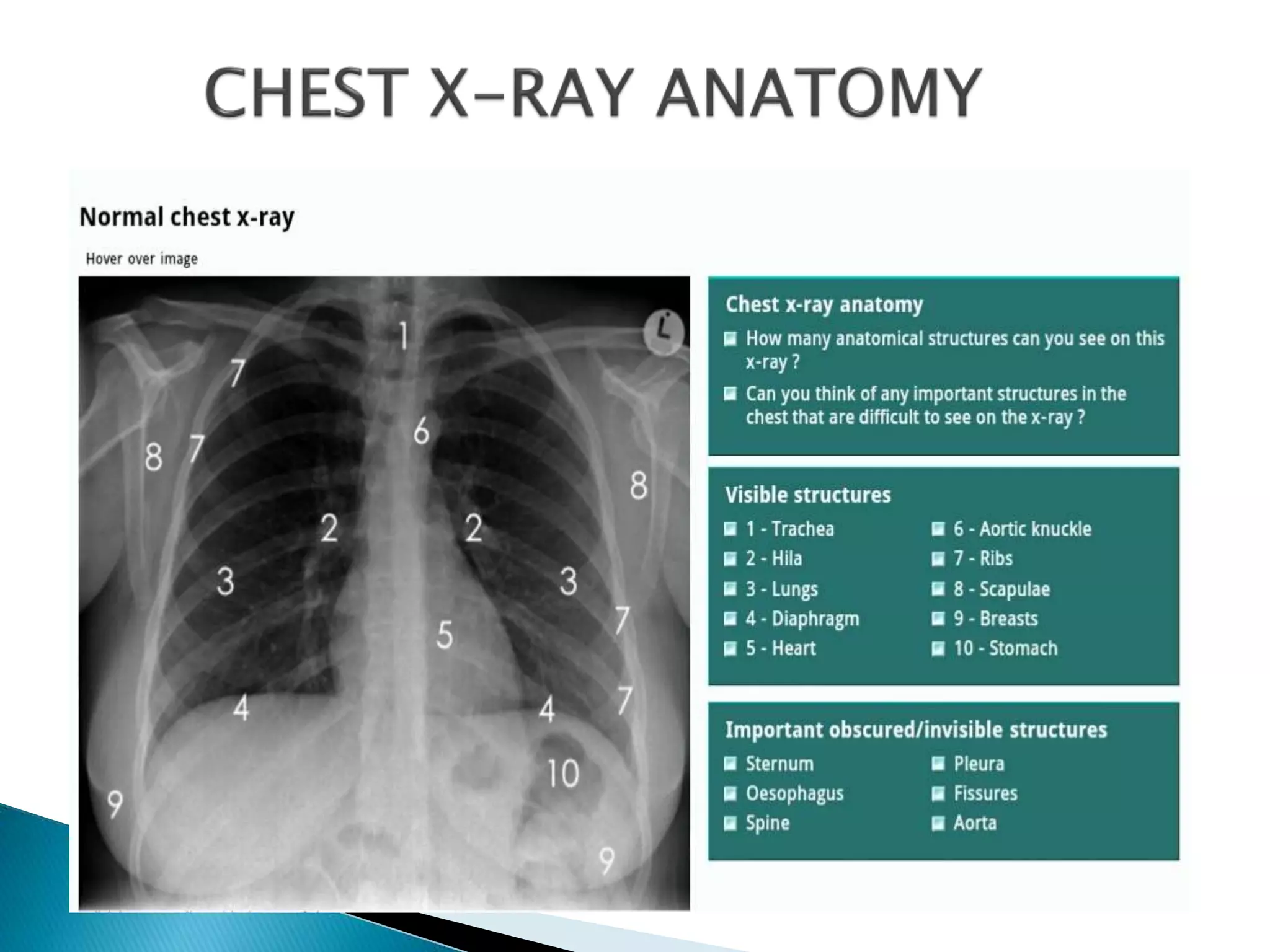
Presented by Prof Frank Peters
2018
Chest X-Ray is one of the most frequently requested
hospital investigations.
It is readily available and inexpensive in comparison
to other imaging studies.
The basic interpretation is of utmost importance in
answering several clinical questions at hand.
It is an important tool to complement both history
and initial clinical examination.
A. Patient details
 Name of the patient
 Age
 Date
B. Quality
• Image quality influences interpretation
• Quality is influenced by radiographic technique
and patient factors.
• First determine if the clinical question can be
answered.
• Check the image for – Projection, rotation,
inspiration, penetration and artefacts.
1. Projection
 Look to see if the film is antero-posterior (AP) or
postero-anterior (PA) view
 With an AP view the X-ray beam is in front the
patient and the X-Ray placed at the back, and the
other way round for PA.
 The standard CXR is PA but many emergency CXRs
are AP.
 The CXR projection has an important bearing on
the interpretation of the structures.
2. Orientation
 Identify the left/right markings
 Identify the anatomical structures, erect/supine.
 Do not always assume that the heart will always be
on the left because certain pathologies can result
with mediastinal shift, dextrocardia can also be a
possibility.
 You do not have to solely rely on just the CXR
markings.
3. Rotation
 Identify the medial ends of the clavicles and select
one of the thoracic vertebra spinous processes that
falls between them.
 The medial ends of the clavicles should be
equidistant from the spinous process, if that’s not
the case then the X-Ray is rotated.
4. Inspiration (Degree of inspiration)
 To judge the degree of inspiration, count the number of
ribs above the diaphragm.
 The midpoint of the right hemi-diaphragm should be
between the 5th and 7th ribs anteriorly.
 The anterior end of the 6th rib should be above the
diaphragm as should the posterior end of the 10th rib.
 If more ribs are visible the patient is hyperinflated
 If fewer it indicates inadequate inspiration
 Poor inspiration will make the heart look larger, give
appearance of basal shadowing and cause the trachea to
appear deviated to the right
5. Penetration
• To check the penetration, look at the lower part of the
cardiac shadow
• The vertebral bodies should be barely visible through the
cardiac shadow at this point.
• If they are clearly visible then the film is over penetrated and
you may miss low density lesion.
• If you cannot see them at all then the film is under penetrated
and the lung fields will appear falsely opaque (white).
• The left hemidiaphragm should be visible to the edge of the
spine
• When comparing X-Rays first determine if the level of
penetration is similar.
1. TRACHEA
 It should be central or slightly deviated to the right.
- In case of deviation decide if is due to rotation or
pathology
 View the carina, angle should be between 60 –100
degrees.
 Because it contains air, it appears darker
(blacker/radiolucent).
 Trachea normally narrows at the vocal cords (T3/T4)
2. HILAR STRUCTURES
• Also called lung root, consists of the major
bronchi and pulmonary vessels (veins/arteries).
• The hila are not symmetrical but consist of the
same basic structures.
• The lymph nodes are also present but no visible
unless abnormal.
3. LUNGS
• The lungs occupies the largest portion of the thoracic
cavity.
• The lungs are assessed and described by dividing them
into upper, middle and lower zones.
• The lung zones do not equate to lung lobes e.g. The
lower zone on the right consists of middle and lower
lobes.
• Compare left with right.
• Compare an area of abnormality with the rest of the lung
on the same side.
• If there is any asymmetry decide which side is abnormal
4. PLEURA AND PLEURAL SPACES
• The pleura are only visible when there is an
abnormality present.
• This can be due to pleural thickening and fluid
or air accumulating in the pleural spaces.
• Lung markings should reach the thoracic wall
5. COSTOPHRENIC ANGLE AND RECESS
• The costophrenic recesses are formed by
hemidiaphragms and chest wall.
• They contain the rim of the lung bases which lie
over the dome of each hemidiaphragm.
• These angles are known as the costophrenic
angles.
• Costophrenic angles should form acute angles
that are sharp to the point.
6. HEMIDIAPHRAGM
7. HEART
• The heart lies more to the left of the thoracic cavity.
• The heart is assessed by means of the cardio-
thoracic ratio (CTR).
• CTR = Cardiac width : Thoracic width
• CTR > 50% is abnormal – PA view only
• The left hemidiaphragm should be visible behind the
heart.
• The hemidiaphrams do not represent the lowest
point of the lungs.
8. THE MEDIASTINUM
• The mediastinum contains the heart and great vessels
(Middle mediatinum) and potential spaces in front of the
heart (anterior mediastinum), behind the heart (Posterior
mediastinum) and above the heart (superior
mediastinum).
• These potential spaces are not defined on a normal CXR,
but their awareness can help in describing location of
disease processes.
• There are several structures in the superior mediastinum
that should always be checked. These include aortic
knuckle, aorto-pulmonary window and the right para-
tracheal stripe.
9. SOFT TISSUE
• Normal fat planes are clearly defined in the soft
tissues.
• They appear as smooth layers of low density (black),
between layers of relatively dense (whiter) muscles.
• Irregular low density within soft tissues may be as a
result of tracking air as a result of injury to the
airways or pleura.
• This is known as surgical emphysema and produces
the distinctive clinical sign of palpable subcutaneous
‘bubble wrap’.
10. BONES
• The most dense tissue visible on CXR.
• Look for fractures, dislocation, subluxation,
osteoblastic or osteolytic lesions etc.
a. The CXR is an important tool to complement both
history and initial clinical examination.
b. Low density structures appear
dark(black/radiolucent) and high density are
whitish (opaque).
c. Abnormalities need to be described in detail.
d. Identify the most striking abnormality first.
However, once you are done with this, it is vital to
check the rest of the image.
Describing abnormalities
Systematic CXR interpretation is important
The NB! Question is ‘ Can the clinical question be
answered?’
1. Radiology masterclass.[Online] Accessed [30
May 15]. Available
from:http://www.radiologymasterclass.co.uk
2. Corne J, Pointon K. Chest X-Ray Made Easy 3rd
Ed. Churchill Livingstone. 2010

Chest x-ray.zp162335 (1)

  • 1.
    Presented by ProfFrank Peters 2018
  • 2.
    Chest X-Ray isone of the most frequently requested hospital investigations. It is readily available and inexpensive in comparison to other imaging studies. The basic interpretation is of utmost importance in answering several clinical questions at hand. It is an important tool to complement both history and initial clinical examination.
  • 3.
    A. Patient details Name of the patient  Age  Date
  • 4.
    B. Quality • Imagequality influences interpretation • Quality is influenced by radiographic technique and patient factors. • First determine if the clinical question can be answered. • Check the image for – Projection, rotation, inspiration, penetration and artefacts.
  • 5.
    1. Projection  Lookto see if the film is antero-posterior (AP) or postero-anterior (PA) view  With an AP view the X-ray beam is in front the patient and the X-Ray placed at the back, and the other way round for PA.  The standard CXR is PA but many emergency CXRs are AP.  The CXR projection has an important bearing on the interpretation of the structures.
  • 9.
    2. Orientation  Identifythe left/right markings  Identify the anatomical structures, erect/supine.  Do not always assume that the heart will always be on the left because certain pathologies can result with mediastinal shift, dextrocardia can also be a possibility.  You do not have to solely rely on just the CXR markings.
  • 10.
    3. Rotation  Identifythe medial ends of the clavicles and select one of the thoracic vertebra spinous processes that falls between them.  The medial ends of the clavicles should be equidistant from the spinous process, if that’s not the case then the X-Ray is rotated.
  • 12.
    4. Inspiration (Degreeof inspiration)  To judge the degree of inspiration, count the number of ribs above the diaphragm.  The midpoint of the right hemi-diaphragm should be between the 5th and 7th ribs anteriorly.  The anterior end of the 6th rib should be above the diaphragm as should the posterior end of the 10th rib.  If more ribs are visible the patient is hyperinflated  If fewer it indicates inadequate inspiration  Poor inspiration will make the heart look larger, give appearance of basal shadowing and cause the trachea to appear deviated to the right
  • 16.
    5. Penetration • Tocheck the penetration, look at the lower part of the cardiac shadow • The vertebral bodies should be barely visible through the cardiac shadow at this point. • If they are clearly visible then the film is over penetrated and you may miss low density lesion. • If you cannot see them at all then the film is under penetrated and the lung fields will appear falsely opaque (white). • The left hemidiaphragm should be visible to the edge of the spine • When comparing X-Rays first determine if the level of penetration is similar.
  • 19.
    1. TRACHEA  Itshould be central or slightly deviated to the right. - In case of deviation decide if is due to rotation or pathology  View the carina, angle should be between 60 –100 degrees.  Because it contains air, it appears darker (blacker/radiolucent).  Trachea normally narrows at the vocal cords (T3/T4)
  • 20.
    2. HILAR STRUCTURES •Also called lung root, consists of the major bronchi and pulmonary vessels (veins/arteries). • The hila are not symmetrical but consist of the same basic structures. • The lymph nodes are also present but no visible unless abnormal.
  • 22.
    3. LUNGS • Thelungs occupies the largest portion of the thoracic cavity. • The lungs are assessed and described by dividing them into upper, middle and lower zones. • The lung zones do not equate to lung lobes e.g. The lower zone on the right consists of middle and lower lobes. • Compare left with right. • Compare an area of abnormality with the rest of the lung on the same side. • If there is any asymmetry decide which side is abnormal
  • 23.
    4. PLEURA ANDPLEURAL SPACES • The pleura are only visible when there is an abnormality present. • This can be due to pleural thickening and fluid or air accumulating in the pleural spaces. • Lung markings should reach the thoracic wall
  • 25.
    5. COSTOPHRENIC ANGLEAND RECESS • The costophrenic recesses are formed by hemidiaphragms and chest wall. • They contain the rim of the lung bases which lie over the dome of each hemidiaphragm. • These angles are known as the costophrenic angles. • Costophrenic angles should form acute angles that are sharp to the point.
  • 27.
  • 29.
    7. HEART • Theheart lies more to the left of the thoracic cavity. • The heart is assessed by means of the cardio- thoracic ratio (CTR). • CTR = Cardiac width : Thoracic width • CTR > 50% is abnormal – PA view only • The left hemidiaphragm should be visible behind the heart. • The hemidiaphrams do not represent the lowest point of the lungs.
  • 32.
    8. THE MEDIASTINUM •The mediastinum contains the heart and great vessels (Middle mediatinum) and potential spaces in front of the heart (anterior mediastinum), behind the heart (Posterior mediastinum) and above the heart (superior mediastinum). • These potential spaces are not defined on a normal CXR, but their awareness can help in describing location of disease processes. • There are several structures in the superior mediastinum that should always be checked. These include aortic knuckle, aorto-pulmonary window and the right para- tracheal stripe.
  • 36.
    9. SOFT TISSUE •Normal fat planes are clearly defined in the soft tissues. • They appear as smooth layers of low density (black), between layers of relatively dense (whiter) muscles. • Irregular low density within soft tissues may be as a result of tracking air as a result of injury to the airways or pleura. • This is known as surgical emphysema and produces the distinctive clinical sign of palpable subcutaneous ‘bubble wrap’.
  • 38.
    10. BONES • Themost dense tissue visible on CXR. • Look for fractures, dislocation, subluxation, osteoblastic or osteolytic lesions etc.
  • 39.
    a. The CXRis an important tool to complement both history and initial clinical examination. b. Low density structures appear dark(black/radiolucent) and high density are whitish (opaque). c. Abnormalities need to be described in detail. d. Identify the most striking abnormality first. However, once you are done with this, it is vital to check the rest of the image.
  • 40.
  • 46.
    Systematic CXR interpretationis important The NB! Question is ‘ Can the clinical question be answered?’
  • 48.
    1. Radiology masterclass.[Online]Accessed [30 May 15]. Available from:http://www.radiologymasterclass.co.uk 2. Corne J, Pointon K. Chest X-Ray Made Easy 3rd Ed. Churchill Livingstone. 2010