This document discusses the generations of computed tomography (CT) scanning technology and their key features. It begins with a brief history of CT scanning's development. The generations covered include first generation translate-rotate systems with a single detector, second generation systems adding multiple detectors, third generation pure rotational systems, fourth generation using a stationary detector ring, and fifth generation stationary-stationary systems used for cardiac imaging. Each generation is characterized by its scanning geometry and improvements such as faster scanning times, larger detector arrays, and elimination of mechanical motions. The document also discusses developments like spiral/helical CT, multi-slice CT, and advances in detectors and x-ray tubes.
principle of ct scanner
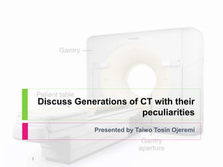
generations
scanning motion
EMI unit
xray beam
x ray tube
advantages
disadvantages
in this you PPT got clear idea about generation of ct
if you have any doubt text me
insta ID - ___sadham_____
this slide sharer contents are basic principle of CT fluoroscopy , software and hardware parts of equipment and image aqua cation and radiation dose comparison and videos related to equipment .
This slide best explains the introduction of CT, basis and types of CT image reconstructions with detailed explanation about Interpolation, convolution, Fourier slice theorem, Fourier transformation and brief explanation about the image domain i.e digital image processing.
this power-point slide presentation includes lots of information like how MRI coil works. what is shimming, magnet, fringe, and design of mri coil and also magnet. this will help a lot for radiologist and technician radiographers.. thanks.
Basic physics of multidetector computed tomography ( CT Scan) - how ct scan works, different generations of ct, how image is generated and displayed and image artifacts related to CT Scan.
Recent advancements in modern x ray tubeSantosh Ojha
All the advancements in X-ray tubes till date are done to increase the Tube heat storage capacity thus increasing the lifetime of x -ray tubes. This slide explains about these recent advancements in x-ray tubes.
principle of ct scanner
generations
scanning motion
EMI unit
xray beam
x ray tube
advantages
disadvantages
in this you PPT got clear idea about generation of ct
if you have any doubt text me
insta ID - ___sadham_____
this slide sharer contents are basic principle of CT fluoroscopy , software and hardware parts of equipment and image aqua cation and radiation dose comparison and videos related to equipment .
This slide best explains the introduction of CT, basis and types of CT image reconstructions with detailed explanation about Interpolation, convolution, Fourier slice theorem, Fourier transformation and brief explanation about the image domain i.e digital image processing.
this power-point slide presentation includes lots of information like how MRI coil works. what is shimming, magnet, fringe, and design of mri coil and also magnet. this will help a lot for radiologist and technician radiographers.. thanks.
Basic physics of multidetector computed tomography ( CT Scan) - how ct scan works, different generations of ct, how image is generated and displayed and image artifacts related to CT Scan.
Recent advancements in modern x ray tubeSantosh Ojha
All the advancements in X-ray tubes till date are done to increase the Tube heat storage capacity thus increasing the lifetime of x -ray tubes. This slide explains about these recent advancements in x-ray tubes.
generations of CT, explains each generations of CT, muti detector computer tomography, slip ring technology, main terminologies such as FOV , pitch, voxel and matrix, pixel size equation. EBCT, Basic configuration of CT, Data acquisition systems DAS, multi-slice CT
its about the CT scan and generations in the form of PPT explaining each of first generation , second generation, third generation, fourth generation, fith generation and sith generation
Flu Vaccine Alert in Bangalore Karnatakaaddon Scans
As flu season approaches, health officials in Bangalore, Karnataka, are urging residents to get their flu vaccinations. The seasonal flu, while common, can lead to severe health complications, particularly for vulnerable populations such as young children, the elderly, and those with underlying health conditions.
Dr. Vidisha Kumari, a leading epidemiologist in Bangalore, emphasizes the importance of getting vaccinated. "The flu vaccine is our best defense against the influenza virus. It not only protects individuals but also helps prevent the spread of the virus in our communities," he says.
This year, the flu season is expected to coincide with a potential increase in other respiratory illnesses. The Karnataka Health Department has launched an awareness campaign highlighting the significance of flu vaccinations. They have set up multiple vaccination centers across Bangalore, making it convenient for residents to receive their shots.
To encourage widespread vaccination, the government is also collaborating with local schools, workplaces, and community centers to facilitate vaccination drives. Special attention is being given to ensuring that the vaccine is accessible to all, including marginalized communities who may have limited access to healthcare.
Residents are reminded that the flu vaccine is safe and effective. Common side effects are mild and may include soreness at the injection site, mild fever, or muscle aches. These side effects are generally short-lived and far less severe than the flu itself.
Healthcare providers are also stressing the importance of continuing COVID-19 precautions. Wearing masks, practicing good hand hygiene, and maintaining social distancing are still crucial, especially in crowded places.
Protect yourself and your loved ones by getting vaccinated. Together, we can help keep Bangalore healthy and safe this flu season. For more information on vaccination centers and schedules, residents can visit the Karnataka Health Department’s official website or follow their social media pages.
Stay informed, stay safe, and get your flu shot today!
Ozempic: Preoperative Management of Patients on GLP-1 Receptor Agonists Saeid Safari
Preoperative Management of Patients on GLP-1 Receptor Agonists like Ozempic and Semiglutide
ASA GUIDELINE
NYSORA Guideline
2 Case Reports of Gastric Ultrasound
New Drug Discovery and Development .....NEHA GUPTA
The "New Drug Discovery and Development" process involves the identification, design, testing, and manufacturing of novel pharmaceutical compounds with the aim of introducing new and improved treatments for various medical conditions. This comprehensive endeavor encompasses various stages, including target identification, preclinical studies, clinical trials, regulatory approval, and post-market surveillance. It involves multidisciplinary collaboration among scientists, researchers, clinicians, regulatory experts, and pharmaceutical companies to bring innovative therapies to market and address unmet medical needs.
- Video recording of this lecture in English language: https://youtu.be/lK81BzxMqdo
- Video recording of this lecture in Arabic language: https://youtu.be/Ve4P0COk9OI
- Link to download the book free: https://nephrotube.blogspot.com/p/nephrotube-nephrology-books.html
- Link to NephroTube website: www.NephroTube.com
- Link to NephroTube social media accounts: https://nephrotube.blogspot.com/p/join-nephrotube-on-social-media.html
TEST BANK for Operations Management, 14th Edition by William J. Stevenson, Ve...kevinkariuki227
TEST BANK for Operations Management, 14th Edition by William J. Stevenson, Verified Chapters 1 - 19, Complete Newest Version.pdf
TEST BANK for Operations Management, 14th Edition by William J. Stevenson, Verified Chapters 1 - 19, Complete Newest Version.pdf
Couples presenting to the infertility clinic- Do they really have infertility...Sujoy Dasgupta
Dr Sujoy Dasgupta presented the study on "Couples presenting to the infertility clinic- Do they really have infertility? – The unexplored stories of non-consummation" in the 13th Congress of the Asia Pacific Initiative on Reproduction (ASPIRE 2024) at Manila on 24 May, 2024.
Title: Sense of Taste
Presenter: Dr. Faiza, Assistant Professor of Physiology
Qualifications:
MBBS (Best Graduate, AIMC Lahore)
FCPS Physiology
ICMT, CHPE, DHPE (STMU)
MPH (GC University, Faisalabad)
MBA (Virtual University of Pakistan)
Learning Objectives:
Describe the structure and function of taste buds.
Describe the relationship between the taste threshold and taste index of common substances.
Explain the chemical basis and signal transduction of taste perception for each type of primary taste sensation.
Recognize different abnormalities of taste perception and their causes.
Key Topics:
Significance of Taste Sensation:
Differentiation between pleasant and harmful food
Influence on behavior
Selection of food based on metabolic needs
Receptors of Taste:
Taste buds on the tongue
Influence of sense of smell, texture of food, and pain stimulation (e.g., by pepper)
Primary and Secondary Taste Sensations:
Primary taste sensations: Sweet, Sour, Salty, Bitter, Umami
Chemical basis and signal transduction mechanisms for each taste
Taste Threshold and Index:
Taste threshold values for Sweet (sucrose), Salty (NaCl), Sour (HCl), and Bitter (Quinine)
Taste index relationship: Inversely proportional to taste threshold
Taste Blindness:
Inability to taste certain substances, particularly thiourea compounds
Example: Phenylthiocarbamide
Structure and Function of Taste Buds:
Composition: Epithelial cells, Sustentacular/Supporting cells, Taste cells, Basal cells
Features: Taste pores, Taste hairs/microvilli, and Taste nerve fibers
Location of Taste Buds:
Found in papillae of the tongue (Fungiform, Circumvallate, Foliate)
Also present on the palate, tonsillar pillars, epiglottis, and proximal esophagus
Mechanism of Taste Stimulation:
Interaction of taste substances with receptors on microvilli
Signal transduction pathways for Umami, Sweet, Bitter, Sour, and Salty tastes
Taste Sensitivity and Adaptation:
Decrease in sensitivity with age
Rapid adaptation of taste sensation
Role of Saliva in Taste:
Dissolution of tastants to reach receptors
Washing away the stimulus
Taste Preferences and Aversions:
Mechanisms behind taste preference and aversion
Influence of receptors and neural pathways
Impact of Sensory Nerve Damage:
Degeneration of taste buds if the sensory nerve fiber is cut
Abnormalities of Taste Detection:
Conditions: Ageusia, Hypogeusia, Dysgeusia (parageusia)
Causes: Nerve damage, neurological disorders, infections, poor oral hygiene, adverse drug effects, deficiencies, aging, tobacco use, altered neurotransmitter levels
Neurotransmitters and Taste Threshold:
Effects of serotonin (5-HT) and norepinephrine (NE) on taste sensitivity
Supertasters:
25% of the population with heightened sensitivity to taste, especially bitterness
Increased number of fungiform papillae
Lung Cancer: Artificial Intelligence, Synergetics, Complex System Analysis, S...Oleg Kshivets
RESULTS: Overall life span (LS) was 2252.1±1742.5 days and cumulative 5-year survival (5YS) reached 73.2%, 10 years – 64.8%, 20 years – 42.5%. 513 LCP lived more than 5 years (LS=3124.6±1525.6 days), 148 LCP – more than 10 years (LS=5054.4±1504.1 days).199 LCP died because of LC (LS=562.7±374.5 days). 5YS of LCP after bi/lobectomies was significantly superior in comparison with LCP after pneumonectomies (78.1% vs.63.7%, P=0.00001 by log-rank test). AT significantly improved 5YS (66.3% vs. 34.8%) (P=0.00000 by log-rank test) only for LCP with N1-2. Cox modeling displayed that 5YS of LCP significantly depended on: phase transition (PT) early-invasive LC in terms of synergetics, PT N0—N12, cell ratio factors (ratio between cancer cells- CC and blood cells subpopulations), G1-3, histology, glucose, AT, blood cell circuit, prothrombin index, heparin tolerance, recalcification time (P=0.000-0.038). Neural networks, genetic algorithm selection and bootstrap simulation revealed relationships between 5YS and PT early-invasive LC (rank=1), PT N0—N12 (rank=2), thrombocytes/CC (3), erythrocytes/CC (4), eosinophils/CC (5), healthy cells/CC (6), lymphocytes/CC (7), segmented neutrophils/CC (8), stick neutrophils/CC (9), monocytes/CC (10); leucocytes/CC (11). Correct prediction of 5YS was 100% by neural networks computing (area under ROC curve=1.0; error=0.0).
CONCLUSIONS: 5YS of LCP after radical procedures significantly depended on: 1) PT early-invasive cancer; 2) PT N0--N12; 3) cell ratio factors; 4) blood cell circuit; 5) biochemical factors; 6) hemostasis system; 7) AT; 8) LC characteristics; 9) LC cell dynamics; 10) surgery type: lobectomy/pneumonectomy; 11) anthropometric data. Optimal diagnosis and treatment strategies for LC are: 1) screening and early detection of LC; 2) availability of experienced thoracic surgeons because of complexity of radical procedures; 3) aggressive en block surgery and adequate lymph node dissection for completeness; 4) precise prediction; 5) adjuvant chemoimmunoradiotherapy for LCP with unfavorable prognosis.
3. Introduction
Computerized tomography scan is a
diagnostic radiological imaging
procedure that uses x-rays to build
cross sectional images(slices) of the
visualized body parts.
Density of the object studied is
extrapolated from their attenuation
coefficient.
Unlike x-ray radiography, cross
section images are taken from
different angles and this allows for
information on its depth.
Mathematical algorithm are used for
image reconstruction
CT has undergone several evolutions
with MSCT for better clinical
application
courtesy :
radiology key
3
4. Brief History
April 1972- G Hounsfield constructed new imaging
technique he called computerized axial transverse
scanning.
1975- He built a whole body scanner.
1979 Hounsfield and cormack got Nobel prize in
physiology and medicine.
4
5. Definition of Generation
.
● Classification of computed
tomography (CT) based upon:
arrangement of components and
mechanical motion for data
acquisition.
● “Generation” the order in which CT
scanner design has been
introduced, and each has a number
associated with it
5
6. First Generation
Design:
● XRAY TUBE: single X-ray
source.
● DETECTOR: one for
single slice
● GEOMETRY: translate,
rotate
● Duration of scan:
average 25-30mins
● Beam: Pencil beam
● translated across
patient to obtain set of
parallel projection
measurements at one
angle
Courtesy internet.
Courtesy :: kau.edu.sa
6
7. First generation
Translation and rotation
process, this geometry is
referred to as a
translate/rotate scanner.
Process is repeated once
for each projection angle
until 180 projections ,
across a 24 cm FOV.
Linear translation 160.
Total projection 28800
Source : M.
mongkolsuk
7
8. First CT
.
• EMI Mark I scanner (1973)
• Earliest versions:4.5 minutes for a single
scan and thus were restricted to some
regions (patient motion controlled)
• Later versions: procedures = series of
scans
– procedure time reduced some what by using
two detectors so that two parallel sections
were acquired in one scan
• Contrast resolution of internal
structures was unprecedented,
8
12. Second Generation CT
Design:
● XRAY TUBE: single
X-ray source.
● DETECTOR: multilple
(up to 30)
● GEOMETRY:
translate, rotate
● Duration of scan: less
than 90s
● Beam: Fan shaped
beam
..
Courtesy :: kau.edu.sa
12
13. ● Design: multiple
detectors
● X-ray source emits
radiation over a large
angle, the efficiency of
measuring projections
was greatly improved
Source : M.
mongkolsuk
13
14. Second CT
.
• Early versions: 3 detectors each displaced
by1°
– Since each detector viewed the x-ray tube at a different angle , a
single translation produced 3 projections
– The system could rotate 3° to the next projection rather than 1°
– make only 60 translations instead of 180 to acquire a complete
section
– Scan times were reduced X 3
• Later versions: up to 53 detectors
– Fast enough (tens of seconds)to permit acquisition during a single
breath hold
– First designs to permit scans of the trunk
– Because rotating anode tubes could not
14
15. PROS
Fewer linear
translation needed.
• Reducing scan time
By adding detectors
angularly displaced ,
several projections
could be obtained in a
single translation
• The trunk could be
imaged
15
16. THIRD GENERATION
Design:
● XRAY TUBE: single
X-ray source single
● DETECTOR: multiple
(288 originally up to
700 in an arc)
● GEOMETRY: rotate,
rotate
● Duration of scan:
approximately 5s
● Beam: Fan shaped x-
ray beam
.
Courtesy :: kau.edu.sa
16
17. Third Generation CT
“Slam-bang translational
motion” was replaced with
smooth rotational motion
X-ray tube is collimated to
a wide x-ray
beam(fan-shaped )
• Directed toward an arc-
shaped row of detectors
• Tube and detector array
rotate around patient
Source : M.
mongkolsuk
17
18. Third Generation ; Rotate -rotate
.
• Design: larger array Of detectors
– (300-700detectors, usually circular
– Shorter scanning time (2-5 sec)
– Designers: pure rotational scanning motion could be used , then
it would be possible to use higher-power ,rotating anode x-ray
tubes and thus improve scan speeds in thicker body parts
– higher-output rotating anode x-ray tubes could be used
– greatly reducing scan times
• Different projections are obtained during rotation
by pulsing x-ray source or by sampling the
detectors at a very high rate
18
19. ● Pros
● Imaging process is
significantly faster than
1st or 2nd generation
systems
● Angle of fan beam
increased to cover entire
patient
● Eliminated need for
translational motion
● Improvement in detector
and data acquisition
technology
● detector array with enough, high
spatial resolution cells to allow
measurement of a fan-beam
● Cons:
● Ring artefacts due to
electron drift between many
detectors.
● very high performance
detectors are needed to
avoid ring artefacts and the
system is more sensitive to
aliasing than 1st or 2nd
generation scanners
19
20. FOURTH GENERATION
Design:
● XRAY TUBE: single
X-ray source
● DETECTOR: multiple
(more than 2000
arranged in fixed
outer ring)
● GEOMETRY: rotate,
stationary
● Duration of scan: very
few seconds
● Beam: Fan shaped x-
ray beam
.
Courtesy :: kau.edu.sa
20
21. FOURTH GENERATION CT SCAN
Design: stationary
detector ring & rotating X-
ray tube
Source : M.
mongkolsuk
21
22. Fourth CT
Pros
Stationary detector has a
larger acceptance angle for
radiation.
● Larger fan beam
● Shorter scanning time
● eliminated translate-
rotate motion
● Circular array of FIXED
detectors
● Source only rotates within a
stationary ring of detectors
Cons
Require larger number of
detector cells and electronic
channels (higher cost) to
achieve the same spatial
resolution and dose
efficiency as a 3rd
generation system
● less efficient use of
detectors , less than 1/4 are
used at any point during
scanning.
● Larger acceptance angle
hence more sensitive to
scatter.
● Stationary dectectors
addressed ring artefacts
22
25. More Categories
.
● Several other CT scanner geometries
which have been developed and
marketed do not precisely fit the above
categories
25
26. FIFTH Generation ; stationary-stationary
● . Design: x-ray
tube is a large ring
that circles
patient, opposed
to detector ring
● Use: for cardiac
tomographic imaging
“cine CT”
● X - rays produced = high -
energy electron beam
● No moving parts to this
scanner gantry
● It is capable of 50 -
millisecond scan times
and can produce 17
CT slices/second. Source : M.
mongkolsuk
26
28. X-Ray Tube
.
● Vacuum
● Accelerating electrons
● e- will travel faster
● Filament
● Alternating current
● Thermal electrons
● Cathode (-)
● Filament plate with a tiny slit
● Connected to high voltage battery
source
● Target/Anode (+)
● Electrons collide with target
● Produce x-ray
● Must have high melting point
28
29. The cine CT system
.
● No mechanical scanning motion
● X-ray detector and tube anode are stationary
● Anode, is a very large semicircular ring that
forms an arc around the patient scan circle
● Source of X-rays is moved around the
same path as a fourth generation CT
scanner by steering an electron beam
around the X-ray anode
● Terms millisecond CT, ultrafast CT and
electron beam CT have also been used,
although the latter can be confusing since
the term suggests that the patient is
exposed to an electron beam
29
30. Cont.
. ● Very fast scanner ,data collection for 1slice is
50-100 ms
● Requires no mechanical motion to acquire
data
● Sweeps an intense electron beam across
a large, stationary anode target which
surrounds the patient
● X-rays are emitted from the point where
electrons strike target
● X-rays transmitted through object are
measured by a stationary array of
detectors
● Cine CT systems, have higher noise level
and lower spatial resolution but are ideal
30
31. Pros
No mechanically moving
parts
Fastest commercial CT
temporal resolution
Cons
High cost and maintanance
Increased signal to noise
ratio and spatial resolution
compared to conventional
CT
31
32. Generation Source Source Collimation Detector
1st Single X-ray Tube Pencil Beam Single
2nd Single X-ray Tube Fan Beam (not enough to cover FOV) Multiple
3rd Single X-ray Tube Fan Beam (enough to cover FOV) Many
4th Single X-ray Tube Fan Beam covers FOV
Stationary
Ring of
Detectors
5th
Many tungsten
anodes in single
large tube Fan Beam
Stationary
Ring of
Detectors
32
33. Spiral/Helical CT
● Design: x-ray tube
rotates as patient is
moved smoothly into
x-ray scan field
● Simultaneous source
rotation, table
translation and data
acquisition
● Produces one
continuous volume set
of data for entire region
● Data for multiple slices
from patient acquired at
1sec/slice
.
33
34. Advantages of Spiral
● . Speed: patient movement continuos ,shorter exam time
; entire abdomen or chest: 30 sec
● Improved detections
● Improved contrast: image a region in a short period,
contrast can be timed
● Improved reconstruction & manipulation: volume
of data collected, transverse data can be
reconstructed in any plane- strip away skin, muscles,
etc….
34
35. Spiral/Helical CT
. Three technological
developments:
1. Slip-ring gantry
designs
2. Very high power x-
ray tubes
3. Interpolation
algorithms to handle
projection data
.
35
36. 1. Slip-Ring Technology
● . Alternative to cabling system =
slip-ring
● 1989 Kalender
● Electromechanical
devices:
circular electrical
conductive rings
and brushes
● Transmit electrical energy
across a moving interface
● All power and control signals
from the stationary parts of
the scanner system are
communicated to the rotating
frame through slip ring
● Allow scan frame to rotate
continuously with no need to
stop between rotations to
rewind system cables
.
36
37. 2. High Power X-ray Tube
. ● Thermal load in CT
● 1st and 2nd, stationary tube(low heat, slow scans)
● Oil cooling thermal systems around tube, fast scans
● scan time vs. Heat capacity increased x 5
● thermal demands on the x-ray tube
● Tubes with much higher thermal capacities were
required to withstand continuous operation over
multiple rotations
● New design: ceramic insulators ,oil cooling of
bearing, compact metal envelop
● Expected life of tube 10,000-40,000 hrs vs. 1000
regular one
37
38. 3. Interpolation Algorithms
.
● Kalender developed interpolation
methods to generate projections in a
single plane
● Overlapping sections generated by
math, not beam, improve z-axis with
no increase in dose
● Improved image quality
38
40. Multi-slice CT
● Single row had its limitation.
● Design: multiple detector array.
● Cone Beam & multiple parallel rows
of detectors
● The collimator spacing is wider and
more of the x-rays that are
produced by the tube are used in
producing image data
● Opening up the collimator in a single
array scanner increases slice thickness,
reducing spatial resolution in the slice
thickness dimension
● With multiple detector array
scanners, slice thickness is
determined by detector size, not
by the collimator
.
40
41. MSCT
1998 all major manufacturers introduced multislice CT, simultaneous
acquisition of four slices, providing a great improvement.
4 slice, not fast enough to avoid venous overlay assuming a cerebral
circulation time of less than 5 sec
In 2000, 8 slice CT were presented, followed by 16 slice in 2001
Most modern generation of MSCT is 320 slices per rotation,
enabling a whole body CTA with 1,500mm scan range in 22-
25seconds
Components: tubes and detector measurement systems, large
influence on performance
Development in software and computer capacity lead to processing
and reconstruction in a short time
41
42. MSCT advantages
● “turbo-charged” spiral
● Up to 8 rows of
detectors
● 4 rows, large volume of
patient scanned 1 BH
● (thorax, abdomen,
pelvis) at once
● Allows 1mm sections
though chest in 20 sec
● Improvement in details
● Problem with PACS,
stain on storage system
.
42
43. ● Widened (z-direction) x
ray beam & detector
array to acquire multiple
(4-64 slices
simultaneously)
● Advantage: reducing scan
time/ increase z-resolution
● Disadvantage: less scatter
rejection compared to
single slice, very
expensive
.
43
There are 4 bassic densities-air=-1000, fat =-60 to -120, water= 0, compact bone=+1000
Source/detector rotate slightly and a subsequent set of measurements are obtained during a translation past patient
Source and array of detectors are translated as in a first generation system
but since beam measured by each detector is at a slightly different angle with respect to object, each translation step generates multiple parallel ray projections
Multiple projections obtained during each traversal past the patient
this scanner is significantly more efficient and faster than 1st one
.
Source and array of detectors are translated as in a first generation system
but since beam measured by each detector is at a slightly different angle with respect to object, each translation step generates multiple parallel ray projections
Multiple projections obtained during each traversal past the patient
this scanner is significantly more efficient and faster than 1st one
Sampling considerations required scanning an additional arc of one fan angle beyond 180°, although most scanners rotate 360° for each scan.
Current helical scanners are based on modifications of rotate- rotate designs
Scan times = few seconds or less, and recent versions are capable of subsecond scan times
Number of detectors increased substantially (to more than 800 detectors)
Mechanically joined x-ray tube and detector array rotate together
Newer systems have scan times of ½ second
Design: also Early versions: had some 600 detectors
Later versions: had up to 4,800
Only the x-ray generator and tube rotate at 360 ,thus shortening the scanning time even more
10 times faster than true scan speed of a 256slice CT(500ms)
5 ttimes faster than dual ssoure CT (166ms)
Radiation dose time times lower than that of 64slice CT
Due to its complexity and cost effectivenes aand the rapid advancement of MDCT, EBCT is under shadoweed.
1990,Significant advancement in technology
Allowed 3D image acquisition within a single breath hold
gantry rotation
Fast data acquisition system
High speed image reconstruction
High rating x-ray tube
Multiple reconstruction technique
Relates to the technique of double or triple rotation of the tube and detectors around the same axial plane
Provides double of triple the volume per slice, upon which the final image can be derived
In practice each rotation produces its own bank of raw data,
Hence motion which may occur during one rotation can be averaged out from data of the remaining two rotations
Multiscanning therefore reduces motion artifacts and consequently improves image quality