SlideShare a Scribd company logo
Presented by: Sandip Mandal
(B.Sc.MIT)
(BPKIHS,DHARAN,NEPAL)
CT TUBE AND DETECTOR
INTRODUCTION AND BRIEF HISTORY OF X-RAY TUBE
X-ray tube is a device in which energy
conversion takes place i.e. kinetic energy of fast moving electrons is
converted into x-ray (1%) and heat (99%).
There has been a series of advancement in the X-ray tube during the
course of time;
1895- Roentgen discovered x-rays (using crook’s type of tube)
1913- The Coolidge hot cathode x-ray tube
1929- Rotating anode tube
1932- Grid controlled stationary anode tube
1937- Grid controlled rotating anode tube
 1959- High speed tube
 1962-Rhenium alloyed tungsten composite anode tube
 1967-First dedicated mammography unit with molybdenum anode
 1971-Glass metal tube with molybdenum anode
 1973- Three layered anode (W-Re) + Mo or (W-Re) + (Ti-Zn-Mo)
1979- Metal ceramic tube
1981- Three focus tube
1989- Direct anode cooling with noiseless rotor
X-RAY TUBES USED IN CT
Since CT require longer continuous exposure time at higher KV and mA than needed
for general radiography hence; the general radiographic X-ray tubes cannot be used
for CT.It Should supply monochromatic X-ray beam for accurate reconstruction.
X-ray tubes for CT have been charged with heavy duty rotating anode tube with
higher thermal capacity and smaller focal spot (up to 0.6mm). These tubes are air
cooled with current value up to 600mA.
COMPARISION OF CT TUBE WITH VARIETY OF X-RAY TUBES AVAILABLE
BRIEF HISTORY AND ADVANCEMENTS IN CT X-RAY TUBE
CT X-ray tubes which are located in the heart of the gantry Provide
radiation source for CT.
• Early experimental models used radionuclide to supply such a radiation beam.
• First and second generation scanner used fixed anode (relatively large
(2x16mm)focal spot operated at 120 kVp & 30 mA & heavily filtered) , oil-cooled x-
ray tube but with the demand for increased output, gradually rotating anode
tubes become common in CT scanner.
• The introduction of spiral/helical CT with the continuous rotation scanner has
placed new demands on CT tubes.
Several technical advances in component design have been made to deal with
the problem of heat generation, heat storage and heat dissipation. For
example,
i)anode assembly including anode rotation- Anode is of larger diameter
with graphite backing, which allows the anode to absorb& dissipate large amounts of heat.
ii)target design- Anode target angle is made 7-10 degrees to diminish heel effect.
iii)cathode assembly- Bigger filament size, increased effective focal spot. Focal
spot size smaller (0.6 mm).
iv)the tube envelop-Although the borosilicate glass envelope in early CT tube
provides good thermal and electrical insulation, electrical arching result from tungsten
deposits on the glass caused by vaporization. To solve above problem, tubes with metal
envelop and ceramic insulator are now common e.g. Metal ceramic x-ray tube
METAL CERAMIC X-RAY TUBE USED IN CT
• Glass envelope has been replaced by metal casing and ceramic is used as insulation
of high voltage cable.
• A more recent development in x ray tube construction is metal ceramic tube,it’s
construction includes alloy of chromium and iron cylinder brazed to alumina
ceramic (aluminium oxide) insulators at each end. These insulators carry the anode
and cathode assemblies .
• The metal ceramic tubes are smaller and more robust than their glass equivalents.
They have another advantage, in that they enable more flexibility in the electrical
circuitary associated with the tube.
• Metal envelope grounded offers no chance of arcing of x-ray tube and hence
increase tube life.
• Anode rotates on an axle with bearing at each end providing greater stability and
reduced stress on shaft permits massive anode approx-2kg.
• Ceramic insulator (Al oxide) are used to insulate high voltage parts of x-ray tube
from metal envelope allowing more compact tube design.
• Metal ceramic tube offers Higher tube loading, Reduce off focus radiation, Allow
high tube current.
8-Feb-17 5
METAL CERAMIC X-RAY TUBE USED IN SPIRAL CT
8-Feb-17 7
Fig. Schematic diagram o f a Super Rotalix ceramic x-
ray tube: 1.Metal casing, 2. Anode, 3/6. Ball bearings,
4/8. Ceramic insulators, 5. Cathode, 7. Stator
windings, 9. Anode shaft, 10. Beryllium window
SUPER ROTALIX CERAMIC X-RAY TUBE IN CT
--It is also the type of metal ceramic X-ray tube
Detector X-ray
beam
X-ray
tube
• Higher Tube Loading
– Allows higher tube currents to be used because of larger heat
storage capacity of anode
• Longer Tube Life
– Deposition of tungsten on the glass wall acts as electrode
causing arcing bet. Glass and filament shortening tube life.
When metal enclosure is grounded, this deposition will not
alter grounding thus increasing its life
• Reduced off Focus Radiation
– Electrons back scattered from the anode may strike anode
again producing x-rays from areas other than focal spot. The
metal enclosure decreases off focus radiation by attracting off
focus electrons to the grounded metal wall relatively Positive
as compared to electrons. Low atomic no. of metal may
produce few and low energy x-rays.
10
Advantages Of Metal ceramic Tube
OTHER TYPES OF X-RAY TUBES USED IN CT
MRC(MAXIMUM ROTALIX CERAMIC) X-RAY TUBE
o In 1989 Phillips became the first company to introduce MRC.IT
is based on the technology of spiral groove bearing using liquid
metal alloy as lubricant. Focuses Significant improvement in
rotating anode x-ray tube.
FEATURES
o Higher output and longer tube life
o 200 mm graphite backed anode
o Anode heat storage capacity(8MHU)
o Tube voltage - 90 to 140 KV
o Tube current - 20 to 500 mA
o Anode angle - 7 degree
o Directly cooled anode
OTHER TYPES OF X-RAY TUBES USED IN CT
MRC X-RAY TUBE
Advantages
o Noiselessly rotating anode that could be switched on the
morning and switched off in the evening.
o Avoid waiting time during and between examination.
o Possible to achieve dose saving filter technique in
angiography.
USES
o Cardiovascular imaging
o MDCT
OTHER TYPES OF X-RAY TUBES USED IN CT
AQUILION X-RAY TUBE
o High capacity multi-slice CT tube
o Heat storage capacity 7.5 MHU
o Cooling rate 1.7 MHU/min
o Anode grounded
o Focal spot 1.4mm × 1.4 mm
o Air cooled
Aquilion vs conventional tube
TYPES OF X-RAY TUBE USED IN CT
NEW STRATON TUBE
o New construction
o Focused and deflected beam
of thermal electron
o whole tube and anode assembly rotates
o Bearing located out side
o Oil cooled
FEATURES
o Zero heat storage capacity
o Cooling rate 4.7 MHU/min
o Cooled down within 20 sec
o Enables gantry speed of 0.37 sec per rotation
o Tube current 500 mA
o Based on RET (rotating envelope tube) technology
o The electron beam in the tube is shaped and controlled
by magnetic deflection coil
o Focal spot – tungsten and rhenium
• One of the more interesting developments is the Siemens Straton
x-ray tube, which is currently available as an option on Sensation
16 scanners (Fig ).
• The tube itself is a radical new design, where the entire tube body
rotates, rather than just the anode, as is the case with conventional
designs. This change allows all the bearings to be located outside
the evacuated tube, and enables the anode to be cooled more
efficiently.
• The Straton has a low inherent heat capacity of 0.8 MHU, but an
extremely fast cooling rate of 5 MHU / min.
16
RECENT DEVELOPMENT IN X-RAY TUBE
• This compares with typical figures of 7-8 MHU and up to 1.4
MHU / min for existing tubes.
• The heat capacity and cooling rate combine to produce a tube
which Siemens claim is ‘0 MHU', implying that tube cooling
considerations are a thing of the past. Sensation 16 scanners fitted
with the Straton tube now have a fastest scan time of 0.37 seconds.
17
18
Conventional VS Straton
CT DETECTOR
o CT detector capture the radiation beam from the
patient and convert it into electrical signal, which
subsequently converted into binary coded
information
o Detector characteristic
o A. efficiency:- refers to the ability to capture,
absorb and convert x-ray photons to electrical
signal
Detectors measure the intensity of radiation transmitted
through the patient
• B. Stability:- refers to the steadiness of the
detector response, if not stable frequent
calibrations are required to render the signals
useful
• C. Response time:- refers to the speed with
which the detector can detect an x-ray event and
recover to detect another event (should be very
short in micro second)
• D. Dynamic range:- refers to the ratio of the
largest signal to be measured to the precision of
the smallest signal to be discriminated
o E . After glow:- refers to the persistence of the
image even after the radiation has been turned off
o TYPES OF DETECTOR
Two types:-
1. Gas ionization detector
Convert x-ray energy directly into electrical signal
2. Scintillation detectors
Convert x-ray energy into light
1. GAS IONIZATION DETECTOR
o Use high pressure ( about 25 atm)
nonradioactive xenon gas, in long thin cells
between two metal plates
o Based on the principle of ionization
o Consists of a series of individual gas chamber,
usually separated by tungsten plate carefully
positioned to act as electron collection plates
with voltage applied across it.
o When x-ray fall on the individual chamber, ionization
of gas result and produces positive and negative
ions. The positive ions migrate to the negatively
charged plate, whereas the negative ions at
positively charged plate
o The migration of ions causes a small signal current
that varies directly with the number of photons
absorbs
o QDE is only 50 – 60 percent
o Can be used for 3rd generation scanner only
Fig. Shows a typical gas-filled ionization detector
Charges
8-Feb-17 25
2. SCINTILLATION DETECTORS
o Solid state detectors that consist of a scintillation
crystal coupled to a photodiode tube
o When x-ray falls onto the crystals, flashes of
light are produced. The light is then directed to
PM tubes. Which then releases electrons and
these electrons cascade through a series of
dynodes that are carefully arranged and
maintained at diff. potential to result in small out
put signal
o Early scanner used sodium iodide crystals
coupled to PM tube. Due to afterglow problem
and the limited dynamic range of sodium iodide,
other crystals such as calcium fluoride and
bismuth germinate used in later scanner
o Now a days, solid state photodiode multiplier
scintillation crystal detectors are used
o The photodiode is a semiconductor whose p-n
junction allows current flow when exposed to
light
o Photodiodes are normally used with amplifier
because of the low output from the diode
o Response time of photodiode is extremely fast
(about 0.5 -250 nanosecond)
o Scintillation materials currently used with
photodiodes are cadmium tungsten and a ceramic
material made of high purity, rare earth oxides
based on doped rare earth compounds such as
yttrium and gadolinium oxysulphide, these crystals
are optically bonded to the photodiode
o The conversion efficiency and photon capture
efficiency of cadmium tungsten are 25-30% and
45% respectively and the dynamic range is 1
million to 1
X-ray beam X-ray photon
8-Feb-17 30
Isotropically
emitted lightPhotodiode
Electrical signals
Fig. Solid-state detector. The photodiode is coated with a scintillator. When the remnant x-ray photons activate
the scintillator, light photons are emitted and detected by the photodiode. The photodiode then gives off an
electrical signal.
DETECTOR CONFIGURATION
o One major problem with single slice, single row
detector is related to the length of time needed
to acquire data
o CT scanner now use multi-row detector to image
multi-slice during a 360 degree rotation
o DUAL ROW/DUAL SLICE DETECTOR
 In 1992, Elscint introduced the first dual slice
volume CT scanner
o This technology uses a dual-row, solid state detector
array coupled with a special x-ray tube based on a
double dynamic focus system
o The dynamic focal spot is where the position of focal
spot is switched by a computer controlled electron-
optic system during each scan to double the
sampling density and total no. of measurement
o Twin beam technology results in the simultaneous
scan of two contiguous slices with excellent
resolution
MULTI-ROW/MULTI -SLICE DETECTOR
o The goal of MR/MS detector is to increase the
volume coverage speed
o MR/MS detector are solid state detectors that
can acquire 4 to 64 to 320 slices per 360 degree
rotation
 Multi-row detector falls into three groups
o A . MATRIX ARRAY DETECTOR
o Referred to as a fixed array detectors
o Contains channels or cells, they are often referred
to isotropic, that are equal in all dimensions
o B . ADAPTIVE ARRAY DETECTORS
o The adaptive array detectors are anisotropic in
design
o The cells are not equal but rather they have diff.
sizes
• C . HYBRID DETECTORS
o Has a no. of narrow detector elements in the
centre of the detector and different no. of
wider detector (usually double the width of
the narrow detector) on both sides of the
span of narrow detector
o The no. of narrow and wider detector can vary
R.R. YADAV (CT INSTRUMENTATION)
AREA DETECTORS
o Currently undergoing testing
o Two such CT scanners based on area detector
technology are the 256 slice ct scanner
prototype(Toshiba Acquillion) and the flat panel CT
scanner prototypes ( one from siemens and another
from koning corporation)
o The 256-slice CT detector is a wide area multi-row
array detector that has 912 channels * 256
segments and a beam width of 128 mm
o Possible to scan entire heart in a single rotation
o Flat panel detectors are similar to the ones used
in digital radiography are being investigated for
the use in CT imaging
o The detector is a flat panel type and is based on
the CsI indirect conversion digital radiography
detectors
o Recently, flat panel detectors are being
investigated for use in breast CT
Ultra Fast Ceramic UFC) Detectors
• The afterglow of the UFC detector material developed
in decays 400 times faster than yttrium gadolinium
oxide.
• Aside from the rare earth element gadolinium, the UFC
detectors also includes sulfur and other additives.
8-Feb-17 41
Scintillation/Solid-state Detectors
Detector performance
8-Feb-17 42
Detector Array configuration
8-Feb-17 43
CROOK’S X-RAY TUBE
COOLIDGE X-RAY TUBE
EARLY LOW VOLTAGE THERAPY TUBE
STATIONARY ANODE X-RAY TUBE
HOODED ANODE X-RAY TUBE
Hooded Anode
ROTATING ANODE X-RAY TUBE
Rotating Anode X-ray Tube
Anode Pitting
YOUARE SAFE NOTBECAUSEOF THE ABSENCEOF DANGER
BUT
BECAUSEOF THE PRESENCEOF RADIOLOGIC TECHNOLOGIST

More Related Content

What's hot

MDCT (2)
MDCT (2)MDCT (2)
MDCT (2)
SHASHI BHUSHAN
 
Lec2 Ali 5.Lecture 5 - CT Scan Data Acquisition System.pptx
Lec2 Ali 5.Lecture 5 - CT Scan Data Acquisition System.pptxLec2 Ali 5.Lecture 5 - CT Scan Data Acquisition System.pptx
Lec2 Ali 5.Lecture 5 - CT Scan Data Acquisition System.pptx
Ali Ayaz
 
CR and DR ppt
CR and DR pptCR and DR ppt
CR and DR ppt
shonimaindiaultrasound
 
Automatic exposure control
Automatic exposure controlAutomatic exposure control
Automatic exposure control
BeuniquewithNehaSing
 
Ct instrument
Ct instrumentCt instrument
Ct instrument
Riadh Al-Haidari
 
Ct Generations
Ct  GenerationsCt  Generations
Ct Generations
Dr. Mohit Goel
 
Recent advancements in modern x ray tube
Recent advancements in modern x ray tubeRecent advancements in modern x ray tube
Recent advancements in modern x ray tube
Santosh Ojha
 
X ray generators
X ray generatorsX ray generators
X ray generators
kartheek guthikonda
 
CT Artifacts
CT ArtifactsCT Artifacts
CT Artifacts
Kyle Rousseau
 
Post processing of computed tomography
Post processing of computed tomographyPost processing of computed tomography
Post processing of computed tomography
BeuniquewithNehaSing
 
Mobile & portable radiography
Mobile & portable radiographyMobile & portable radiography
Mobile & portable radiography
Priyanka Parimala
 
Xray beam restrictors 1436711280968
Xray beam restrictors 1436711280968Xray beam restrictors 1436711280968
Xray beam restrictors 1436711280968
Sarin Vidya
 
CT ARTIFACTS.pptx
CT ARTIFACTS.pptxCT ARTIFACTS.pptx
CT ARTIFACTS.pptx
dypradio
 
CT detector technology
CT detector technologyCT detector technology
CT detector technology
TamanKumar1
 
Advances in ct technology
Advances in ct technologyAdvances in ct technology
Advances in ct technology
Mitusha Verma
 
Usg artifacts
Usg artifactsUsg artifacts
Usg artifacts
Bhaskar Sangamreddy
 
Flouroscopic imging
Flouroscopic imgingFlouroscopic imging
Flouroscopic imgingSahith Reddy
 
CT fluoroscopy1 (1).pptx
CT fluoroscopy1 (1).pptxCT fluoroscopy1 (1).pptx
CT fluoroscopy1 (1).pptx
VANI PUSHPA MUDAVATH
 

What's hot (20)

MDCT (2)
MDCT (2)MDCT (2)
MDCT (2)
 
Lec2 Ali 5.Lecture 5 - CT Scan Data Acquisition System.pptx
Lec2 Ali 5.Lecture 5 - CT Scan Data Acquisition System.pptxLec2 Ali 5.Lecture 5 - CT Scan Data Acquisition System.pptx
Lec2 Ali 5.Lecture 5 - CT Scan Data Acquisition System.pptx
 
CR and DR ppt
CR and DR pptCR and DR ppt
CR and DR ppt
 
Automatic exposure control
Automatic exposure controlAutomatic exposure control
Automatic exposure control
 
X ray generators
X ray generatorsX ray generators
X ray generators
 
Ct instrument
Ct instrumentCt instrument
Ct instrument
 
Ct Generations
Ct  GenerationsCt  Generations
Ct Generations
 
Recent advancements in modern x ray tube
Recent advancements in modern x ray tubeRecent advancements in modern x ray tube
Recent advancements in modern x ray tube
 
X ray generators
X ray generatorsX ray generators
X ray generators
 
CT Artifacts
CT ArtifactsCT Artifacts
CT Artifacts
 
Post processing of computed tomography
Post processing of computed tomographyPost processing of computed tomography
Post processing of computed tomography
 
Mobile & portable radiography
Mobile & portable radiographyMobile & portable radiography
Mobile & portable radiography
 
Basics in ct
Basics in ctBasics in ct
Basics in ct
 
Xray beam restrictors 1436711280968
Xray beam restrictors 1436711280968Xray beam restrictors 1436711280968
Xray beam restrictors 1436711280968
 
CT ARTIFACTS.pptx
CT ARTIFACTS.pptxCT ARTIFACTS.pptx
CT ARTIFACTS.pptx
 
CT detector technology
CT detector technologyCT detector technology
CT detector technology
 
Advances in ct technology
Advances in ct technologyAdvances in ct technology
Advances in ct technology
 
Usg artifacts
Usg artifactsUsg artifacts
Usg artifacts
 
Flouroscopic imging
Flouroscopic imgingFlouroscopic imging
Flouroscopic imging
 
CT fluoroscopy1 (1).pptx
CT fluoroscopy1 (1).pptxCT fluoroscopy1 (1).pptx
CT fluoroscopy1 (1).pptx
 

Viewers also liked

Physics of Multidetector CT Scan
Physics of Multidetector CT ScanPhysics of Multidetector CT Scan
Physics of Multidetector CT Scan
Dr Varun Bansal
 
CT detectors
CT detectorsCT detectors
CT detectors
Ovais Ur Rehman
 
CT ITS BASIC PHYSICS
CT ITS BASIC PHYSICSCT ITS BASIC PHYSICS
CT ITS BASIC PHYSICS
DEEPAK
 
Computer Tomography (CT Scan)
Computer Tomography (CT Scan)Computer Tomography (CT Scan)
Computer Tomography (CT Scan)
Likan Patra
 
Computed tomography basics
Computed tomography basicsComputed tomography basics
Computed tomography basics
Maulik Shah
 
SNOMED CT-A technologist's Perspective
SNOMED CT-A technologist's PerspectiveSNOMED CT-A technologist's Perspective
SNOMED CT-A technologist's Perspective
Apollo Hospitals Group and ATNF
 
Computed Tomography
Computed TomographyComputed Tomography
Computed Tomography
Kadriye Doğan
 
MR physics 101
 MR physics 101 MR physics 101
MR physics 101
Sairam Geethanath
 
Mri physics-uk
Mri physics-ukMri physics-uk
Mri physics-uk
Arun Alanallur A
 
Slice profile ieee2011_siu
Slice profile ieee2011_siuSlice profile ieee2011_siu
Slice profile ieee2011_siulinlinc
 
Gradient echo pulse sequence and its application
Gradient echo pulse sequence and its applicationGradient echo pulse sequence and its application
Gradient echo pulse sequence and its application
Jayanti Gyawali
 
Ct physics – II
Ct physics – IICt physics – II
Ct physics – II
Archana Koshy
 
Two Dimensional Image Reconstruction Algorithms
Two Dimensional Image Reconstruction AlgorithmsTwo Dimensional Image Reconstruction Algorithms
Two Dimensional Image Reconstruction Algorithms
mastersrihari
 
L10 Patient Dose
L10 Patient DoseL10 Patient Dose
L10 Patient Doselidgor
 
Mri physics
Mri physicsMri physics
Mri physics
Archana Koshy
 
Mri physics ii
Mri physics iiMri physics ii
Mri physics ii
Archana Koshy
 
MRI Physics RV
MRI Physics RVMRI Physics RV
MRI Physics RV
Roshan Valentine
 

Viewers also liked (20)

Physics of Multidetector CT Scan
Physics of Multidetector CT ScanPhysics of Multidetector CT Scan
Physics of Multidetector CT Scan
 
CT detectors
CT detectorsCT detectors
CT detectors
 
CT ITS BASIC PHYSICS
CT ITS BASIC PHYSICSCT ITS BASIC PHYSICS
CT ITS BASIC PHYSICS
 
Computer Tomography (CT Scan)
Computer Tomography (CT Scan)Computer Tomography (CT Scan)
Computer Tomography (CT Scan)
 
Computed tomography basics
Computed tomography basicsComputed tomography basics
Computed tomography basics
 
CT History
CT HistoryCT History
CT History
 
SNOMED CT-A technologist's Perspective
SNOMED CT-A technologist's PerspectiveSNOMED CT-A technologist's Perspective
SNOMED CT-A technologist's Perspective
 
Computed Tomography
Computed TomographyComputed Tomography
Computed Tomography
 
MR physics 101
 MR physics 101 MR physics 101
MR physics 101
 
Mri physics-uk
Mri physics-ukMri physics-uk
Mri physics-uk
 
Slice profile ieee2011_siu
Slice profile ieee2011_siuSlice profile ieee2011_siu
Slice profile ieee2011_siu
 
Gradient echo pulse sequence and its application
Gradient echo pulse sequence and its applicationGradient echo pulse sequence and its application
Gradient echo pulse sequence and its application
 
Ct physics – II
Ct physics – IICt physics – II
Ct physics – II
 
Two Dimensional Image Reconstruction Algorithms
Two Dimensional Image Reconstruction AlgorithmsTwo Dimensional Image Reconstruction Algorithms
Two Dimensional Image Reconstruction Algorithms
 
Mri Pulse Seqs
Mri Pulse SeqsMri Pulse Seqs
Mri Pulse Seqs
 
L10 Patient Dose
L10 Patient DoseL10 Patient Dose
L10 Patient Dose
 
Mri physics
Mri physicsMri physics
Mri physics
 
Mri physics ii
Mri physics iiMri physics ii
Mri physics ii
 
MRI Physics RV
MRI Physics RVMRI Physics RV
MRI Physics RV
 
Dose reduction
Dose reductionDose reduction
Dose reduction
 

Similar to Ct tube and detectors

CT TUBES AND ITS TYPES.pptx...............
CT TUBES AND ITS TYPES.pptx...............CT TUBES AND ITS TYPES.pptx...............
CT TUBES AND ITS TYPES.pptx...............
justinfan550
 
X-Ray Tube.pptx
X-Ray Tube.pptxX-Ray Tube.pptx
X-Ray Tube.pptx
Rohit Bansal
 
X ray tube
X ray tube X ray tube
X ray tube
DrMansi Mehta
 
X ray tube
X ray tubeX ray tube
X ray tube, cassette and screens
X ray tube, cassette and screensX ray tube, cassette and screens
X ray tube, cassette and screens
Tarun Goyal
 
X ray tubes types and advancements
X  ray tubes types and advancementsX  ray tubes types and advancements
X ray tubes types and advancements
AnwesaSardar1
 
X ray machine- ppt
X ray machine- pptX ray machine- ppt
X ray machine- ppt
SUCHITHRAKS3
 
Components of X-Ray Tube
Components of X-Ray TubeComponents of X-Ray Tube
Components of X-Ray Tube
Alisha Karmali
 
Introduction-to-Rotating-Anode-Tube.pptx
Introduction-to-Rotating-Anode-Tube.pptxIntroduction-to-Rotating-Anode-Tube.pptx
Introduction-to-Rotating-Anode-Tube.pptx
babaaijaz01
 
Faults in x ray tube and its care
Faults in x ray tube and its careFaults in x ray tube and its care
Faults in x ray tube and its care
SaruGosain
 
Production of x rays & generators
Production of x rays & generatorsProduction of x rays & generators
Production of x rays & generators
Rakesh Ca
 
xraytub3.pdf
xraytub3.pdfxraytub3.pdf
xraytub3.pdf
AkhmadiUSG
 
X ray tube
X ray tubeX ray tube
X ray tube
Aiims New Delhi
 
Conventional X-RAY Tube
 Conventional X-RAY Tube Conventional X-RAY Tube
Conventional X-RAY Tube
VibhaJoshi12
 
Basics of CT- Lecture 4.ppt
Basics of CT- Lecture 4.pptBasics of CT- Lecture 4.ppt
Basics of CT- Lecture 4.ppt
Magde Gad
 
CT SCAN equipment.pptx
CT SCAN equipment.pptxCT SCAN equipment.pptx
CT SCAN equipment.pptx
Rohit Bansal
 
medical equipment lec 2 x-rays
medical equipment lec 2 x-raysmedical equipment lec 2 x-rays
medical equipment lec 2 x-rays
cairo university
 
Production of x rays
Production of x rays Production of x rays
Production of x rays
DrMeghaBahal
 
Carbonnanotubes
Carbonnanotubes Carbonnanotubes
Carbonnanotubes
PeerMohamed63
 
Production of x rays
Production of x raysProduction of x rays
Production of x rays
aslam bs
 

Similar to Ct tube and detectors (20)

CT TUBES AND ITS TYPES.pptx...............
CT TUBES AND ITS TYPES.pptx...............CT TUBES AND ITS TYPES.pptx...............
CT TUBES AND ITS TYPES.pptx...............
 
X-Ray Tube.pptx
X-Ray Tube.pptxX-Ray Tube.pptx
X-Ray Tube.pptx
 
X ray tube
X ray tube X ray tube
X ray tube
 
X ray tube
X ray tubeX ray tube
X ray tube
 
X ray tube, cassette and screens
X ray tube, cassette and screensX ray tube, cassette and screens
X ray tube, cassette and screens
 
X ray tubes types and advancements
X  ray tubes types and advancementsX  ray tubes types and advancements
X ray tubes types and advancements
 
X ray machine- ppt
X ray machine- pptX ray machine- ppt
X ray machine- ppt
 
Components of X-Ray Tube
Components of X-Ray TubeComponents of X-Ray Tube
Components of X-Ray Tube
 
Introduction-to-Rotating-Anode-Tube.pptx
Introduction-to-Rotating-Anode-Tube.pptxIntroduction-to-Rotating-Anode-Tube.pptx
Introduction-to-Rotating-Anode-Tube.pptx
 
Faults in x ray tube and its care
Faults in x ray tube and its careFaults in x ray tube and its care
Faults in x ray tube and its care
 
Production of x rays & generators
Production of x rays & generatorsProduction of x rays & generators
Production of x rays & generators
 
xraytub3.pdf
xraytub3.pdfxraytub3.pdf
xraytub3.pdf
 
X ray tube
X ray tubeX ray tube
X ray tube
 
Conventional X-RAY Tube
 Conventional X-RAY Tube Conventional X-RAY Tube
Conventional X-RAY Tube
 
Basics of CT- Lecture 4.ppt
Basics of CT- Lecture 4.pptBasics of CT- Lecture 4.ppt
Basics of CT- Lecture 4.ppt
 
CT SCAN equipment.pptx
CT SCAN equipment.pptxCT SCAN equipment.pptx
CT SCAN equipment.pptx
 
medical equipment lec 2 x-rays
medical equipment lec 2 x-raysmedical equipment lec 2 x-rays
medical equipment lec 2 x-rays
 
Production of x rays
Production of x rays Production of x rays
Production of x rays
 
Carbonnanotubes
Carbonnanotubes Carbonnanotubes
Carbonnanotubes
 
Production of x rays
Production of x raysProduction of x rays
Production of x rays
 

Recently uploaded

Mammalian Pineal Body Structure and Also Functions
Mammalian Pineal Body Structure and Also FunctionsMammalian Pineal Body Structure and Also Functions
Mammalian Pineal Body Structure and Also Functions
YOGESH DOGRA
 
What is greenhouse gasses and how many gasses are there to affect the Earth.
What is greenhouse gasses and how many gasses are there to affect the Earth.What is greenhouse gasses and how many gasses are there to affect the Earth.
What is greenhouse gasses and how many gasses are there to affect the Earth.
moosaasad1975
 
Comparing Evolved Extractive Text Summary Scores of Bidirectional Encoder Rep...
Comparing Evolved Extractive Text Summary Scores of Bidirectional Encoder Rep...Comparing Evolved Extractive Text Summary Scores of Bidirectional Encoder Rep...
Comparing Evolved Extractive Text Summary Scores of Bidirectional Encoder Rep...
University of Maribor
 
Seminar of U.V. Spectroscopy by SAMIR PANDA
 Seminar of U.V. Spectroscopy by SAMIR PANDA Seminar of U.V. Spectroscopy by SAMIR PANDA
Seminar of U.V. Spectroscopy by SAMIR PANDA
SAMIR PANDA
 
SCHIZOPHRENIA Disorder/ Brain Disorder.pdf
SCHIZOPHRENIA Disorder/ Brain Disorder.pdfSCHIZOPHRENIA Disorder/ Brain Disorder.pdf
SCHIZOPHRENIA Disorder/ Brain Disorder.pdf
SELF-EXPLANATORY
 
(May 29th, 2024) Advancements in Intravital Microscopy- Insights for Preclini...
(May 29th, 2024) Advancements in Intravital Microscopy- Insights for Preclini...(May 29th, 2024) Advancements in Intravital Microscopy- Insights for Preclini...
(May 29th, 2024) Advancements in Intravital Microscopy- Insights for Preclini...
Scintica Instrumentation
 
general properties of oerganologametal.ppt
general properties of oerganologametal.pptgeneral properties of oerganologametal.ppt
general properties of oerganologametal.ppt
IqrimaNabilatulhusni
 
Body fluids_tonicity_dehydration_hypovolemia_hypervolemia.pptx
Body fluids_tonicity_dehydration_hypovolemia_hypervolemia.pptxBody fluids_tonicity_dehydration_hypovolemia_hypervolemia.pptx
Body fluids_tonicity_dehydration_hypovolemia_hypervolemia.pptx
muralinath2
 
in vitro propagation of plants lecture note.pptx
in vitro propagation of plants lecture note.pptxin vitro propagation of plants lecture note.pptx
in vitro propagation of plants lecture note.pptx
yusufzako14
 
Deep Behavioral Phenotyping in Systems Neuroscience for Functional Atlasing a...
Deep Behavioral Phenotyping in Systems Neuroscience for Functional Atlasing a...Deep Behavioral Phenotyping in Systems Neuroscience for Functional Atlasing a...
Deep Behavioral Phenotyping in Systems Neuroscience for Functional Atlasing a...
Ana Luísa Pinho
 
PRESENTATION ABOUT PRINCIPLE OF COSMATIC EVALUATION
PRESENTATION ABOUT PRINCIPLE OF COSMATIC EVALUATIONPRESENTATION ABOUT PRINCIPLE OF COSMATIC EVALUATION
PRESENTATION ABOUT PRINCIPLE OF COSMATIC EVALUATION
ChetanK57
 
GBSN - Microbiology (Lab 4) Culture Media
GBSN - Microbiology (Lab 4) Culture MediaGBSN - Microbiology (Lab 4) Culture Media
GBSN - Microbiology (Lab 4) Culture Media
Areesha Ahmad
 
insect taxonomy importance systematics and classification
insect taxonomy importance systematics and classificationinsect taxonomy importance systematics and classification
insect taxonomy importance systematics and classification
anitaento25
 
role of pramana in research.pptx in science
role of pramana in research.pptx in sciencerole of pramana in research.pptx in science
role of pramana in research.pptx in science
sonaliswain16
 
Citrus Greening Disease and its Management
Citrus Greening Disease and its ManagementCitrus Greening Disease and its Management
Citrus Greening Disease and its Management
subedisuryaofficial
 
erythropoiesis-I_mechanism& clinical significance.pptx
erythropoiesis-I_mechanism& clinical significance.pptxerythropoiesis-I_mechanism& clinical significance.pptx
erythropoiesis-I_mechanism& clinical significance.pptx
muralinath2
 
In silico drugs analogue design: novobiocin analogues.pptx
In silico drugs analogue design: novobiocin analogues.pptxIn silico drugs analogue design: novobiocin analogues.pptx
In silico drugs analogue design: novobiocin analogues.pptx
AlaminAfendy1
 
Orion Air Quality Monitoring Systems - CWS
Orion Air Quality Monitoring Systems - CWSOrion Air Quality Monitoring Systems - CWS
Orion Air Quality Monitoring Systems - CWS
Columbia Weather Systems
 
THE IMPORTANCE OF MARTIAN ATMOSPHERE SAMPLE RETURN.
THE IMPORTANCE OF MARTIAN ATMOSPHERE SAMPLE RETURN.THE IMPORTANCE OF MARTIAN ATMOSPHERE SAMPLE RETURN.
THE IMPORTANCE OF MARTIAN ATMOSPHERE SAMPLE RETURN.
Sérgio Sacani
 
platelets- lifespan -Clot retraction-disorders.pptx
platelets- lifespan -Clot retraction-disorders.pptxplatelets- lifespan -Clot retraction-disorders.pptx
platelets- lifespan -Clot retraction-disorders.pptx
muralinath2
 

Recently uploaded (20)

Mammalian Pineal Body Structure and Also Functions
Mammalian Pineal Body Structure and Also FunctionsMammalian Pineal Body Structure and Also Functions
Mammalian Pineal Body Structure and Also Functions
 
What is greenhouse gasses and how many gasses are there to affect the Earth.
What is greenhouse gasses and how many gasses are there to affect the Earth.What is greenhouse gasses and how many gasses are there to affect the Earth.
What is greenhouse gasses and how many gasses are there to affect the Earth.
 
Comparing Evolved Extractive Text Summary Scores of Bidirectional Encoder Rep...
Comparing Evolved Extractive Text Summary Scores of Bidirectional Encoder Rep...Comparing Evolved Extractive Text Summary Scores of Bidirectional Encoder Rep...
Comparing Evolved Extractive Text Summary Scores of Bidirectional Encoder Rep...
 
Seminar of U.V. Spectroscopy by SAMIR PANDA
 Seminar of U.V. Spectroscopy by SAMIR PANDA Seminar of U.V. Spectroscopy by SAMIR PANDA
Seminar of U.V. Spectroscopy by SAMIR PANDA
 
SCHIZOPHRENIA Disorder/ Brain Disorder.pdf
SCHIZOPHRENIA Disorder/ Brain Disorder.pdfSCHIZOPHRENIA Disorder/ Brain Disorder.pdf
SCHIZOPHRENIA Disorder/ Brain Disorder.pdf
 
(May 29th, 2024) Advancements in Intravital Microscopy- Insights for Preclini...
(May 29th, 2024) Advancements in Intravital Microscopy- Insights for Preclini...(May 29th, 2024) Advancements in Intravital Microscopy- Insights for Preclini...
(May 29th, 2024) Advancements in Intravital Microscopy- Insights for Preclini...
 
general properties of oerganologametal.ppt
general properties of oerganologametal.pptgeneral properties of oerganologametal.ppt
general properties of oerganologametal.ppt
 
Body fluids_tonicity_dehydration_hypovolemia_hypervolemia.pptx
Body fluids_tonicity_dehydration_hypovolemia_hypervolemia.pptxBody fluids_tonicity_dehydration_hypovolemia_hypervolemia.pptx
Body fluids_tonicity_dehydration_hypovolemia_hypervolemia.pptx
 
in vitro propagation of plants lecture note.pptx
in vitro propagation of plants lecture note.pptxin vitro propagation of plants lecture note.pptx
in vitro propagation of plants lecture note.pptx
 
Deep Behavioral Phenotyping in Systems Neuroscience for Functional Atlasing a...
Deep Behavioral Phenotyping in Systems Neuroscience for Functional Atlasing a...Deep Behavioral Phenotyping in Systems Neuroscience for Functional Atlasing a...
Deep Behavioral Phenotyping in Systems Neuroscience for Functional Atlasing a...
 
PRESENTATION ABOUT PRINCIPLE OF COSMATIC EVALUATION
PRESENTATION ABOUT PRINCIPLE OF COSMATIC EVALUATIONPRESENTATION ABOUT PRINCIPLE OF COSMATIC EVALUATION
PRESENTATION ABOUT PRINCIPLE OF COSMATIC EVALUATION
 
GBSN - Microbiology (Lab 4) Culture Media
GBSN - Microbiology (Lab 4) Culture MediaGBSN - Microbiology (Lab 4) Culture Media
GBSN - Microbiology (Lab 4) Culture Media
 
insect taxonomy importance systematics and classification
insect taxonomy importance systematics and classificationinsect taxonomy importance systematics and classification
insect taxonomy importance systematics and classification
 
role of pramana in research.pptx in science
role of pramana in research.pptx in sciencerole of pramana in research.pptx in science
role of pramana in research.pptx in science
 
Citrus Greening Disease and its Management
Citrus Greening Disease and its ManagementCitrus Greening Disease and its Management
Citrus Greening Disease and its Management
 
erythropoiesis-I_mechanism& clinical significance.pptx
erythropoiesis-I_mechanism& clinical significance.pptxerythropoiesis-I_mechanism& clinical significance.pptx
erythropoiesis-I_mechanism& clinical significance.pptx
 
In silico drugs analogue design: novobiocin analogues.pptx
In silico drugs analogue design: novobiocin analogues.pptxIn silico drugs analogue design: novobiocin analogues.pptx
In silico drugs analogue design: novobiocin analogues.pptx
 
Orion Air Quality Monitoring Systems - CWS
Orion Air Quality Monitoring Systems - CWSOrion Air Quality Monitoring Systems - CWS
Orion Air Quality Monitoring Systems - CWS
 
THE IMPORTANCE OF MARTIAN ATMOSPHERE SAMPLE RETURN.
THE IMPORTANCE OF MARTIAN ATMOSPHERE SAMPLE RETURN.THE IMPORTANCE OF MARTIAN ATMOSPHERE SAMPLE RETURN.
THE IMPORTANCE OF MARTIAN ATMOSPHERE SAMPLE RETURN.
 
platelets- lifespan -Clot retraction-disorders.pptx
platelets- lifespan -Clot retraction-disorders.pptxplatelets- lifespan -Clot retraction-disorders.pptx
platelets- lifespan -Clot retraction-disorders.pptx
 

Ct tube and detectors

  • 1. Presented by: Sandip Mandal (B.Sc.MIT) (BPKIHS,DHARAN,NEPAL) CT TUBE AND DETECTOR
  • 2. INTRODUCTION AND BRIEF HISTORY OF X-RAY TUBE X-ray tube is a device in which energy conversion takes place i.e. kinetic energy of fast moving electrons is converted into x-ray (1%) and heat (99%). There has been a series of advancement in the X-ray tube during the course of time; 1895- Roentgen discovered x-rays (using crook’s type of tube) 1913- The Coolidge hot cathode x-ray tube 1929- Rotating anode tube 1932- Grid controlled stationary anode tube 1937- Grid controlled rotating anode tube  1959- High speed tube  1962-Rhenium alloyed tungsten composite anode tube  1967-First dedicated mammography unit with molybdenum anode  1971-Glass metal tube with molybdenum anode  1973- Three layered anode (W-Re) + Mo or (W-Re) + (Ti-Zn-Mo) 1979- Metal ceramic tube 1981- Three focus tube 1989- Direct anode cooling with noiseless rotor
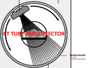
  • 3. X-RAY TUBES USED IN CT Since CT require longer continuous exposure time at higher KV and mA than needed for general radiography hence; the general radiographic X-ray tubes cannot be used for CT.It Should supply monochromatic X-ray beam for accurate reconstruction. X-ray tubes for CT have been charged with heavy duty rotating anode tube with higher thermal capacity and smaller focal spot (up to 0.6mm). These tubes are air cooled with current value up to 600mA. COMPARISION OF CT TUBE WITH VARIETY OF X-RAY TUBES AVAILABLE
  • 4. BRIEF HISTORY AND ADVANCEMENTS IN CT X-RAY TUBE CT X-ray tubes which are located in the heart of the gantry Provide radiation source for CT. • Early experimental models used radionuclide to supply such a radiation beam. • First and second generation scanner used fixed anode (relatively large (2x16mm)focal spot operated at 120 kVp & 30 mA & heavily filtered) , oil-cooled x- ray tube but with the demand for increased output, gradually rotating anode tubes become common in CT scanner. • The introduction of spiral/helical CT with the continuous rotation scanner has placed new demands on CT tubes. Several technical advances in component design have been made to deal with the problem of heat generation, heat storage and heat dissipation. For example, i)anode assembly including anode rotation- Anode is of larger diameter with graphite backing, which allows the anode to absorb& dissipate large amounts of heat. ii)target design- Anode target angle is made 7-10 degrees to diminish heel effect. iii)cathode assembly- Bigger filament size, increased effective focal spot. Focal spot size smaller (0.6 mm). iv)the tube envelop-Although the borosilicate glass envelope in early CT tube provides good thermal and electrical insulation, electrical arching result from tungsten deposits on the glass caused by vaporization. To solve above problem, tubes with metal envelop and ceramic insulator are now common e.g. Metal ceramic x-ray tube
  • 5. METAL CERAMIC X-RAY TUBE USED IN CT • Glass envelope has been replaced by metal casing and ceramic is used as insulation of high voltage cable. • A more recent development in x ray tube construction is metal ceramic tube,it’s construction includes alloy of chromium and iron cylinder brazed to alumina ceramic (aluminium oxide) insulators at each end. These insulators carry the anode and cathode assemblies . • The metal ceramic tubes are smaller and more robust than their glass equivalents. They have another advantage, in that they enable more flexibility in the electrical circuitary associated with the tube. • Metal envelope grounded offers no chance of arcing of x-ray tube and hence increase tube life. • Anode rotates on an axle with bearing at each end providing greater stability and reduced stress on shaft permits massive anode approx-2kg. • Ceramic insulator (Al oxide) are used to insulate high voltage parts of x-ray tube from metal envelope allowing more compact tube design. • Metal ceramic tube offers Higher tube loading, Reduce off focus radiation, Allow high tube current. 8-Feb-17 5
  • 6. METAL CERAMIC X-RAY TUBE USED IN SPIRAL CT
  • 7. 8-Feb-17 7 Fig. Schematic diagram o f a Super Rotalix ceramic x- ray tube: 1.Metal casing, 2. Anode, 3/6. Ball bearings, 4/8. Ceramic insulators, 5. Cathode, 7. Stator windings, 9. Anode shaft, 10. Beryllium window SUPER ROTALIX CERAMIC X-RAY TUBE IN CT --It is also the type of metal ceramic X-ray tube
  • 8.
  • 10. • Higher Tube Loading – Allows higher tube currents to be used because of larger heat storage capacity of anode • Longer Tube Life – Deposition of tungsten on the glass wall acts as electrode causing arcing bet. Glass and filament shortening tube life. When metal enclosure is grounded, this deposition will not alter grounding thus increasing its life • Reduced off Focus Radiation – Electrons back scattered from the anode may strike anode again producing x-rays from areas other than focal spot. The metal enclosure decreases off focus radiation by attracting off focus electrons to the grounded metal wall relatively Positive as compared to electrons. Low atomic no. of metal may produce few and low energy x-rays. 10 Advantages Of Metal ceramic Tube
  • 11. OTHER TYPES OF X-RAY TUBES USED IN CT MRC(MAXIMUM ROTALIX CERAMIC) X-RAY TUBE o In 1989 Phillips became the first company to introduce MRC.IT is based on the technology of spiral groove bearing using liquid metal alloy as lubricant. Focuses Significant improvement in rotating anode x-ray tube. FEATURES o Higher output and longer tube life o 200 mm graphite backed anode o Anode heat storage capacity(8MHU) o Tube voltage - 90 to 140 KV o Tube current - 20 to 500 mA o Anode angle - 7 degree o Directly cooled anode
  • 12. OTHER TYPES OF X-RAY TUBES USED IN CT MRC X-RAY TUBE Advantages o Noiselessly rotating anode that could be switched on the morning and switched off in the evening. o Avoid waiting time during and between examination. o Possible to achieve dose saving filter technique in angiography. USES o Cardiovascular imaging o MDCT
  • 13. OTHER TYPES OF X-RAY TUBES USED IN CT AQUILION X-RAY TUBE o High capacity multi-slice CT tube o Heat storage capacity 7.5 MHU o Cooling rate 1.7 MHU/min o Anode grounded o Focal spot 1.4mm × 1.4 mm o Air cooled Aquilion vs conventional tube
  • 14. TYPES OF X-RAY TUBE USED IN CT NEW STRATON TUBE o New construction o Focused and deflected beam of thermal electron o whole tube and anode assembly rotates o Bearing located out side o Oil cooled
  • 15. FEATURES o Zero heat storage capacity o Cooling rate 4.7 MHU/min o Cooled down within 20 sec o Enables gantry speed of 0.37 sec per rotation o Tube current 500 mA o Based on RET (rotating envelope tube) technology o The electron beam in the tube is shaped and controlled by magnetic deflection coil o Focal spot – tungsten and rhenium
  • 16. • One of the more interesting developments is the Siemens Straton x-ray tube, which is currently available as an option on Sensation 16 scanners (Fig ). • The tube itself is a radical new design, where the entire tube body rotates, rather than just the anode, as is the case with conventional designs. This change allows all the bearings to be located outside the evacuated tube, and enables the anode to be cooled more efficiently. • The Straton has a low inherent heat capacity of 0.8 MHU, but an extremely fast cooling rate of 5 MHU / min. 16 RECENT DEVELOPMENT IN X-RAY TUBE
  • 17. • This compares with typical figures of 7-8 MHU and up to 1.4 MHU / min for existing tubes. • The heat capacity and cooling rate combine to produce a tube which Siemens claim is ‘0 MHU', implying that tube cooling considerations are a thing of the past. Sensation 16 scanners fitted with the Straton tube now have a fastest scan time of 0.37 seconds. 17
  • 19. CT DETECTOR o CT detector capture the radiation beam from the patient and convert it into electrical signal, which subsequently converted into binary coded information o Detector characteristic o A. efficiency:- refers to the ability to capture, absorb and convert x-ray photons to electrical signal Detectors measure the intensity of radiation transmitted through the patient
  • 20. • B. Stability:- refers to the steadiness of the detector response, if not stable frequent calibrations are required to render the signals useful • C. Response time:- refers to the speed with which the detector can detect an x-ray event and recover to detect another event (should be very short in micro second) • D. Dynamic range:- refers to the ratio of the largest signal to be measured to the precision of the smallest signal to be discriminated
  • 21. o E . After glow:- refers to the persistence of the image even after the radiation has been turned off o TYPES OF DETECTOR Two types:- 1. Gas ionization detector Convert x-ray energy directly into electrical signal 2. Scintillation detectors Convert x-ray energy into light
  • 22. 1. GAS IONIZATION DETECTOR o Use high pressure ( about 25 atm) nonradioactive xenon gas, in long thin cells between two metal plates o Based on the principle of ionization o Consists of a series of individual gas chamber, usually separated by tungsten plate carefully positioned to act as electron collection plates with voltage applied across it.
  • 23. o When x-ray fall on the individual chamber, ionization of gas result and produces positive and negative ions. The positive ions migrate to the negatively charged plate, whereas the negative ions at positively charged plate o The migration of ions causes a small signal current that varies directly with the number of photons absorbs o QDE is only 50 – 60 percent o Can be used for 3rd generation scanner only
  • 24.
  • 25. Fig. Shows a typical gas-filled ionization detector Charges 8-Feb-17 25
  • 26. 2. SCINTILLATION DETECTORS o Solid state detectors that consist of a scintillation crystal coupled to a photodiode tube o When x-ray falls onto the crystals, flashes of light are produced. The light is then directed to PM tubes. Which then releases electrons and these electrons cascade through a series of dynodes that are carefully arranged and maintained at diff. potential to result in small out put signal
  • 27. o Early scanner used sodium iodide crystals coupled to PM tube. Due to afterglow problem and the limited dynamic range of sodium iodide, other crystals such as calcium fluoride and bismuth germinate used in later scanner o Now a days, solid state photodiode multiplier scintillation crystal detectors are used o The photodiode is a semiconductor whose p-n junction allows current flow when exposed to light
  • 28. o Photodiodes are normally used with amplifier because of the low output from the diode o Response time of photodiode is extremely fast (about 0.5 -250 nanosecond) o Scintillation materials currently used with photodiodes are cadmium tungsten and a ceramic material made of high purity, rare earth oxides based on doped rare earth compounds such as yttrium and gadolinium oxysulphide, these crystals are optically bonded to the photodiode
  • 29. o The conversion efficiency and photon capture efficiency of cadmium tungsten are 25-30% and 45% respectively and the dynamic range is 1 million to 1
  • 30. X-ray beam X-ray photon 8-Feb-17 30 Isotropically emitted lightPhotodiode Electrical signals Fig. Solid-state detector. The photodiode is coated with a scintillator. When the remnant x-ray photons activate the scintillator, light photons are emitted and detected by the photodiode. The photodiode then gives off an electrical signal.
  • 31. DETECTOR CONFIGURATION o One major problem with single slice, single row detector is related to the length of time needed to acquire data o CT scanner now use multi-row detector to image multi-slice during a 360 degree rotation o DUAL ROW/DUAL SLICE DETECTOR  In 1992, Elscint introduced the first dual slice volume CT scanner
  • 32. o This technology uses a dual-row, solid state detector array coupled with a special x-ray tube based on a double dynamic focus system o The dynamic focal spot is where the position of focal spot is switched by a computer controlled electron- optic system during each scan to double the sampling density and total no. of measurement o Twin beam technology results in the simultaneous scan of two contiguous slices with excellent resolution
  • 33. MULTI-ROW/MULTI -SLICE DETECTOR o The goal of MR/MS detector is to increase the volume coverage speed o MR/MS detector are solid state detectors that can acquire 4 to 64 to 320 slices per 360 degree rotation  Multi-row detector falls into three groups o A . MATRIX ARRAY DETECTOR o Referred to as a fixed array detectors
  • 34. o Contains channels or cells, they are often referred to isotropic, that are equal in all dimensions o B . ADAPTIVE ARRAY DETECTORS o The adaptive array detectors are anisotropic in design o The cells are not equal but rather they have diff. sizes
  • 35.
  • 36. • C . HYBRID DETECTORS o Has a no. of narrow detector elements in the centre of the detector and different no. of wider detector (usually double the width of the narrow detector) on both sides of the span of narrow detector o The no. of narrow and wider detector can vary R.R. YADAV (CT INSTRUMENTATION)
  • 37.
  • 38. AREA DETECTORS o Currently undergoing testing o Two such CT scanners based on area detector technology are the 256 slice ct scanner prototype(Toshiba Acquillion) and the flat panel CT scanner prototypes ( one from siemens and another from koning corporation) o The 256-slice CT detector is a wide area multi-row array detector that has 912 channels * 256 segments and a beam width of 128 mm o Possible to scan entire heart in a single rotation
  • 39. o Flat panel detectors are similar to the ones used in digital radiography are being investigated for the use in CT imaging o The detector is a flat panel type and is based on the CsI indirect conversion digital radiography detectors o Recently, flat panel detectors are being investigated for use in breast CT
  • 40.
  • 41. Ultra Fast Ceramic UFC) Detectors • The afterglow of the UFC detector material developed in decays 400 times faster than yttrium gadolinium oxide. • Aside from the rare earth element gadolinium, the UFC detectors also includes sulfur and other additives. 8-Feb-17 41 Scintillation/Solid-state Detectors
  • 46. EARLY LOW VOLTAGE THERAPY TUBE
  • 54. YOUARE SAFE NOTBECAUSEOF THE ABSENCEOF DANGER BUT BECAUSEOF THE PRESENCEOF RADIOLOGIC TECHNOLOGIST