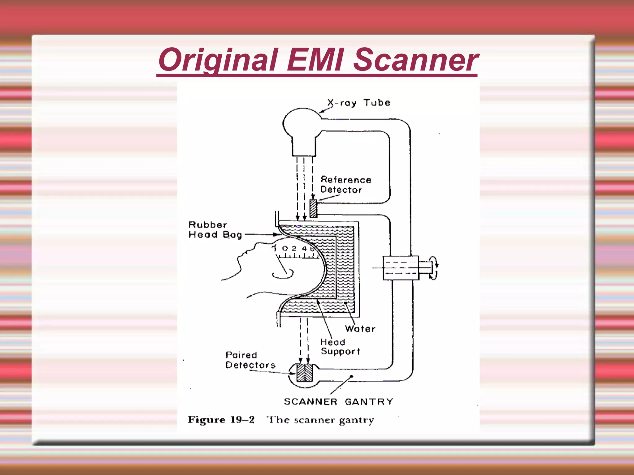
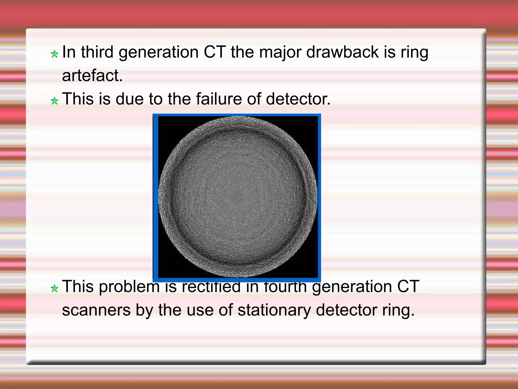
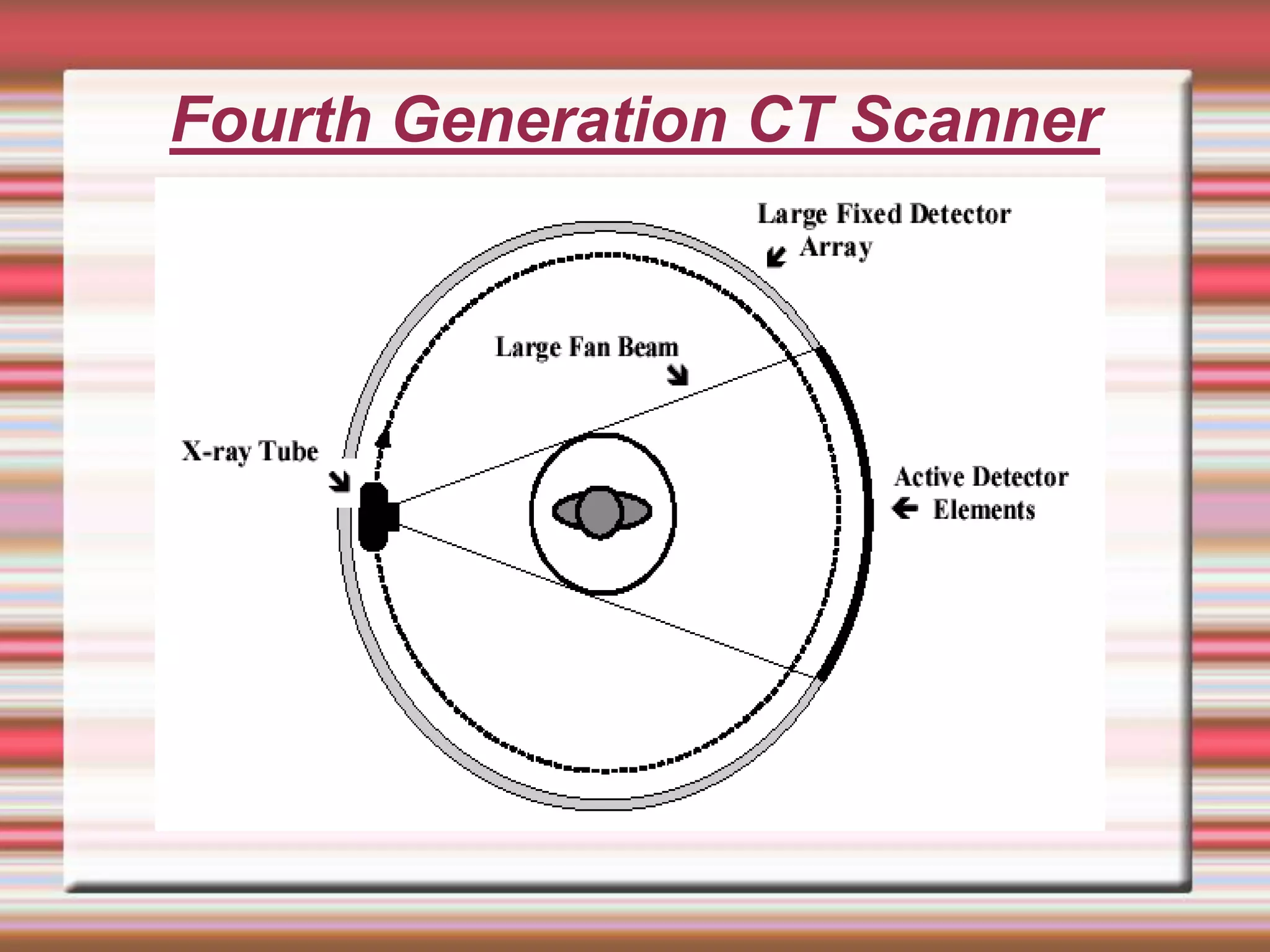
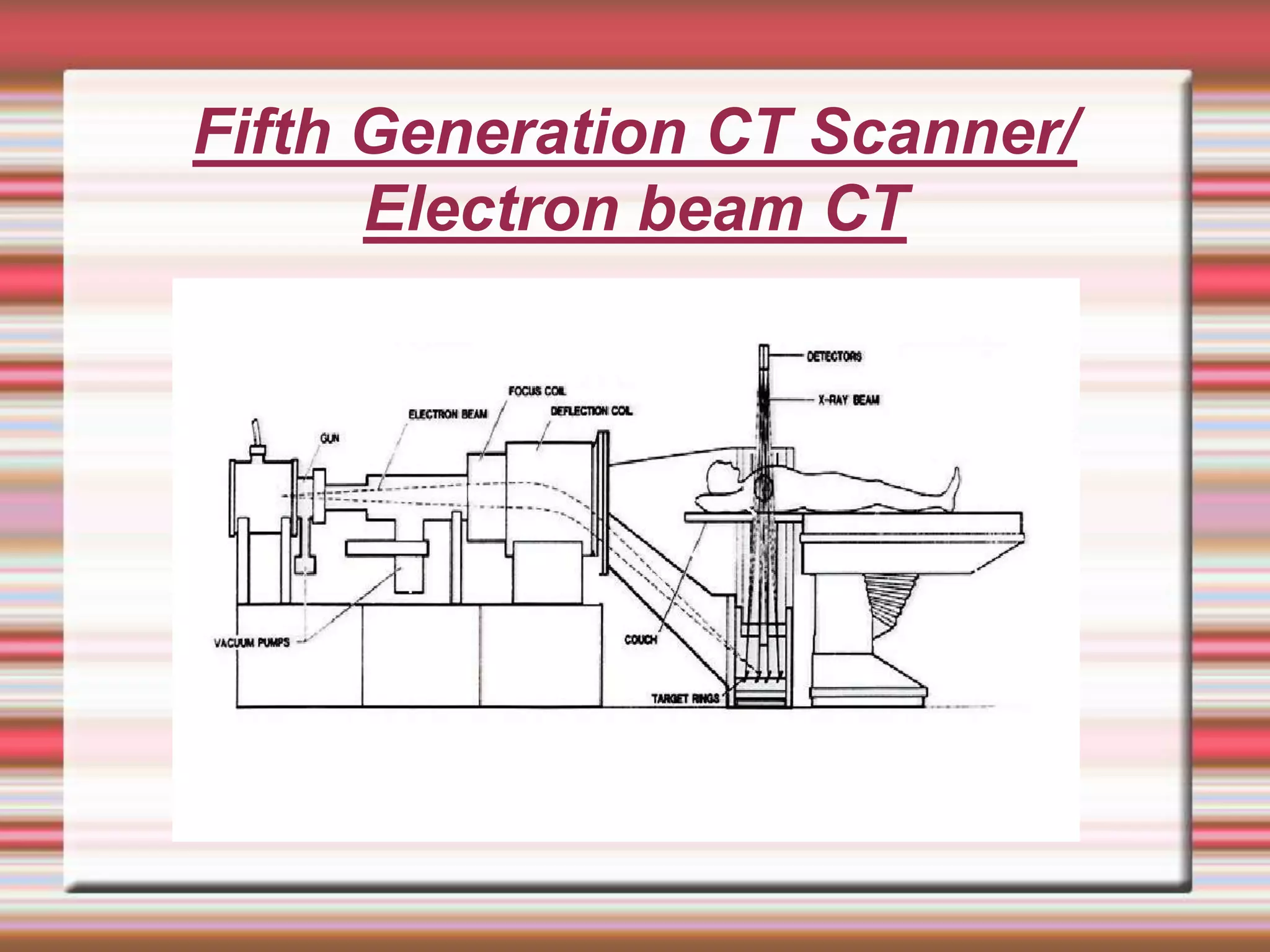
CT- GENERATIONS
Adavala.Mallikarjuna
45th DRP
Guide: Shri K.K.Sanu
Introduction of CT scan solves the major drawbacks
of conventional radiography Viz. superposition of
overlaying structures and scatter.
Both these reduces image contrast.
In the early1970s Godfrey Hounsfield developed a
scanner and demonstrated.
Cormack is a physicist who developed mathematics
for the reconstruction of image from multiple
projections.
The basic principle of computed tomography is
reconstruction of image from multiple projections.
In1979 Hounsfield and Cormack shared Noble prize
in medicine for this invention.
CT scanners are classified into different
categories
Based on source detector configuration and
their relative movement.
Original EMI Scanner
The original EMI scanner consists of an X-ray tube
and a pair of detectors.
Tube and detectors are mounted on a gantry
opposite to each other.
A reference detector is placed in the path of X-ray
beam before it reaches to the patient.
The source-detector system makes linear and
rotatory motions.
The linear motion was repeated over and over for
180 times.
Between each of these linear movements the gantry
rotated 10 .
The axis of rotation passes through the centre of the
patient head.
The X-ray tube was on during linear motion and off in
rotatory motion.
The transmitted X-rays was measured for 160 times
during the linear motion.
Total 28,800 projections are counted.
The total scan time was 5 min and reconstruction time
is 1 min.
For 10 slices the scan time is about 25 to 30 min.
First Generation CT Scanner
The original EMI scanner belongs to first generation
CT scanner.
In all other first generation CT scanners only one
detector per slice.
Detector used in this CT scanners was NaI(Tl).
Image quality is excellent in this type of CT
scanners.
Second Generation CT Scanner
Second Generation CT has Rotate-Translate
movement.
In this narrow fan beam and an array of detectors are
used. About 30 detectors are arranged along an arc.
Due to the increased utilization of X-ray output (about
30 times more than the first generation) scan time was
reduced.
Scan time per slice reduces to 10 to 90 sec.
Inclusion of scatter component is more in this
generation compared to first generation.
Third Generation CT Scanner
In third generation CT scanners, translational motion
is completely removed.
X-ray tube and detector system makes rotatory
motion around the patient for acquiring information,
hence it is called Rotate-Rotate movement.
In this, a wide fan beam is used to cover the entire
body and more no of detectors (more than 800) are
used.
In this system, scan time reduces to about 5 seconds.
Newer systems have scan time of about 1.5 sec.
A single detector towards the end of array acts as the
reference detector.
In third generation CT the major drawback is ring
artefact.
This is due to the failure of detector.
This problem is rectified in fourth generation CT
scanners by the use of stationary detector ring.
Fourth Generation CT Scanner
Rotate – Stationary movement.
In this system a detector ring is used around
the patient. X-ray tube is rotating inside the
detector ring.
Each detector acts as its own reference
detector.
Scan time is reduced to about 1 sec.
Fifth Generation CT Scanner/
Electron beam CT
No moving parts.
In this a large arc of tungsten is used as X-ray target .
Detector ring is positioned directly opposite to the
tungsten arc.
An electron beam steered through a wave guide to
strike the tungsten arc through a bending magnet to
produce x-rays.
It is not very common type.
During the X-ray production patient couch is
moved to image different body cross-sections.
Capable of 50 m sec scan time.
It can produce fast-frame-rate CT moves of
beating heart.
It has special applications in cardiology
Slip ring technology
In previous generations, the rotation of source -
detector system around the patient was carried out
by the movement of the system through 3600 rotation
in one direction for a scan and in opposite direction
for the next scan.
Continuous rotation of source-detector system
became possible with the introduction of Slip-ring
technology.
Slip-ring technology is used in modern CT scanners
Spiral / Helical CT
In spiral CT source and detector system makes
continuous motion around the patient, the couch too
moves continuously into the axial direction.
As a result x-ray beam follows a spiral/helical trajectory
hence the name is spiral/helical CT.
Speed of rotation of the gantry and speed of table
determines the pitch of the helix.
Pitch =couch movement per 3600 / Slice thickness.
During the spiral CT scan image data is received
continuously
However the data obtained during spiral scan may not be
sufficient for reconstruction of images of all slices along
the axial direction.
Therefore image reconstruction in spiral CT is done by
the interpolation of data available at 3600 apart.
Entire scan can be done in a breath hold time.
Axial resolution decreases with increase in pitch.Smaller
pitch increase patient dose.
Longer processing time as more data have to be
handled.
Spiral CT is advantageous in CT Fluoroscopy as use of
contrast media can be minimised .
Reconstruction of images is possible in different planes
3D image too can be reconstructed.
Applications of Spiral/helical CT
MULTI SLICE CT
 The principle in multi slice CT is similar to the
helical / spiral CT but there are more number of
detector channels.
 No. of channels : 4, 8, 16, 32, 40 & 64.
 use of cone beam
 The major advantage of multi slice CT is more
volume is covered in a single scan .
 Effective utilization of x-ray beam.
Generation Configuration Detectors Beam Min Scan Time
First Translate-rotate 1~2 Pencil thin 2.5 min
Second Translate-rotate 3~52 Narrow fan 10 sec
Third Rotate-rotate 256~1000 Wide fan 0.5 sec
Fourth Rotate-fixed 600~4800 Wide fan 1 sec
Fifth Electron beam 1284 detectors
Wide fan
beam
33 ms
Examination Typical effective dose (m Sv)
Chest X-ray 0.1
Head CT 1.5
Abdomen CT 5.3
Chest CT 5.8
Chest, Abdomen and Pelvis
CT
9.9
Cardiac CT angiogram 6.7-13
CT colonography (virtual
colonoscopy)
3.6 - 8.8
Thank you

CT- GENERATIONS.ppt

  • 1.
  • 2.
    Introduction of CTscan solves the major drawbacks of conventional radiography Viz. superposition of overlaying structures and scatter. Both these reduces image contrast. In the early1970s Godfrey Hounsfield developed a scanner and demonstrated. Cormack is a physicist who developed mathematics for the reconstruction of image from multiple projections. The basic principle of computed tomography is reconstruction of image from multiple projections. In1979 Hounsfield and Cormack shared Noble prize in medicine for this invention.
  • 3.
    CT scanners areclassified into different categories Based on source detector configuration and their relative movement.
  • 4.
  • 5.
    The original EMIscanner consists of an X-ray tube and a pair of detectors. Tube and detectors are mounted on a gantry opposite to each other. A reference detector is placed in the path of X-ray beam before it reaches to the patient. The source-detector system makes linear and rotatory motions. The linear motion was repeated over and over for 180 times. Between each of these linear movements the gantry rotated 10 .
  • 6.
    The axis ofrotation passes through the centre of the patient head. The X-ray tube was on during linear motion and off in rotatory motion. The transmitted X-rays was measured for 160 times during the linear motion. Total 28,800 projections are counted. The total scan time was 5 min and reconstruction time is 1 min. For 10 slices the scan time is about 25 to 30 min.
  • 7.
    First Generation CTScanner The original EMI scanner belongs to first generation CT scanner. In all other first generation CT scanners only one detector per slice. Detector used in this CT scanners was NaI(Tl). Image quality is excellent in this type of CT scanners.
  • 8.
  • 9.
    Second Generation CThas Rotate-Translate movement. In this narrow fan beam and an array of detectors are used. About 30 detectors are arranged along an arc. Due to the increased utilization of X-ray output (about 30 times more than the first generation) scan time was reduced. Scan time per slice reduces to 10 to 90 sec. Inclusion of scatter component is more in this generation compared to first generation.
  • 10.
  • 11.
    In third generationCT scanners, translational motion is completely removed. X-ray tube and detector system makes rotatory motion around the patient for acquiring information, hence it is called Rotate-Rotate movement. In this, a wide fan beam is used to cover the entire body and more no of detectors (more than 800) are used. In this system, scan time reduces to about 5 seconds. Newer systems have scan time of about 1.5 sec. A single detector towards the end of array acts as the reference detector.
  • 12.
    In third generationCT the major drawback is ring artefact. This is due to the failure of detector. This problem is rectified in fourth generation CT scanners by the use of stationary detector ring.
  • 13.
  • 14.
    Rotate – Stationarymovement. In this system a detector ring is used around the patient. X-ray tube is rotating inside the detector ring. Each detector acts as its own reference detector. Scan time is reduced to about 1 sec.
  • 15.
    Fifth Generation CTScanner/ Electron beam CT
  • 16.
    No moving parts. Inthis a large arc of tungsten is used as X-ray target . Detector ring is positioned directly opposite to the tungsten arc. An electron beam steered through a wave guide to strike the tungsten arc through a bending magnet to produce x-rays. It is not very common type.
  • 17.
    During the X-rayproduction patient couch is moved to image different body cross-sections. Capable of 50 m sec scan time. It can produce fast-frame-rate CT moves of beating heart. It has special applications in cardiology
  • 18.
    Slip ring technology Inprevious generations, the rotation of source - detector system around the patient was carried out by the movement of the system through 3600 rotation in one direction for a scan and in opposite direction for the next scan. Continuous rotation of source-detector system became possible with the introduction of Slip-ring technology. Slip-ring technology is used in modern CT scanners
  • 19.
  • 20.
    In spiral CTsource and detector system makes continuous motion around the patient, the couch too moves continuously into the axial direction. As a result x-ray beam follows a spiral/helical trajectory hence the name is spiral/helical CT. Speed of rotation of the gantry and speed of table determines the pitch of the helix. Pitch =couch movement per 3600 / Slice thickness. During the spiral CT scan image data is received continuously
  • 21.
    However the dataobtained during spiral scan may not be sufficient for reconstruction of images of all slices along the axial direction. Therefore image reconstruction in spiral CT is done by the interpolation of data available at 3600 apart. Entire scan can be done in a breath hold time. Axial resolution decreases with increase in pitch.Smaller pitch increase patient dose. Longer processing time as more data have to be handled.
  • 22.
    Spiral CT isadvantageous in CT Fluoroscopy as use of contrast media can be minimised . Reconstruction of images is possible in different planes 3D image too can be reconstructed. Applications of Spiral/helical CT
  • 23.
    MULTI SLICE CT The principle in multi slice CT is similar to the helical / spiral CT but there are more number of detector channels.  No. of channels : 4, 8, 16, 32, 40 & 64.  use of cone beam  The major advantage of multi slice CT is more volume is covered in a single scan .  Effective utilization of x-ray beam.
  • 25.
    Generation Configuration DetectorsBeam Min Scan Time First Translate-rotate 1~2 Pencil thin 2.5 min Second Translate-rotate 3~52 Narrow fan 10 sec Third Rotate-rotate 256~1000 Wide fan 0.5 sec Fourth Rotate-fixed 600~4800 Wide fan 1 sec Fifth Electron beam 1284 detectors Wide fan beam 33 ms
  • 26.
    Examination Typical effectivedose (m Sv) Chest X-ray 0.1 Head CT 1.5 Abdomen CT 5.3 Chest CT 5.8 Chest, Abdomen and Pelvis CT 9.9 Cardiac CT angiogram 6.7-13 CT colonography (virtual colonoscopy) 3.6 - 8.8
  • 27.