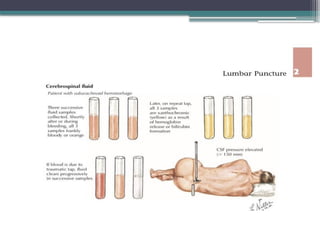
DIAGNOSTIC EVALUATION OF
CEREBROSPINAL FLUID
Dr Innocent Agaba (MBBS)
Department of Paediatrics,
Modibbo Adama University Teaching Hospital Yola
14/02/24
“… I punctured the subarachnoid space in the lumbar
area, passing a very fine cannula two centimeter deep
between the third and fourth lumbar spinal arches and
drop by drop I drained a few cubic centimeters of
watery clear fluid.”
Quincke,
April 1891
OUTLINE
• Introduction
• Historical Background
• Relevant anatomy and physiology
• Evaluation
- Obtaining the CSF
- Specimen Processing
- Normal CSF
- Investigative modalities
• CSF findings in Diseases
• Future perspectives
• Conclusion
• References
INTRODUCTION
• Cerebrospinal Fluid (CSF) is a dynamic metabolically active fluid
surrounding the brain and the spinal cord.
• It is a sterile, crystal clear, colourless and odourless body fluid produced
in the choroid plexus of the brain.
• CSF obtained by lumbar puncture is the main diagnostic tool for
neurological disorders.
• CSF analysis is not necessarily diagnostic but can be useful in the
evaluation of several neurologic conditions.
• In the last 30 years, great progress has been achieved in diseases like
Dementia & Alzheimer’s Disease using biological markers
HISTORICAL PERSPECTIVES
• The first description of the meningis, ventricles, and CSF appeared in the Edwin
Smith Surgical Papyrus believed to be written in the 17th century BC.
• In 1664, Galen described “a liquid” in the aqueduct of Sylvius that connects the
ventricles, noting that in “epidemic fever,” the consistency of the “liquid” is altered
• In 1902 the term “xanthochromia” was first used to describe a yellowish
pigmentation of CSF from a patient with SAH.
• Two years later, Henri Dufour (1904) turned diagnostic cytology of the CSF
possible with the development of technology to identify neoplastic cells.
• In 1912, William Mestrezat made the first detailed description of the chemical
composition of CSF.
HISTORICAL PERSPECTIVES
RELEVANT ANATOMY & PHYSIOLOGY
EVALUATION
• Aim: Diagnosis, Therapy, Monitoring, prognostication
• Parameters : Quantity, pressure and components.
• Invasive
- Lumbar puncture (LP)
-Subdural puncture
- Cisternal puncture
- Lateral cervical puncture
- ICP monitoring devices
• Non-invasive
-Transfontannel USS
-CT scanning
-Neuroimaging
-Radionuclide tracers
-Phase-contrast MR
-FLAIR sequences
Obtaining the CSF – Lumbar puncture
• Proper understanding of the underlying anatomy and careful positioning of the
patient are the two main keys to performing a successful LP.
• Positioning
1. Lateral recumbent
-The neck, hips, and knees should be flexed,
-the entire spine should be parallel to the table,
-the coronal plane of the trunk should form a
right angle to the floor, and
-the patient’s shoulders should be perpendicular to the
table
2. Sitting
- Preferable for sick neonates
- Should be returned to lateral decubitus before measurement of
opening pressure
Obtaining the CSF – Lumbar puncture
• Because the conus medullaris extends below L1–L2 in infants and
children, the lowest possible interspace should always be used in young
patients, with needle insertion preferably no higher than at L4–L5.
• Once the appropriate interspace has been identified and marked, the
overlying skin should be sterilized and draped using standard aseptic
technique.
• The bevel of the needle is inserted in the midline, parallel to the
longitudinal fibers of the Dura, and directed toward the umbilicus.
• Once fluid appears at the needle hub, a manometer with a three-way
stopcock should be gently connected to measure the opening pressure.
Obtaining the CSF – Lumbar puncture
Peri-procedural considerations
• Cranial CT or magnetic resonance imaging (MRI) scan is obligatory for any
patient who presents with focal neurological deficits, new-onset seizures, altered
level of consciousness, and/or papilledema before an LP can safely be performed.
• The above must include a fundoscopic exam to search for any evidence of
papilledema
• Anticoagulation therapy should be stopped or reversed prior to performance of an
LP whenever possible
• Finally, careful thought should always be given to the sample volume that will be
needed to perform all the desired assays
• For patient with an uncertain diagnosis, it is advised to set aside a few extra
milliliters of CSF in a clinical refrigerator whenever possible
Obtaining CSF – Lumbar Puncture
CSF ASSAY SAMPLE VOLUME NEEDED
Cell count & Differential 1ml each in tube #1 and #4
Glucose & Protein concentration 1ml
Bacterial Gram stain & Culture Minimum of 1ml
Viral PCRs 1ml per viral PCR
AFB smear & Culture 5ml
Flow Cytometry & Cytopathology Minimum of 5ml, preferably 10 – 15ml
Oligoclonal bands, Myelin Basic
proteins,IgG index
0.5ml per test
Contraindications
• Localised infection – such as cellulitis, sacral decubitis ulcers, or
known epidural abscess – overlying the lumbar spine
• Intracranial space-occupying brain lesions that result in midline shift
or pressure on the contents of the posterior fossa.
• Known coagulopathy related to anticoagulant medications, liver
failure, or thrombocytopenia
• INR greater than 1.4 or a platelet count of less than 20 x 109
/l
• Critical illness with cardiopulmonary compromise
Complications
• Brain herniation
• Bleeding Spinal (subarachnoid, subdural, epidural) Intracranial
(subarachnoid, subdural) Traumatic puncture (“bloody tap”)
• Infection
• Headache
• Backache/radicular symptoms
• Other Cranial neuropathies (diplopia, hearing loss)
• Implantation of an epidermoid tumor
• Spurious laboratory data
• Unique complications of LP in infants include hypoxemia due
to improper patient positioning
Specimen processing
• Should be transported without delay as cells can lyse leading
to falsely abnormal biochemical analytes.
• At room temp, cell count depreciate by 32% in 1 hr, 50% in
2 hrs.
• 1st
tube collected should be the one sent for culture.
• Preferably 10 – 15ml. Minimum should be 2ml esp for Fungi
& mycobacterium.
• Gram staining less sensitive than Acridine orange for
detecting bacteria.
Normal CSF macroscopy
• Under normal circumstances, CSF should appear clear and colorless.
• An aliquot of CSF should be visually inspected in direct comparison to
an equal volume of water at the time of acquisition, preferably using
clear glass tubes.
• Normal opening pressure : 10 – 100mmH20 in newborns,60 –
180mmH20 in young children & 12 – 120mmH20 in older children.
• Turbidity resulting from increased cellularity can be seen under
standard lighting conditions at counts starting around 400 cells/mm3
• CSF will appear pink at counts of 500–6,000 RBCs/mm3
. The fluid
becomes frankly bloody above 6,000 RBC/mm3
.
Normal CSF Cellularity
• Techniques :
- Direct microscopic examination.
- Automated cell-counting methodologies.
- Immunofluorescent staining of cells and
analysis by flow cytometry.
• There is general consensus that normal CSF should not contain
more than 5 WBCs/mm3 and 5 RBCs per ul. Neonates in contrast
may have up to 20wbcs.
• Neutrophils are always abnormal in a child but 1-2/mm3
may be
present in healthy neonates.
Normal CSF Cellularity
• Normal CSF leukocytes are about 80% T cells (CD4+ >
CD8+ T cells).
• Monocytes constitute only about 5% of normal CSF
cells, and fewer than 1% are B cells.
• Normal CSF may also rarely contain cells that derive
from tissues in physical contact with the subarachnoid
space, including the choroid plexus, the ependymal
lining of the ventricles, and the arachnoid membrane
Normal CSF proteins
Normal CSF proteins
• Measured using a spectrophotometer
• About 10% of the total protein is IgG
• With a traumatic tap, CSF protein is increased by
approximately 1mg/dl for every 1000 RBCs/mm3.
Normal CSF glucose
• The CSF should be tested for glucose as quickly as possible as
glucose levels begin to fall after 20 minutes.
• Measured by Colorimetry
• Approximately two – thirds of serum levels
• CSF glucose levels of 40–45 mg/dl are often abnormal, and that
values below 40 mg/dl always reflect a pathological state.
• Thus, it is advisable to simultaneously monitor the CSF:serum
glucose ratio in addition to absolute glucose concentrations.
Normal CSF
Other constituents of Normal CSF
• Electrolytes
• Amino acids
• Neurotransmitters
• Enzymes and enzyme inhibitors
• Lipids
• Cytokines
• Hormones
• Trace Metals
• Vitamins
• Metabolic by products
Investigative modalities
• A “standard” profile of :
• Opening pressure,
• Cell count and diff
• Glucose
• Protein concentrations
• Bacterial Gram stain and culture status.
• Acid-fast bacilli (AFB) using a smear
• Is there a place for these?
• Urinalysis strips
• Glucometer strips
Investigative modalities
• As indicated :
• Countercurrent Immunoelectrophoresis (CIE)
- Almost as sensitive as culture
• Coagglutination Test (COAG)
• Latex agglutination test (LAT)
• ELISA
• Polymerase chain reaction
• 4-3-3 protein
• Oligoclonal bands,
• Elevated immunoglobulin (Ig) G levels (IgG index)
• Flow cytometric and cytopathologic analyses
• Paraneoplastic antibodies
CSF FINDINGS
IN DISEASES
• Infections of the nervous system
• Neoplastic & Paraneoplastic disorders
• Inflammatory & Demyelinating disorders
• Cerebrovascular disorders
• Isolated seizures & epileptic disorders
Others are
• Neurodegenerative and movement disorders
• Parasitic, spirochetal disorders
• Transmissible spongiform Encephalopathies
• Nutritional and metabolic disorders
• Developmental disorders
• Headache syndromes
• Psychiatric disorders
• Neuromuscular disorders
1. Bacterial Meningitis
Opening pressure
• Raised i.e >200 mmH20
Macroscopy
• Clear in early disease
• Cloudy or purulent
• Occasionally, pus gets encapsulated , as in pneumococcal meningitis in the frontal lobe area. In such
cases, even though the CSF may remain clear the patient does not respond adequately to therapy and may
eventually succumb to the illnes
Cellularity
• Pleocytosis is the rule with WBC 100 – 10,000/mm3
• PMNs dominate (>90% of total).
• WBC > 50,000/mm3
– suspect Brain abscess.
• Pleocytosis may be absent in patients with overwhelming sepsis & meningitis.
CSF microscopy - Neisseria meningitides
CSF microscopy – Haemophilus influenzae
CSF microscopy – Streptococcus pneumonia
Bacterial Meningitis cont…
Biochemistry
• Protein is usually
• Glucose is less than 2/3rd
random serum glucose. May be absent.
Microscopy
• Gram stain is positive in 70 – 90% of patients with untreated bacterial
meningitis
Culture
• Ideally inoculated on sheep blood agar & enriched chocolate agar and
incubated for 4 days at 35 – 37% in 5% Co2.
NAATS
2. Partially treated (Bacterial) Meningitis
• 25-50% of children being evaluated for bacterial meningitis are receiving oral antibiotics
when their CSF is obtained
Cellularity
• Pleocytosis with neutrophil predominance.
Biochemistry
• Elevated protein levels and hypoglycorrhachia usually persists for several days after
administration of appropriate IV antibiotics.
Microscopy
• Gram staining may be negative .
Culture
• May yield no growth.
• Bacterial Antigen testing is indicated .
3. Viral Meningoencephalitis
Macroscopy
• Clear
• Ocassionally slightly turbid
Cellularity
• PMNs- predominate ;Later - Mononuclear cells
Biochemistry:
• Protein is normal or slightly elevated. Concentrations may be very high if brain destruction
is extensive e.g HSV Encephalitis
• Glucose is usually normal (In mumps, hypoglycorrhacia may be observed)
Culture
• Isolating a virus is most likely early in the illness. If not isolated, Serology is advised 2 -3
wks later to demonstate rising titers
• Take concomitant samples from Nasopharyngeal swabs, faeces & urine to increase yield
• Antibody or PCR studies
Viral meningoencephalitis cont…
• COVID 19 WITH CNS INVOLVMENT
• Has been reported to affect – directly & indirectly –the nervous
system in a substantial number of cases.
Biochemistry :
• Total protein is elevated
• An elevated CSF/serum ratio for albumin
• Cytoalbumin dissociation was found in half of patients involved in a
large scale multicenter study in 2022
• Important to avoid misinterpretation of treatable coexisting
neurological disorders as complications of COVID 19
4. Mycobacterial Meningitis
Opening pressure
• Usually raised
Macroscopy
• Turbid
• Cobweb appearance after 10 – 20min
Cellularity
• During early stage 1, the CSF can resemble that of viral aseptic meningitis only to
progress to the more-severe CSF profile over several weeks.
Biochemistry:
• The CSF glucose is typically less than 40mg/dl but rarely <20mg/dl
• The protein level is elevated and may be markedly high (400-5,000 mg/dL) i.e 100
– 300 in early stage. Often >1000 in Late stages.
Mycobacterial meningitis cont…
Microscopy
• Staining with Ziehl-Neelsen stain yields acid fast bacilli in up to 30% of cases.
•
Culture:
• Culture in Lowenstein Jensen medium is positive in 40-60% of cases.
• The success of the microscopic examination of acid-fast–stained CSF and
mycobacterial culture is related directly to the volume of the CSF sample.
• Examinations or culture of small amounts of CSF are unlikely to demonstrate
M. tuberculosis. When 5-10 mL of lumbar CSF can be obtained, the acid-fast
stain of the CSF sediment is positive in up to 30% of cases and the culture is
positive in 50-70% of cases.
• Polymerase chain reaction (PCR) testing of the CSF can improve diagnosis.
Cultures of other body fluids can help confirm the diagnosis
4 . Fungal infection of the nervous system
Cellularity
• CSF WBC count in Cryptococcal meningitis can range from 0 to 3,700
cells/mm3.
• Levels tend to be significantly lower in HIV-seropositive individuals,
where up to 50% of patients with proven CM can actually have normal
CSF WBC counts at the time of diagnosis.
Biochemistry
• Total protein levels are usually elevated while CSF glucose
concentrations can either be normal or depressed.
Fungal infections of Nervous system cont…
Microscopy
• An India ink examination of the CSF shows yeast forms in 70–90% of untreated AIDS
patients with CM due to higher pathogen loads, but may be positive in only 50% of the
non-AIDS population
Serology & antigen detection
• Immunological detection of cryptococcal polysaccharide antigen in serum and CSF by
rapid latex agglutination tests or enzyme immunoassays has a sensitivity in excess of 90%.
Culture studies
• Cryptococcus neoformans isolated from blood or CSF produces white mucoid colonies on
many types of culture media, often within 72 h after plating
5. Parasitic infestation of the nervous system
• Causes eosinophilic meningitis i.e eosinophil count >10/mm3
• a) CEREBRAL MALARIA
Cellularity
• Nearly all of the cells present in the CSF during cerebral malaria are lymphocytes,
and the presence of neutrophils should suggest an alternative diagnosis.
Biochemistry
• The total protein content may be slightly elevated, sometimes reaching a level of
150 mg/dl but rarely more than that.
• The CSF:serum glucose ratio is invariably normal.
Parasitic infestations of the Nervous system
cont…
• b) CEREBRAL TOXOPLASMOSIS
Cellularity
• CSF shows a mild mononuclear cell pleocytosis,
Biochemistry
• Elevated protein content
• Normal glucose level.
Serology
• The diagnosis is made by showing rising serum and CSF anti-Toxoplasma
6. Spirochetal infection of nervous system
• Symptomatic involvement of the CNS by Treponema pallidum can occur
any time beyond the primary stage of infection.
• Various forms : Asymptomatic Neurosyphilis, Syphilitic meningitis,
Meningovascular syphilis, Parenchymatous neurosyphilis, Gummatous
neurosyphiliis,Neurosyphilis in HIV infected individuals.
• Current standards advocate that patients with any two of the following
features be treated for presumptive neurosyphilis:
(a) CSF pleocytosis of >8 WBC/mm3,
(b) CSF protein level >50 mg/dl, and
(c) reactive CSF VDRL.
7. Drug and chemical induced Meningitis
• NSAIDS, Antibiotics, IV immunoglobulins are known causes of aseptic meningitis
• The usual clinical scenario is that signs and symptoms suspicious of such an event
begin within hours after drug ingestion, but cases have been reported as far out as
several weeks following exposure.
Cellularity
• Predominance of either PMNs or mononuclear cells, and not infrequently has a
measurable proportion of eosinophils.
• Total cell counts can range from 10 to 10,000 WBC/mm3 although most cases
have 200–400 WBC/mm3 detected
Biochemistry
• Protein content ranges from normal up to as high as 500 mg/dl
8. Neoplastic disorders
• Cartilage cells and cells derived from the bone marrow (presumably due to trauma
from the spinal needle) have also been rarely identified in CSF samples and these
cells are commonly mistaken for malignant cells.
• Presence of malignant cells aid in the diagnosis and staging of Burkit Lymphoma,
Leukemias e.t.c
• CARCINOMATOUS MENINGITIS
Biochemistry
• Low CSF glucose
• Increased CSF protein
Cellularity
• Slightly elevated to very high 0-100 or more; mononuclear or blast cells 50-1,000
Cytology
• May be positive
9. Transmissible spongiform encephalopathies
Cellularity
• Normal
Biochemistry
• Normal
• Measurement of CSF NSE levels could be useful as a confirmatory
test in the setting of a suspected sporadic CJD diagnosis
• Elevated CSF levels of 14-3-3 protein.
10. Cerebrovascular disorders
SUBARACHNOID HAEMORRHAGE
• Indicated if only if CT scan is normal.
• Typical Xanthochromic appearance
• Xanthochromia may be absent in in bleeds
• May not be seen<12hrs after the incident particularly when
laboratories rely on visual inspection rather than spectroscopy.
Neuromuscular Diseases
• a) GULLAIN BARRE SYNDROME
• The classic triad of findings in GBS – weakness, areflexia and elevated CSF protein – were
first reported by Guillain, Barré in 1916
• The CSF changes in this disorder is termed “cytoalbuminologic dissociation,” reflecting an
elevation of CSF protein, frequently greater than 100 mg/dl, with minimal or no elevation
in the number of CSF leukocytes.
• The initial CSF results should be interpreted cautiously, however, as only 50% of patients
with GBS have an elevated CSF protein concentration at presentation.
• The mean CSF protein level reaches a maximum at 16–30 days from symptom onset,
ranging from 140 to 213 mg/dl in one cohort, at which time 90% of patients demonstrate an
elevation of CSF protein.
• An exception to the cytoalbuminologic dissociation occurs in HIV-associated GBS, where
there may be up to 50 leukocytes/mm3 in the CSF
11. Neuromuscular diseases
• b) Poliomyelitis
• A cause of aseptic meningitis
• 1-2% of all cases of poliomyelitis
Cellularity
• Lymphocytic pleocytosis
• Biochemistry
• Rise in protein
• Normal sugar content
12. Nutritional & Metabolic Disorders
• a) PROTEIN – ENERGY MALNUTRITION
• Ammonia levels were elevated in the CSF of children
with either type of protein-calorie malnutrition
• Activities of two particular enzymes, glutamic-
oxalacetic transaminase and lactate dehydrogenase,
were elevated in the CSF of children with marasmus
or kwashiorko
Nutritional and Metabolic Disorders cont…
• b) HEPATIC ENCEPHALOPATHY
• Cases of acute liver failure are commonly associated with an
elevated Opening pressure if LP is performed in this setting
• Elevated levels of ammonia, glutamine, alpha-ketoglutaramate,
and lactate.
• High CSF and brain levels of both glutamine and ammonia
cause visible swelling of astrocytes.
13. Inflammatory & demyelinating disorders
• a) Multiple Sclerosis
• The CSF cell count is typically normal or only mildly
elevated in most MS patients
• Diagnosis based on clinical features with
- IEF evidence of ≥2 Oligoclonal Bands,
- Elevated IgG index
14. Connective tissue disorders
• SYSTEMIC LUPUS ERYTHEMATOSUS
Cellularity
• Slightly elevated 0-500; PMNs usually predominate; lymphocytes may be
present
Biochemistry
• Normal or slightly decreased glucose
Culture
• No organisms on smear or culture.
• Positive neuronal and ribosomal P protein antibodies in CSF
15. Psychiatric Disorders
AUTISTIC SPECTRUM DISORDER
• Hx : impaired social, behavioral, and communication skills. It can be
associated with mental retardation and epilepsy
• CSF shows high levels of Multiple inflammatory mediators
including ;
-monocyte chemoattractant protein (MCP)-1
- interferon-gamma
- transforming growth factor (TGF)-beta2
-Interleukin (IL)-8)
16. Neurodegenerative and Movement
Disorders
• a) Huntington’s disease
• Autosomal Dominant
• Characterised by chorea, behavioral disturbances, dementia, and motor
impersistence.
• A diagnosis is made by genetic testing that reveals an expansion of a CAG repeat in
the huntington gene found on chromosome 4
CSF Findings
• Levels of the dopamine metabolite homovanillic acid (HVA) were reduced compared
to normal individuals
17. Developmental disorders
b). Pyridoxine dependent Epilepsy
• Rare autosomal recessive disorder,
• Presents with seizures in the neonatal period.
• Unresponsive to the classic anticonvulsants,
• Rapidly cease when intravenous pyridoxine is
administered
CSF findings
• Elevated plasma, urine, and CSF levels of both Pipecolic
Acid
18. Disorders of intracranial pressure and cerebrospinal
fluid circulation
IDIOPATHIC INTRACRANIAL HYPERTENSION (IIH)
• The hallmark of idiopathic intracranial hypertension – previously known as
pseudotumor cerebri – is elevated pressure in the CSF compartment
• CSF Findings : Pcsf less than 20 cmH2O is considered normal, while
readings of greater than 25 cmHO are consistent with IIH.
• Pressures between 20 and 25 cmH2O are ambiguous; if clinical suspicion is
high, treatment can be started with careful follow-up
Future perspectives
• “Omic” approaches including :
- proteomics
- metabolomics
- transcriptomics and
- micro analysis.
Future perspectives
REFERENCES
• Kliegman R. M. et al, Nelson Textbook of Paediatrics. 20th
ed Elsevier 2016
• Azubike JC, Nkangineme KE. Paediatrics and Child Health in a tropical region.
3rd
edition, Educational Printing Press 2016
• Rudolph M., Rudolph CD, Paller,Hostettter MK,Lister G. & Siegel Nj
(2011)Rudolphs Textbook of Paediatrics 22nd
Ed. McGraw Hill
• Nkangineme KE et al, Pathophysiology of Clinical symptoms, signs and
Laboratory parameters, 1st
ed Athena Academic Ltd 2016.
• Regeniter A, Kuhle J, Mehling M, (2009) . A modern approach to CSF analysis:
pathophysiology, clinical application, proof of concept and laboratory reporting.
Clin Neurol Neurosurg. May;111(4)
THANK YOU
FOR
LISTENING

CEREBROSPINAL FLUID EVALUATION IN DISEASESpptx

  • 1.
    DIAGNOSTIC EVALUATION OF CEREBROSPINALFLUID Dr Innocent Agaba (MBBS) Department of Paediatrics, Modibbo Adama University Teaching Hospital Yola 14/02/24
  • 2.
    “… I puncturedthe subarachnoid space in the lumbar area, passing a very fine cannula two centimeter deep between the third and fourth lumbar spinal arches and drop by drop I drained a few cubic centimeters of watery clear fluid.” Quincke, April 1891
  • 3.
    OUTLINE • Introduction • HistoricalBackground • Relevant anatomy and physiology • Evaluation - Obtaining the CSF - Specimen Processing - Normal CSF - Investigative modalities • CSF findings in Diseases • Future perspectives • Conclusion • References
  • 4.
    INTRODUCTION • Cerebrospinal Fluid(CSF) is a dynamic metabolically active fluid surrounding the brain and the spinal cord. • It is a sterile, crystal clear, colourless and odourless body fluid produced in the choroid plexus of the brain. • CSF obtained by lumbar puncture is the main diagnostic tool for neurological disorders. • CSF analysis is not necessarily diagnostic but can be useful in the evaluation of several neurologic conditions. • In the last 30 years, great progress has been achieved in diseases like Dementia & Alzheimer’s Disease using biological markers
  • 5.
    HISTORICAL PERSPECTIVES • Thefirst description of the meningis, ventricles, and CSF appeared in the Edwin Smith Surgical Papyrus believed to be written in the 17th century BC. • In 1664, Galen described “a liquid” in the aqueduct of Sylvius that connects the ventricles, noting that in “epidemic fever,” the consistency of the “liquid” is altered • In 1902 the term “xanthochromia” was first used to describe a yellowish pigmentation of CSF from a patient with SAH. • Two years later, Henri Dufour (1904) turned diagnostic cytology of the CSF possible with the development of technology to identify neoplastic cells. • In 1912, William Mestrezat made the first detailed description of the chemical composition of CSF.
  • 6.
  • 7.
  • 8.
  • 9.
  • 10.
  • 11.
  • 12.
    EVALUATION • Aim: Diagnosis,Therapy, Monitoring, prognostication • Parameters : Quantity, pressure and components. • Invasive - Lumbar puncture (LP) -Subdural puncture - Cisternal puncture - Lateral cervical puncture - ICP monitoring devices • Non-invasive -Transfontannel USS -CT scanning -Neuroimaging -Radionuclide tracers -Phase-contrast MR -FLAIR sequences
  • 13.
    Obtaining the CSF– Lumbar puncture • Proper understanding of the underlying anatomy and careful positioning of the patient are the two main keys to performing a successful LP. • Positioning 1. Lateral recumbent -The neck, hips, and knees should be flexed, -the entire spine should be parallel to the table, -the coronal plane of the trunk should form a right angle to the floor, and -the patient’s shoulders should be perpendicular to the table 2. Sitting - Preferable for sick neonates - Should be returned to lateral decubitus before measurement of opening pressure
  • 15.
    Obtaining the CSF– Lumbar puncture • Because the conus medullaris extends below L1–L2 in infants and children, the lowest possible interspace should always be used in young patients, with needle insertion preferably no higher than at L4–L5. • Once the appropriate interspace has been identified and marked, the overlying skin should be sterilized and draped using standard aseptic technique. • The bevel of the needle is inserted in the midline, parallel to the longitudinal fibers of the Dura, and directed toward the umbilicus. • Once fluid appears at the needle hub, a manometer with a three-way stopcock should be gently connected to measure the opening pressure.
  • 16.
    Obtaining the CSF– Lumbar puncture
  • 17.
    Obtaining the CSF– Lumbar puncture Peri-procedural considerations • Cranial CT or magnetic resonance imaging (MRI) scan is obligatory for any patient who presents with focal neurological deficits, new-onset seizures, altered level of consciousness, and/or papilledema before an LP can safely be performed. • The above must include a fundoscopic exam to search for any evidence of papilledema • Anticoagulation therapy should be stopped or reversed prior to performance of an LP whenever possible • Finally, careful thought should always be given to the sample volume that will be needed to perform all the desired assays • For patient with an uncertain diagnosis, it is advised to set aside a few extra milliliters of CSF in a clinical refrigerator whenever possible
  • 18.
    Obtaining CSF –Lumbar Puncture CSF ASSAY SAMPLE VOLUME NEEDED Cell count & Differential 1ml each in tube #1 and #4 Glucose & Protein concentration 1ml Bacterial Gram stain & Culture Minimum of 1ml Viral PCRs 1ml per viral PCR AFB smear & Culture 5ml Flow Cytometry & Cytopathology Minimum of 5ml, preferably 10 – 15ml Oligoclonal bands, Myelin Basic proteins,IgG index 0.5ml per test
  • 19.
    Contraindications • Localised infection– such as cellulitis, sacral decubitis ulcers, or known epidural abscess – overlying the lumbar spine • Intracranial space-occupying brain lesions that result in midline shift or pressure on the contents of the posterior fossa. • Known coagulopathy related to anticoagulant medications, liver failure, or thrombocytopenia • INR greater than 1.4 or a platelet count of less than 20 x 109 /l • Critical illness with cardiopulmonary compromise
  • 20.
    Complications • Brain herniation •Bleeding Spinal (subarachnoid, subdural, epidural) Intracranial (subarachnoid, subdural) Traumatic puncture (“bloody tap”) • Infection • Headache • Backache/radicular symptoms • Other Cranial neuropathies (diplopia, hearing loss) • Implantation of an epidermoid tumor • Spurious laboratory data • Unique complications of LP in infants include hypoxemia due to improper patient positioning
  • 21.
    Specimen processing • Shouldbe transported without delay as cells can lyse leading to falsely abnormal biochemical analytes. • At room temp, cell count depreciate by 32% in 1 hr, 50% in 2 hrs. • 1st tube collected should be the one sent for culture. • Preferably 10 – 15ml. Minimum should be 2ml esp for Fungi & mycobacterium. • Gram staining less sensitive than Acridine orange for detecting bacteria.
  • 22.
    Normal CSF macroscopy •Under normal circumstances, CSF should appear clear and colorless. • An aliquot of CSF should be visually inspected in direct comparison to an equal volume of water at the time of acquisition, preferably using clear glass tubes. • Normal opening pressure : 10 – 100mmH20 in newborns,60 – 180mmH20 in young children & 12 – 120mmH20 in older children. • Turbidity resulting from increased cellularity can be seen under standard lighting conditions at counts starting around 400 cells/mm3 • CSF will appear pink at counts of 500–6,000 RBCs/mm3 . The fluid becomes frankly bloody above 6,000 RBC/mm3 .
  • 24.
    Normal CSF Cellularity •Techniques : - Direct microscopic examination. - Automated cell-counting methodologies. - Immunofluorescent staining of cells and analysis by flow cytometry. • There is general consensus that normal CSF should not contain more than 5 WBCs/mm3 and 5 RBCs per ul. Neonates in contrast may have up to 20wbcs. • Neutrophils are always abnormal in a child but 1-2/mm3 may be present in healthy neonates.
  • 25.
    Normal CSF Cellularity •Normal CSF leukocytes are about 80% T cells (CD4+ > CD8+ T cells). • Monocytes constitute only about 5% of normal CSF cells, and fewer than 1% are B cells. • Normal CSF may also rarely contain cells that derive from tissues in physical contact with the subarachnoid space, including the choroid plexus, the ependymal lining of the ventricles, and the arachnoid membrane
  • 26.
  • 27.
    Normal CSF proteins •Measured using a spectrophotometer • About 10% of the total protein is IgG • With a traumatic tap, CSF protein is increased by approximately 1mg/dl for every 1000 RBCs/mm3.
  • 28.
    Normal CSF glucose •The CSF should be tested for glucose as quickly as possible as glucose levels begin to fall after 20 minutes. • Measured by Colorimetry • Approximately two – thirds of serum levels • CSF glucose levels of 40–45 mg/dl are often abnormal, and that values below 40 mg/dl always reflect a pathological state. • Thus, it is advisable to simultaneously monitor the CSF:serum glucose ratio in addition to absolute glucose concentrations.
  • 29.
  • 30.
    Other constituents ofNormal CSF • Electrolytes • Amino acids • Neurotransmitters • Enzymes and enzyme inhibitors • Lipids • Cytokines • Hormones • Trace Metals • Vitamins • Metabolic by products
  • 31.
    Investigative modalities • A“standard” profile of : • Opening pressure, • Cell count and diff • Glucose • Protein concentrations • Bacterial Gram stain and culture status. • Acid-fast bacilli (AFB) using a smear • Is there a place for these? • Urinalysis strips • Glucometer strips
  • 32.
    Investigative modalities • Asindicated : • Countercurrent Immunoelectrophoresis (CIE) - Almost as sensitive as culture • Coagglutination Test (COAG) • Latex agglutination test (LAT) • ELISA • Polymerase chain reaction • 4-3-3 protein • Oligoclonal bands, • Elevated immunoglobulin (Ig) G levels (IgG index) • Flow cytometric and cytopathologic analyses • Paraneoplastic antibodies
  • 33.
  • 34.
    • Infections ofthe nervous system • Neoplastic & Paraneoplastic disorders • Inflammatory & Demyelinating disorders • Cerebrovascular disorders • Isolated seizures & epileptic disorders Others are • Neurodegenerative and movement disorders • Parasitic, spirochetal disorders • Transmissible spongiform Encephalopathies • Nutritional and metabolic disorders • Developmental disorders • Headache syndromes • Psychiatric disorders • Neuromuscular disorders
  • 35.
    1. Bacterial Meningitis Openingpressure • Raised i.e >200 mmH20 Macroscopy • Clear in early disease • Cloudy or purulent • Occasionally, pus gets encapsulated , as in pneumococcal meningitis in the frontal lobe area. In such cases, even though the CSF may remain clear the patient does not respond adequately to therapy and may eventually succumb to the illnes Cellularity • Pleocytosis is the rule with WBC 100 – 10,000/mm3 • PMNs dominate (>90% of total). • WBC > 50,000/mm3 – suspect Brain abscess. • Pleocytosis may be absent in patients with overwhelming sepsis & meningitis.
  • 36.
    CSF microscopy -Neisseria meningitides
  • 37.
    CSF microscopy –Haemophilus influenzae
  • 38.
    CSF microscopy –Streptococcus pneumonia
  • 39.
    Bacterial Meningitis cont… Biochemistry •Protein is usually • Glucose is less than 2/3rd random serum glucose. May be absent. Microscopy • Gram stain is positive in 70 – 90% of patients with untreated bacterial meningitis Culture • Ideally inoculated on sheep blood agar & enriched chocolate agar and incubated for 4 days at 35 – 37% in 5% Co2. NAATS
  • 40.
    2. Partially treated(Bacterial) Meningitis • 25-50% of children being evaluated for bacterial meningitis are receiving oral antibiotics when their CSF is obtained Cellularity • Pleocytosis with neutrophil predominance. Biochemistry • Elevated protein levels and hypoglycorrhachia usually persists for several days after administration of appropriate IV antibiotics. Microscopy • Gram staining may be negative . Culture • May yield no growth. • Bacterial Antigen testing is indicated .
  • 41.
    3. Viral Meningoencephalitis Macroscopy •Clear • Ocassionally slightly turbid Cellularity • PMNs- predominate ;Later - Mononuclear cells Biochemistry: • Protein is normal or slightly elevated. Concentrations may be very high if brain destruction is extensive e.g HSV Encephalitis • Glucose is usually normal (In mumps, hypoglycorrhacia may be observed) Culture • Isolating a virus is most likely early in the illness. If not isolated, Serology is advised 2 -3 wks later to demonstate rising titers • Take concomitant samples from Nasopharyngeal swabs, faeces & urine to increase yield • Antibody or PCR studies
  • 42.
    Viral meningoencephalitis cont… •COVID 19 WITH CNS INVOLVMENT • Has been reported to affect – directly & indirectly –the nervous system in a substantial number of cases. Biochemistry : • Total protein is elevated • An elevated CSF/serum ratio for albumin • Cytoalbumin dissociation was found in half of patients involved in a large scale multicenter study in 2022 • Important to avoid misinterpretation of treatable coexisting neurological disorders as complications of COVID 19
  • 43.
    4. Mycobacterial Meningitis Openingpressure • Usually raised Macroscopy • Turbid • Cobweb appearance after 10 – 20min Cellularity • During early stage 1, the CSF can resemble that of viral aseptic meningitis only to progress to the more-severe CSF profile over several weeks. Biochemistry: • The CSF glucose is typically less than 40mg/dl but rarely <20mg/dl • The protein level is elevated and may be markedly high (400-5,000 mg/dL) i.e 100 – 300 in early stage. Often >1000 in Late stages.
  • 44.
    Mycobacterial meningitis cont… Microscopy •Staining with Ziehl-Neelsen stain yields acid fast bacilli in up to 30% of cases. • Culture: • Culture in Lowenstein Jensen medium is positive in 40-60% of cases. • The success of the microscopic examination of acid-fast–stained CSF and mycobacterial culture is related directly to the volume of the CSF sample. • Examinations or culture of small amounts of CSF are unlikely to demonstrate M. tuberculosis. When 5-10 mL of lumbar CSF can be obtained, the acid-fast stain of the CSF sediment is positive in up to 30% of cases and the culture is positive in 50-70% of cases. • Polymerase chain reaction (PCR) testing of the CSF can improve diagnosis. Cultures of other body fluids can help confirm the diagnosis
  • 45.
    4 . Fungalinfection of the nervous system Cellularity • CSF WBC count in Cryptococcal meningitis can range from 0 to 3,700 cells/mm3. • Levels tend to be significantly lower in HIV-seropositive individuals, where up to 50% of patients with proven CM can actually have normal CSF WBC counts at the time of diagnosis. Biochemistry • Total protein levels are usually elevated while CSF glucose concentrations can either be normal or depressed.
  • 46.
    Fungal infections ofNervous system cont… Microscopy • An India ink examination of the CSF shows yeast forms in 70–90% of untreated AIDS patients with CM due to higher pathogen loads, but may be positive in only 50% of the non-AIDS population Serology & antigen detection • Immunological detection of cryptococcal polysaccharide antigen in serum and CSF by rapid latex agglutination tests or enzyme immunoassays has a sensitivity in excess of 90%. Culture studies • Cryptococcus neoformans isolated from blood or CSF produces white mucoid colonies on many types of culture media, often within 72 h after plating
  • 47.
    5. Parasitic infestationof the nervous system • Causes eosinophilic meningitis i.e eosinophil count >10/mm3 • a) CEREBRAL MALARIA Cellularity • Nearly all of the cells present in the CSF during cerebral malaria are lymphocytes, and the presence of neutrophils should suggest an alternative diagnosis. Biochemistry • The total protein content may be slightly elevated, sometimes reaching a level of 150 mg/dl but rarely more than that. • The CSF:serum glucose ratio is invariably normal.
  • 48.
    Parasitic infestations ofthe Nervous system cont… • b) CEREBRAL TOXOPLASMOSIS Cellularity • CSF shows a mild mononuclear cell pleocytosis, Biochemistry • Elevated protein content • Normal glucose level. Serology • The diagnosis is made by showing rising serum and CSF anti-Toxoplasma
  • 49.
    6. Spirochetal infectionof nervous system • Symptomatic involvement of the CNS by Treponema pallidum can occur any time beyond the primary stage of infection. • Various forms : Asymptomatic Neurosyphilis, Syphilitic meningitis, Meningovascular syphilis, Parenchymatous neurosyphilis, Gummatous neurosyphiliis,Neurosyphilis in HIV infected individuals. • Current standards advocate that patients with any two of the following features be treated for presumptive neurosyphilis: (a) CSF pleocytosis of >8 WBC/mm3, (b) CSF protein level >50 mg/dl, and (c) reactive CSF VDRL.
  • 50.
    7. Drug andchemical induced Meningitis • NSAIDS, Antibiotics, IV immunoglobulins are known causes of aseptic meningitis • The usual clinical scenario is that signs and symptoms suspicious of such an event begin within hours after drug ingestion, but cases have been reported as far out as several weeks following exposure. Cellularity • Predominance of either PMNs or mononuclear cells, and not infrequently has a measurable proportion of eosinophils. • Total cell counts can range from 10 to 10,000 WBC/mm3 although most cases have 200–400 WBC/mm3 detected Biochemistry • Protein content ranges from normal up to as high as 500 mg/dl
  • 51.
    8. Neoplastic disorders •Cartilage cells and cells derived from the bone marrow (presumably due to trauma from the spinal needle) have also been rarely identified in CSF samples and these cells are commonly mistaken for malignant cells. • Presence of malignant cells aid in the diagnosis and staging of Burkit Lymphoma, Leukemias e.t.c • CARCINOMATOUS MENINGITIS Biochemistry • Low CSF glucose • Increased CSF protein Cellularity • Slightly elevated to very high 0-100 or more; mononuclear or blast cells 50-1,000 Cytology • May be positive
  • 52.
    9. Transmissible spongiformencephalopathies Cellularity • Normal Biochemistry • Normal • Measurement of CSF NSE levels could be useful as a confirmatory test in the setting of a suspected sporadic CJD diagnosis • Elevated CSF levels of 14-3-3 protein.
  • 53.
    10. Cerebrovascular disorders SUBARACHNOIDHAEMORRHAGE • Indicated if only if CT scan is normal. • Typical Xanthochromic appearance • Xanthochromia may be absent in in bleeds • May not be seen<12hrs after the incident particularly when laboratories rely on visual inspection rather than spectroscopy.
  • 54.
    Neuromuscular Diseases • a)GULLAIN BARRE SYNDROME • The classic triad of findings in GBS – weakness, areflexia and elevated CSF protein – were first reported by Guillain, Barré in 1916 • The CSF changes in this disorder is termed “cytoalbuminologic dissociation,” reflecting an elevation of CSF protein, frequently greater than 100 mg/dl, with minimal or no elevation in the number of CSF leukocytes. • The initial CSF results should be interpreted cautiously, however, as only 50% of patients with GBS have an elevated CSF protein concentration at presentation. • The mean CSF protein level reaches a maximum at 16–30 days from symptom onset, ranging from 140 to 213 mg/dl in one cohort, at which time 90% of patients demonstrate an elevation of CSF protein. • An exception to the cytoalbuminologic dissociation occurs in HIV-associated GBS, where there may be up to 50 leukocytes/mm3 in the CSF
  • 55.
    11. Neuromuscular diseases •b) Poliomyelitis • A cause of aseptic meningitis • 1-2% of all cases of poliomyelitis Cellularity • Lymphocytic pleocytosis • Biochemistry • Rise in protein • Normal sugar content
  • 56.
    12. Nutritional &Metabolic Disorders • a) PROTEIN – ENERGY MALNUTRITION • Ammonia levels were elevated in the CSF of children with either type of protein-calorie malnutrition • Activities of two particular enzymes, glutamic- oxalacetic transaminase and lactate dehydrogenase, were elevated in the CSF of children with marasmus or kwashiorko
  • 57.
    Nutritional and MetabolicDisorders cont… • b) HEPATIC ENCEPHALOPATHY • Cases of acute liver failure are commonly associated with an elevated Opening pressure if LP is performed in this setting • Elevated levels of ammonia, glutamine, alpha-ketoglutaramate, and lactate. • High CSF and brain levels of both glutamine and ammonia cause visible swelling of astrocytes.
  • 58.
    13. Inflammatory &demyelinating disorders • a) Multiple Sclerosis • The CSF cell count is typically normal or only mildly elevated in most MS patients • Diagnosis based on clinical features with - IEF evidence of ≥2 Oligoclonal Bands, - Elevated IgG index
  • 59.
    14. Connective tissuedisorders • SYSTEMIC LUPUS ERYTHEMATOSUS Cellularity • Slightly elevated 0-500; PMNs usually predominate; lymphocytes may be present Biochemistry • Normal or slightly decreased glucose Culture • No organisms on smear or culture. • Positive neuronal and ribosomal P protein antibodies in CSF
  • 60.
    15. Psychiatric Disorders AUTISTICSPECTRUM DISORDER • Hx : impaired social, behavioral, and communication skills. It can be associated with mental retardation and epilepsy • CSF shows high levels of Multiple inflammatory mediators including ; -monocyte chemoattractant protein (MCP)-1 - interferon-gamma - transforming growth factor (TGF)-beta2 -Interleukin (IL)-8)
  • 61.
    16. Neurodegenerative andMovement Disorders • a) Huntington’s disease • Autosomal Dominant • Characterised by chorea, behavioral disturbances, dementia, and motor impersistence. • A diagnosis is made by genetic testing that reveals an expansion of a CAG repeat in the huntington gene found on chromosome 4 CSF Findings • Levels of the dopamine metabolite homovanillic acid (HVA) were reduced compared to normal individuals
  • 62.
    17. Developmental disorders b).Pyridoxine dependent Epilepsy • Rare autosomal recessive disorder, • Presents with seizures in the neonatal period. • Unresponsive to the classic anticonvulsants, • Rapidly cease when intravenous pyridoxine is administered CSF findings • Elevated plasma, urine, and CSF levels of both Pipecolic Acid
  • 63.
    18. Disorders ofintracranial pressure and cerebrospinal fluid circulation IDIOPATHIC INTRACRANIAL HYPERTENSION (IIH) • The hallmark of idiopathic intracranial hypertension – previously known as pseudotumor cerebri – is elevated pressure in the CSF compartment • CSF Findings : Pcsf less than 20 cmH2O is considered normal, while readings of greater than 25 cmHO are consistent with IIH. • Pressures between 20 and 25 cmH2O are ambiguous; if clinical suspicion is high, treatment can be started with careful follow-up
  • 64.
    Future perspectives • “Omic”approaches including : - proteomics - metabolomics - transcriptomics and - micro analysis.
  • 65.
  • 67.
    REFERENCES • Kliegman R.M. et al, Nelson Textbook of Paediatrics. 20th ed Elsevier 2016 • Azubike JC, Nkangineme KE. Paediatrics and Child Health in a tropical region. 3rd edition, Educational Printing Press 2016 • Rudolph M., Rudolph CD, Paller,Hostettter MK,Lister G. & Siegel Nj (2011)Rudolphs Textbook of Paediatrics 22nd Ed. McGraw Hill • Nkangineme KE et al, Pathophysiology of Clinical symptoms, signs and Laboratory parameters, 1st ed Athena Academic Ltd 2016. • Regeniter A, Kuhle J, Mehling M, (2009) . A modern approach to CSF analysis: pathophysiology, clinical application, proof of concept and laboratory reporting. Clin Neurol Neurosurg. May;111(4)
  • 68.