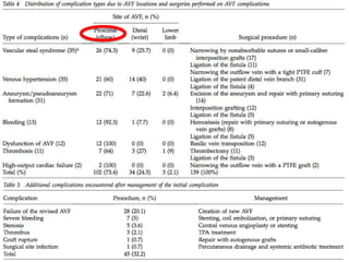
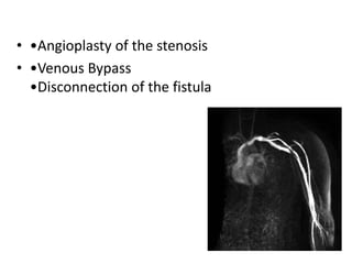
Surgical Interventions for Complications
of Arteriovenous Fistulas
Dicky A.Wartono ,MD FIHA FICA
Cardiac & Vascular Surgery
National Cardiovascular Centre Harapan Kita
Jakarta 2018
Nothing to declare
• 385,000 patients on dialysis in usa
• AV fistula /graft are considered superior to catheter access
• Remarkable success has been achieved in increasing
prevalence of AV fistula
• However, vascular access complications are common and
result in hospitalization, mortality and expense
• Guidelines suggest various methods to maintain patency of
vascular acc
GENERAL PRINCIPLES OF ACCESS
SURGERY
• distal non-dominant arm
• atherosclerotic arteries should be avoided
• chosen site should allow for ease of access for cannulation
• positioned so that patient comfort is assured during
heamodialysis
• vessels should be preserved by avoidance of:
–  Venipuncture
– Intravenous cannulation
– Invasive monitoring lines
Int Surg 2014;99:467–474
• Treatment Goal
• Cure Complication
• Preserving the Access
Failure
• Failure to mature and achieve an adequate flow rate to
maintain dialysis
• Incidence of up to 27%
• Such a complication may be a result of :
– Thrombosis !! 17–25%
– DM
– Techniqal problems in constructing the anastomosis
– A sclerotic vein segment in the forearm because of
previous venisection
– Inadequate venous size
– Cacification of the arterial wall
• Surgical thrombectomy is done by making a small venotomy
and using a fogarty balloon catheter to remove the thrombus
Stenosis & Thrombosis
Infection
• Fever –Erythema -Tenderness
• The second most common cause of AV access failure (0-3%
in AVF and 6%-25%in AV grafts)
• staph aureus -perivascular cellulitis -anatomical
abnormalities - septic pulmonary embolism
• Treatment:
AVFs: Local drainage and antibiotic therapy for 6
weeks
AV grafts: Antibiotic therapy and surgical treatment
(in most cases complete excision of prosthetic graft)
Ischemic changes
• Steal symptoms may occur in around 4% of patients with
autogenous fistula
• The incidence is higher in :
• Diabetic patients
• Atherosclerotic patients
• And in anticubital fistulas
• The symptoms may only manifested during dialysis or at its
worst , gangrene may occur requiring amputation
• Radial AV Fistula 1%
Brachial AV Fistula or Graft 3-6%
• Stage I Retrograde diastolic flow without
complaints; steal phenomenon
• Stage II Pain on exertion and/or during
haemodialysis
• Stage III Rest pain
• Stage IV Ulceration/necrosis/gangrene
• banding procedure
• ligation procedure
• tapered graft insertion
• PAI (Proximalization of the Arterial Inflow)
• DRIL- distal revascularization-interval ligation
• RUDI - revision using distal inflow
• Preferable to use vein
• Increased risk thrombosis PTFE
• Symptoms resolved 33 to 100%
• Improved 17 to 66%
• No change 1 series 11%
• DRIL patency 86 to 100%
Venous hypertension
• the hand distal to the fistula become swollen and uncomfortable
with thickning of the skin and hyperpigmentation
• Sign and symptoms:
• severe upper limb edema
skin discoloration
access dysfunction
peripheral ischaemia with resultant fingertip ulceration.
• In most cases, the underlying venous pathology follows ipsilateral
central venous catheter placement with consequent venous
stenosis
• CT Venography
• •Angioplasty of the stenosis
• •Venous Bypass
•Disconnection of the fistula
Complete resolution, with no mortality
AV f remain patent 100% for 6mo follow up
Morbidity :
1. skin lessions required skin graft
2. graft revision due large collateral
(Pseudo)Aneurysm Formation
• Pseudoaneurysm formation may occur at
(repetitive) puncture sites
• Pseudo aneurysms are hematomas / thrombotic
mass
• True aneurysm are 5%, but have also been
reported in few occasions in the vein distal to the
anastomosis
• Both have Risk of perforation and ulceration
Indications for revision/repair of AV fistula aneurysm:
• The skin overlying the fistula is (ischemic) compromised
• There is a risk of fistula rupture
• Available puncture sites are limited
indications for revision/repair of pseudoaneurysm formation
:
• Symptomatic or threatens the viability of the overlying skin
• Evidence of infection
• Pseudoaneurysm that is enlarging in size or that exceeds
twice the diameter of the graft
• Limited number of cannulation sites
• Cannulation through a pseudoaneurysm must be avoided
• Ligate
• Ressection
• Plication
• Aneurysmoraphy
J Vasc Surg 2011;53:1291-7.
Open Access Surgery 2010:3 9–12
Conclusions
• Fistula First
• Acute complication should be resolve before
patient discharge from OR
• Patency is a priority
• New fistula, is better that compromized fistula
THANK YOU
Surgery for avf complication

Surgery for avf complication

  • 1.
    Surgical Interventions forComplications of Arteriovenous Fistulas Dicky A.Wartono ,MD FIHA FICA Cardiac & Vascular Surgery National Cardiovascular Centre Harapan Kita Jakarta 2018
  • 2.
  • 3.
    • 385,000 patientson dialysis in usa • AV fistula /graft are considered superior to catheter access • Remarkable success has been achieved in increasing prevalence of AV fistula • However, vascular access complications are common and result in hospitalization, mortality and expense • Guidelines suggest various methods to maintain patency of vascular acc
  • 6.
    GENERAL PRINCIPLES OFACCESS SURGERY • distal non-dominant arm • atherosclerotic arteries should be avoided • chosen site should allow for ease of access for cannulation • positioned so that patient comfort is assured during heamodialysis • vessels should be preserved by avoidance of: –  Venipuncture – Intravenous cannulation – Invasive monitoring lines
  • 17.
  • 18.
    • Treatment Goal •Cure Complication • Preserving the Access
  • 20.
    Failure • Failure tomature and achieve an adequate flow rate to maintain dialysis • Incidence of up to 27% • Such a complication may be a result of : – Thrombosis !! 17–25% – DM – Techniqal problems in constructing the anastomosis – A sclerotic vein segment in the forearm because of previous venisection – Inadequate venous size – Cacification of the arterial wall • Surgical thrombectomy is done by making a small venotomy and using a fogarty balloon catheter to remove the thrombus
  • 21.
  • 26.
    Infection • Fever –Erythema-Tenderness • The second most common cause of AV access failure (0-3% in AVF and 6%-25%in AV grafts) • staph aureus -perivascular cellulitis -anatomical abnormalities - septic pulmonary embolism • Treatment: AVFs: Local drainage and antibiotic therapy for 6 weeks AV grafts: Antibiotic therapy and surgical treatment (in most cases complete excision of prosthetic graft)
  • 29.
    Ischemic changes • Stealsymptoms may occur in around 4% of patients with autogenous fistula • The incidence is higher in : • Diabetic patients • Atherosclerotic patients • And in anticubital fistulas • The symptoms may only manifested during dialysis or at its worst , gangrene may occur requiring amputation • Radial AV Fistula 1% Brachial AV Fistula or Graft 3-6%
  • 30.
    • Stage IRetrograde diastolic flow without complaints; steal phenomenon • Stage II Pain on exertion and/or during haemodialysis • Stage III Rest pain • Stage IV Ulceration/necrosis/gangrene
  • 32.
    • banding procedure •ligation procedure • tapered graft insertion • PAI (Proximalization of the Arterial Inflow) • DRIL- distal revascularization-interval ligation • RUDI - revision using distal inflow
  • 34.
    • Preferable touse vein • Increased risk thrombosis PTFE • Symptoms resolved 33 to 100% • Improved 17 to 66% • No change 1 series 11% • DRIL patency 86 to 100%
  • 38.
    Venous hypertension • thehand distal to the fistula become swollen and uncomfortable with thickning of the skin and hyperpigmentation • Sign and symptoms: • severe upper limb edema skin discoloration access dysfunction peripheral ischaemia with resultant fingertip ulceration. • In most cases, the underlying venous pathology follows ipsilateral central venous catheter placement with consequent venous stenosis • CT Venography
  • 41.
    • •Angioplasty ofthe stenosis • •Venous Bypass •Disconnection of the fistula
  • 42.
    Complete resolution, withno mortality AV f remain patent 100% for 6mo follow up Morbidity : 1. skin lessions required skin graft 2. graft revision due large collateral
  • 44.
    (Pseudo)Aneurysm Formation • Pseudoaneurysmformation may occur at (repetitive) puncture sites • Pseudo aneurysms are hematomas / thrombotic mass • True aneurysm are 5%, but have also been reported in few occasions in the vein distal to the anastomosis • Both have Risk of perforation and ulceration
  • 48.
    Indications for revision/repairof AV fistula aneurysm: • The skin overlying the fistula is (ischemic) compromised • There is a risk of fistula rupture • Available puncture sites are limited indications for revision/repair of pseudoaneurysm formation : • Symptomatic or threatens the viability of the overlying skin • Evidence of infection • Pseudoaneurysm that is enlarging in size or that exceeds twice the diameter of the graft • Limited number of cannulation sites • Cannulation through a pseudoaneurysm must be avoided
  • 49.
    • Ligate • Ressection •Plication • Aneurysmoraphy
  • 50.
    J Vasc Surg2011;53:1291-7.
  • 51.
    Open Access Surgery2010:3 9–12
  • 53.
    Conclusions • Fistula First •Acute complication should be resolve before patient discharge from OR • Patency is a priority • New fistula, is better that compromized fistula
  • 54.

Editor's Notes

  • #8 Side to side plg gambang, flow plg tinggi, turbulensi tinggi, flow ke distal, if flo kurang tiggal ligasi distal End to side, aga sulit bisa kingkingm flow rendah
  • #15 Acute.. Perdarahan & hematoma & fail/lowflow
  • #30 Chronic pain • Tissue loss • Non functioning extremity amputation of fingers and/or forearm in 1% of patients There is evidence that the steal syndrome in risk groups may occur in 75–90% of patients after cre- ation of an AVF. This phenomenon remains clinically asymptomatic until the moment when compensatory mechanisms for perfusion by peripheral arteries are ex- hausted [38]. Pathophysiological mechanisms of vascular access- induced distal ischemia are complex and not well known. Symptoms of arterial blood diversion are more common in patients with diabetes mellitus and smokers Data in the literature suggest that symptomatic ischemia develops in 10–25% of patients with brachioce- phalic and brachiobasilic vascular access, 4–6% at the level of the forearm and 1–2% at the radiocephalic level [43]. Due to the increasing age of patients on HD as well as a rising number of comorbidities, the incidence of hand ischemia is significantly increasing. It is assumed that in the future, the frequency of these ischemias will rise due to popularization of vascular access in the prox- imal region of the elbow, as well as the growing number of elderly people on HD [43]. Treatment of this condition is difficult and the risk of amputation of fingers and the forearm is great.
  • #35 DRIL- distal revascularization-interval ligation RUDI - revision using distal inflow