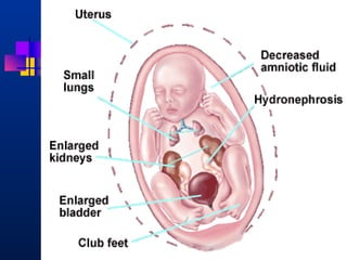
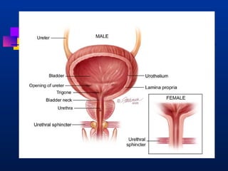
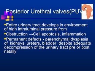
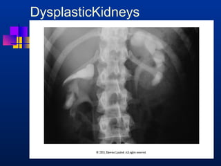
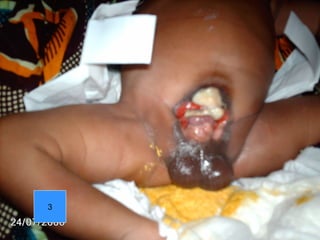
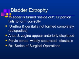
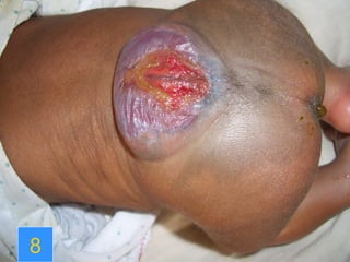
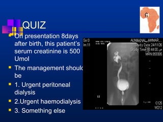
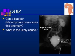
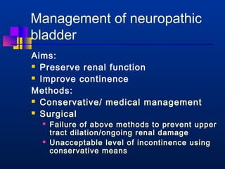
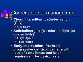
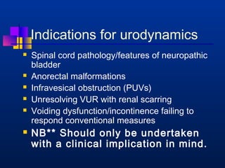
This document provides an overview of chronic kidney disease (CKD) and obstructive uropathy. It defines CKD and obstructive uropathy, discusses the stages of CKD, and outlines the mechanisms by which obstructions can lead to CKD, including both mechanical and non-mechanical obstructions. Specific conditions that can cause obstructions are presented such as posterior urethral valves. Management approaches including surgical and medical treatments are discussed, with an emphasis on long-term monitoring and care to preserve renal function and slow CKD progression.
Obstructive uropathy refers to any obstruction in the urinary tract. It can cause changes including hydronephrosis (distension of the kidneys and ureters) and hydroureteronephrosis (distension of the kidneys, ureters and bladder). The document discusses the classification, causes, pathology, investigations and management of various types of obstructive uropathy including ureteric strictures and retroperitoneal fibrosis. Surgical intervention is often needed to relieve the obstruction and treat associated complications.
This document provides an overview of obstructive uropathy. It begins by defining obstructive uropathy as the functional or anatomic obstruction of urine flow at any level of the urinary tract. It then discusses the prevalence of obstructive uropathy and how it can be classified based on factors like duration and site of obstruction. Potential causes of obstructive uropathy are then reviewed for different parts of the urinary tract. The pathophysiology and hemodynamic changes that occur with obstruction are explained. Cellular and molecular changes that can lead to fibrosis and tubular cell death are described. Management of patients is discussed including diagnostic imaging, issues in patient care like hypertension and pain management, and considerations for surgical intervention.
Acute urinary retention (AUR) is the inability to void despite a full bladder. It can be caused by failure of detrusor contraction or bladder outlet obstruction. Diagnosis involves confirmation of a full bladder on exam and insertion of a urethral catheter. Immediate treatment is catheterization to evacuate the bladder. Further evaluation is then done to identify the underlying cause and provide definitive treatment.
This document summarizes urinary tract obstruction and its causes, signs, symptoms, diagnosis, and treatment. It discusses various types of obstruction including ureteral obstruction, ureterocele, posterior urethral valves, and megaureter. Diagnostic tests include ultrasound, intravenous pyelogram, voiding cystourethrogram, and diuretic renogram. Treatment depends on the location and severity of the obstruction but may include drainage procedures like vesicostomy, reconstruction surgery like pyeloplasty, or excision of the obstructed segment.
Obstructive uropathy presenting as primary enuresis – A case reportApollo Hospitals
An adolescent male presented with persistent nocturnal bedwetting. Confounding family history of nocturnal enuresis lead to delay in seeking medical attention. Careful history and investigations confirmed underlying obstructive uropathy and bladder dysfunction. Primary enuresis presenting beyond childhood period should be investigated to look for underlying organic cause.
Hydronephrosis is the dilation of the renal pelvis and calyces caused by urinary outflow obstruction. It can result from anatomical or functional issues anywhere along the urinary tract. Chronic or severe hydronephrosis can lead to permanent kidney damage if not treated. Treatment depends on the cause and severity but may include ureteral stents, percutaneous nephrostomy tubes, or open surgery to bypass or remove the obstruction. The goal is to relieve obstruction and preserve kidney function.
This document discusses obstructive uropathy, including its definitions, clinical presentation, pathophysiology, clinical implications, and diagnostic evaluation. Obstructive uropathy is resistance to urine flow along the urinary tract that can damage the kidneys. It may present with a wide range of symptoms depending on factors like degree and duration of obstruction. Diagnostic tests include intravenous pyelogram, renal ultrasound, renogram, CT, and MRI to identify the level and severity of obstruction and determine appropriate management.
This document discusses obstructive uropathy in neonates. It begins with an example case of a preterm baby with bilateral hydronephrosis and thickened bladder walls. It then provides general information on obstructive uropathy including causes, presentations, investigations, and treatment principles. Specific conditions discussed in more detail include posterior urethral valves, ureteropelvic junction obstruction, and vesicoureteric reflux.
This document discusses hematuria (blood in the urine) and obstructive uropathy (blockage of urine flow in the urinary tract). It covers evaluating hematuria through urinalysis, imaging tests, and cystoscopy. Common causes of hematuria include infections, stones, tumors, and glomerulonephritis. Obstructive uropathy can be congenital or acquired and cause changes to the urethra, bladder, ureters, and kidneys over time. Relieving the obstruction through surgery, stents, or nephrostomy is the main treatment approach.
Obstructive uropathy can affect people of all ages and has varied presentations depending on the site and degree of obstruction. It can be caused by mechanical factors like congenital anomalies, inflammation, tumors, or stones, as well as functional issues related to neurological or muscular disorders. Common symptoms include pain, hematuria, and hydronephrosis. Diagnostic workup involves lab tests, imaging like ultrasound, CT, IVP, and procedures like cystoscopy. Treatment aims to relieve the obstruction through catheters, stents, or other surgeries, while also treating infections or other complications. Complete or solitary kidney obstructions require urgent intervention to prevent serious issues like renal failure.
Hydronephrosis is the dilatation of the renal pelvis and calyces caused by obstruction of urine flow from the kidney. It can be caused by congenital abnormalities, such as ureteral atresia, or acquired issues like kidney stones. On imaging, the kidney appears enlarged with a distended pelvis and thinning of the renal parenchyma. Treatment depends on the severity and cause of obstruction, ranging from nephrostomy or pyeloplasty for mild hydronephrosis to nephrectomy if the kidney is non-functioning.
Pathophysioogy of urinary tract obstruction bassem presentationfreeburn simunchembu
This document discusses obstructive nephropathy and hydronephrosis. It defines key terms and outlines the prevalence, classifications, causes, pathophysiology, diagnosis and management of obstructive kidney conditions. Specifically, it examines the hemodynamic changes that occur with unilateral and bilateral ureteral obstruction, including triphasic patterns of renal blood flow. It also describes the pathologic findings, compensatory renal growth, diagnostic imaging modalities and approaches to relieving obstruction and treating its underlying causes.
Lectures in urology for undergraduate medical students Elsayed Salih
This document contains lecture notes on various topics in urology from Dr. Elsayed Salih of Al-Azhar University. It includes sections on the diagnosis of urinary tract diseases through symptoms and investigations. It also covers congenital anomalies of the urinary tract including horseshoe kidney and polycystic kidney disease. Specific conditions discussed include renal cysts, vesicoureteral reflux, hydronephrosis, bladder exstrophy, and hypospadias.
1) Hydronephrosis is defined as the dilatation of the pelvi-calyceal system of the kidney due to partial or intermittent blockage of urine flow.
2) Causes include congenital abnormalities, kidney stones, ureteral strictures, or compression from other structures.
3) Treatment depends on the underlying cause and includes procedures to remove obstructions like stones, repair strictures, or decompress the system with nephrostomy tubes. Surgery may be needed for severe hydronephrosis to prevent kidney damage.
This document discusses obstructive uropathy in neonates. It presents a case of a preterm baby with bilateral hydronephrosis and a thick bladder wall. Key points discussed include the causes, presentations, investigations, and management of obstructive uropathy. Posterior urethral valves and ureteropelvic junction obstruction are examined in more detail. Vesicoureteric reflux is also summarized. The document emphasizes relieving obstruction, treating infection, and sorting the primary cause in managing obstructive uropathy.
Acute urinary retention is defined as the painful inability to void with relief after catheterization. It is caused by bladder outlet obstruction from conditions like benign prostatic hyperplasia, urethral stricture, or neurological impairment. Risk factors include older age, larger prostate size, and lower urinary flow rate. For management, watchful waiting is appropriate for mild symptoms while more severe or recurrent cases may require medical treatment with alpha blockers or 5-alpha-reductase inhibitors, or surgical options like transurethral resection of the prostate.
Hydronephrosis is a condition where the kidneys become swollen due to a blockage that prevents urine from exiting properly. It is typically caused by kidney stones, tumors, or narrowing of the ureters. Common symptoms include flank pain, nausea, frequent urination, and fever with infection. Treatment aims to drain urine from the kidneys to relieve pressure and remove the underlying cause of blockage, often through procedures to insert stents or bypass the obstruction site. Without treatment, hydronephrosis can lead to permanent kidney damage or failure.
The document discusses acute urinary retention and acute renal failure. It defines the conditions and outlines their causes, which can be pre-renal, renal, or post-renal in nature. The evaluation and management of the conditions is also described. Causes include issues like decreased perfusion, obstruction, toxicity, and inflammation. Treatment involves supportive care, addressing the underlying cause, and dialysis in some situations to manage electrolyte and acid-base imbalances.
The document discusses various disorders of the genitourinary system including urological obstructions, disorders of the kidney, and disorders of the ureters, urinary bladder and urethra. It covers the etiology, risk factors, clinical presentation, diagnostic evaluation, and management of various conditions like urethral strictures, renal calculi, nephrotic syndrome, acute glomerulonephritis and more. Nursing management is also described which involves monitoring vitals, intake/output, administering medications as ordered, and educating patients.
The document summarizes key information about gastric cancer including:
- The anatomy and blood supply of the stomach.
- Risk factors, sites, pathology, staging, and clinical features of gastric cancer.
- Investigations include endoscopy, imaging, and biopsy for diagnosis.
- Treatment involves a multidisciplinary team and may include endoscopic resection for early cancers, surgery such as gastrectomy with lymph node dissection, and chemotherapy/radiotherapy as adjuvant or palliative treatments.
- Prognosis depends on stage, with early localized cancers having the best outcomes if fully resected.
Hydronephrosis is dilation of the renal pelvis and calyces caused by obstruction of urine flow. It can be caused by intrinsic or extrinsic lesions and may be unilateral or bilateral. Chronic or incomplete obstruction can lead to cortical tubular atrophy, interstitial fibrosis, and thinning of the renal parenchyma over time. Acute obstruction may cause pain, while chronic or partial obstruction may have few symptoms until late stages with impaired urine concentration and renal function decline. Complete bilateral obstruction results in oliguria/anuria and requires relief of obstruction to survive.
The document discusses obstructive uropathy and benign prostatic hyperplasia (BPH). It defines obstructive uropathy as structural or functional hindrance of normal urine flow. BPH is defined as a slowly progressive nodular hyperplasia of the periurethral zone of the prostate. The document outlines causes, symptoms, diagnostic tests and treatments for both conditions. Treatments include medications, minimally invasive surgeries such as transurethral resection of the prostate, and open surgeries like prostatectomy.
This document discusses bladder outlet obstruction (BOO) and its causes such as benign prostatic hyperplasia (BPH). It describes the primary and long term effects of BOO on the bladder, including decreased urinary flow rates and increased voiding pressures. For BPH, it notes the causes include hyperplasia of the prostate gland that typically begins in the third decade. The document outlines the diagnosis, evaluation and treatment of BOO, including medical management with medications like alpha blockers and 5-alpha reductase inhibitors, as well as surgical treatments like transurethral resection of the prostate (TURP).
Evaluation of Obstructive Uropathy with Computed Tomography Urography and Mag...iosrjce
IOSR Journal of Dental and Medical Sciences is one of the speciality Journal in Dental Science and Medical Science published by International Organization of Scientific Research (IOSR). The Journal publishes papers of the highest scientific merit and widest possible scope work in all areas related to medical and dental science. The Journal welcome review articles, leading medical and clinical research articles, technical notes, case reports and others.
Hydronephrosis is the dilation of the renal pelvis and calyces caused by obstruction of urine flow from the kidney. It can be caused by issues in the ureter, bladder, or urethra that limit urine outflow. Unilateral hydronephrosis may cause dull flank pain while bilateral obstruction can lead to decreased urine output. Left untreated, hydronephrosis can damage kidney tissue and impair renal function. Diagnosis is made through imaging tests like intravenous pyelogram. Treatment focuses on resolving the underlying cause of obstruction.
here give the knowledge that you should possess to manage acute and chronic urine retention. the lecture is more concerned about practical patient care and ward setting management. you should minimally be aware about following facts regarding urine retention. the multiple causes of retention will be discussed later in detailed manner. Direction of the lecture seems more toward BPH and acute retention management. beware there are many aspects of a patient present with an AUR. do no harm and always try to keep patient satisfaction. Let me know about your comments an Ideas. try to improve the quality. good luck.
The document discusses obstructive uropathy and benign prostatic hyperplasia (BPH). It defines obstructive uropathy as structural or functional hindrance of normal urine flow. The most common causes of obstruction vary with age, from valves/strictures in children to BPH/prostate cancer in older adults. BPH is a non-cancerous enlargement of the prostate and its symptoms are related to bladder outlet obstruction. Treatment involves eliminating the obstruction either temporarily with stents/tubes or permanently with surgery or procedures like resection or ablation.
This document provides an overview of urinary schistosomiasis caused by the parasite Schistosoma haematobium. It discusses the epidemiology, pathogenesis, clinical manifestations, diagnosis, treatment, and control efforts. Around 90 million people are infected with S. haematobium, which resides in the veins around the bladder and causes urinary tract symptoms. Without treatment, it can lead to bladder cancer or kidney damage from obstruction. Praziquantel is the treatment of choice but control efforts face challenges in some developing areas.
Hepatorenal syndrome is a functional kidney failure that occurs in patients with cirrhosis and advanced liver disease. It is characterized by impaired renal function without structural kidney abnormalities. There are two types - type 1 is a rapidly progressive form with doubling of serum creatinine to over 2.5 mg/dL in less than 2 weeks, while type 2 is a moderate but steady or slowly progressive renal failure. Terlipressin plus albumin is the standard treatment for type 1 HRS and improves renal function and survival compared to albumin alone. For type 2 HRS, terlipressin plus albumin may be better than triple therapy with midodrine, octreotide, and albumin. Liver transplantation is the
Etiopathogenesis, Evaluation & Management of Posterior Urethral ValveShubham Lavania
This document discusses posterior urethral valves (PUV), including their etiology, classification, pathophysiology, clinical presentation, diagnosis, and management. PUV are congenital obstructions of the posterior urethra that commonly cause urinary outflow obstruction in boys. Type I valves are the most common. Initial management involves bladder drainage and antibiotics. Surgical valve ablation is usually curative, but long-term sequelae like renal disease are significant due to the primitive tissue injury caused by the obstruction.
This document discusses acute abdomen in pediatric patients. It defines acute abdomen and provides the main etiologies as inflammatory, traumatic, obstructive, and vascular conditions. It then discusses specific acute abdominal conditions that present at different ages, including neonatal causes like necrotizing enterocolitis, meconium plugs and atresia. Other causes mentioned include gastroenteritis, intussusceptions, malrotation and tumors. Signs and symptoms as well as approaches to specific conditions like appendicitis, meconium ileus and hypertrophic pyloric stenosis are summarized. Radiological images are also included to illustrate various pathologies.
The document discusses acute renal failure (ARF), defining it as the loss of renal function over hours to days resulting in the accumulation of nitrogenous waste products in the blood. It classifies the etiologies of ARF into prerenal, intrinsic renal, and postrenal causes. Prerenal ARF is due to decreased effective blood volume leading to renal vasoconstriction, while postrenal ARF results from urinary tract obstruction. Intrinsic renal ARF includes acute tubular necrosis, acute interstitial nephritis, and glomerulonephritis. Clinical evaluation involves assessing for risk factors and distinguishing between the types of ARF to guide treatment and management.
Hyperkalemia is defined as a plasma potassium level above 5.5 mEq/L. It can be caused by a shift of potassium from intracellular to extracellular space due to acidosis or tissue damage. Other causes include reduced renal excretion due to medications like ACE inhibitors or renal failure. Symptoms range from none to muscle weakness to cardiac arrhythmias. Treatment involves calcium to antagonize cardiac effects, insulin or beta-agonists to shift potassium intracellularly, and cation exchange resins or dialysis to remove excess potassium.
metabolic acidosis develops because of defects in the ability of the renal tubules to perform the normal functions required to maintain acid-base balance.
An abdominal CT scan uses x-rays to create detailed cross-sectional images of the abdomen. During the test, the patient lies still on a table that slides into a scanner, which rotates an x-ray beam around the body. Images are created as "slices" and can be combined to form 3D models. An abdominal CT scan is used to detect various abdominal abnormalities such as masses, tumors, infections, kidney stones, and issues affecting the liver, gallbladder, or pancreas. Abnormal results could indicate cancers, organ problems, appendicitis, aneurysms, or other issues requiring follow-up.
The document provides grooming and personal hygiene standards for hotel staff. It outlines that all staff should maintain high standards of grooming to create a good impression on guests. It then lists detailed standards for hair, facial hair, hands, personal hygiene, shoes, jewelry, and uniforms for both men and women to ensure a professional appearance.
POSTERIOR URETHRAL VALVES- Pediatric Surgery
• Dear viewers,
• Greetings from “ Surgical Educator”
• Today I have uploaded one more video in Pediatric Surgery/Pediatric Urology- “ Posterior Urethral Valves”
• Posterior Urethral Valves is the congenital cause for Bladder Outlet Obstruction, resulting in abnormal development of the kidneys as well as the bladder.
• In this video, I talked about the learning outcomes, introduction, etiopathogenesis, clinical features, investigations, differential diagnosis, treatment, follow-up and prognosis of “ Posterior Urethral Valves”
• I hope you will enjoy the video for its educational value.
• You can watch all my teaching videos in the following links
• surgicaleducator.blogspot.com youtube.com/c/surgicaleducator
• Thank you for watching the video.
This document describes the case of a 4-day old male neonate admitted to the hospital for evaluation of antenatally detected bilateral hydronephrosis. The baby was delivered full-term via normal vaginal delivery and initial examinations were normal. Antenatal ultrasounds showed progressively worsening bilateral hydronephrosis. Postnatal ultrasound confirmed bilateral hydronephrosis more severe on the left side. Laboratory tests and renal function were normal. A micturating cystourethrogram detected bilateral vesicoureteral reflux grade 3 on the right and grade 2 on the left. The baby received antibiotics and was discharged with instructions to follow-up in one month and continue prophylactic medications.
This document discusses the management of fetal hydronephrosis. It begins by defining hydronephrosis as the dilatation of the renal pelvis with or without calyceal dilatation. It then covers the grading of fetal hydronephrosis based on gestational age and renal pelvic diameter. Risk stratification systems like the UTD system are presented. Management depends on factors like severity, laterality, and presence of other anomalies. Most cases resolve spontaneously but severe or progressive cases may require interventions like pyeloplasty.
Posterior urethral valves is a congenital condition caused by abnormal membranes in the proximal urethra that obstruct the flow of urine. It most commonly presents in infancy with failure to pass urine and is diagnosed using imaging like ultrasound and voiding cystourethrography. Treatment involves surgical ablation of the valves via cystoscopy to restore urinary flow and halt renal damage. Prognosis depends on factors like age at diagnosis and degree of renal dysfunction, as patients may develop lifelong complications due to the original renal insults.
Hydronephrosis is the dilation of the renal pelvis and calyces due to partial or intermittent obstruction of urine flow. It is usually caused by blockages or reflux in the urinary tract. Common causes in children include vesicoureteral reflux, ureteropelvic junction obstruction, and posterior urethral valves. Diagnosis involves renal ultrasound and tests to assess kidney function. Treatment depends on the severity and underlying cause, ranging from observation to surgery to correct blockages or reflux. Complications can include urinary tract infections, kidney damage, and renal failure if not properly treated.
Vesicoureteric reflux (VUR) is a common condition in children where urine flows backward from the bladder into the ureters or kidneys. It can lead to urinary tract infections and renal scarring. The document discusses the anatomy, pathophysiology, diagnosis and management of VUR. Management involves continuous antibiotic prophylaxis, endoscopic injection, or open surgical correction depending on the grade of reflux and individual patient factors. Spontaneous resolution is more likely for lower grades of reflux and in younger children. Bowel and bladder dysfunction can complicate management and decrease resolution rates.
Diagnostic test related to congenital disorders of genitourinary system RakhiYadav53
This document discusses congenital disorders of the genitourinary system. It begins by defining key terms like urology, nephrology, and congenital anomalies. It then notes that congenital disorders occur in around 1 in 500 births and discusses common etiologies like genetic and environmental factors. The document proceeds to describe several specific congenital anomalies in detail, including their signs, symptoms, classification, and management approaches. These cover a range of conditions affecting the kidneys, bladder, urethra, and genitals. Various diagnostic tests for congenital disorders are also outlined.
This document discusses various radiological procedures used to examine different parts of the body. It provides information on barium swallow, barium meal, small bowel follow through, barium enema, intravenous urogram, voiding cystourethrogram, retrograde urethrogram, CT enteroclysis, CT urography, hysterosalpingography and their clinical indications and patient preparation requirements.
PUJ obstruction is a restriction of urine flow from the renal pelvis to the ureter. It can be congenital or acquired, with congenital being one of the most common causes of antenatal hydronephrosis. Diagnosis involves ultrasonography, VCUG, diuretic renography and other imaging modalities to determine severity and presence of associated issues. Treatment depends on severity but typically involves surgical intervention like open or laparoscopic pyeloplasty to resect and reanastomose the obstructed segment if drainage is significantly impaired or renal growth is poor. Postoperative follow up with imaging is important to monitor repair.
Antenatally detected hydronephrosis is one of the most common abnormalities detected on prenatal ultrasound. It can identify urinary tract obstructions and reflux before complications develop. The degree of hydronephrosis seen on prenatal ultrasound provides prognostic information, with mild cases often resolving and severe cases more likely to require postnatal intervention. Evaluation after birth depends on the severity and laterality of the hydronephrosis seen prenatally, with more severe or bilateral cases warranting earlier and more extensive testing like dynamic renal scintigraphy to assess kidney function and guide management.
PUV can present in newborns with life-threatening conditions or in older children with minor issues. Initial management involves bladder drainage and valve ablation. Reflux occurs in 50% and often resolves after ablation, though some patients require long-term antibiotics. Persistent hydronephrosis in about half of patients requires further intervention like upper tract diversion, though consensus is initial bladder drainage is preferred. Careful long-term management is needed to preserve renal function and prevent infection.
This document provides an overview of the approach to congenital hydronephrosis. It defines hydronephrosis and describes the most common causes including physiologic hydronephrosis, UPJ obstruction, UVJ obstruction, VUR, Eagle-Barrett Syndrome, PUV, and MCDK. It discusses grading of fetal and neonatal hydronephrosis using RPD, SFU criteria, and UTD classification system. The document outlines the approach including physical exam, imaging studies like VCUG and diuretic renography to diagnose underlying causes and guide treatment.
Antenatal diagnosis of Congenital Anomalies of Kidneys and Urinary Tract (CAKUT)Durre Sabih
Antenatal Diagnosis of Kidney Disease. This presentation gives an overview of the role of ultrasound in the diagnosis of fetal renal disease and congenital renal anomalies
This document provides an overview of the evaluation and management of posterior urethral valves. It begins with an introduction discussing the history and embryology of PUV. It then covers the clinical presentation, diagnostic evaluation, management including in utero and postnatal approaches, complications, and long-term outcomes. Key points include that PUV causes obstructive changes that damage the urinary tract, early diagnosis and relief of obstruction is important to preserve renal function, and bladder dysfunction often persists long-term requiring lifelong management.
1. Despite successful valve ablation in PUV patients, intrinsic bladder dysfunction can lead to deterioration of the upper urinary tracts and incontinence due to detrusor abnormalities, high pressure voiding, or detrusor-sphincter dyssynergia.
2. Anticholinergic drugs like oxybutynin and alpha-blockers like terazosin can help improve bladder function and reduce post-void residuals in PUV patients with bladder dysfunction. Clean intermittent catheterization may also be needed.
3. Longterm follow up of PUV patients is important to monitor renal function, attain urinary continence, and assess if interventions like augmentation cystoplasty or renal
This case study describes a 31-year-old male with a history of Alport syndrome and kidney transplant who presented with acute renal failure secondary to transplant rejection. Key points include: the patient's medical history of ESRD due to Alport syndrome requiring dialysis and kidney transplant; presentation with elevated creatinine and signs of antibody-mediated transplant rejection; treatment involving immunosuppression medication and potential need for dialysis; and nutrition interventions focusing on meeting protein and calorie needs through a renal diet.
Urogenital prolapse and urinary incontinence 22.04.2021Shazia Iqbal
This document discusses urinary incontinence and pelvic organ prolapse. It begins by outlining the objectives and prevalence of these conditions, which commonly affect women. It then describes the different types of urinary incontinence and treatments available, including both surgical and non-surgical options. The document also discusses pelvic organ prolapse, including causes, symptoms, assessment methods, and treatment approaches. Overall, it provides an overview of these common conditions and reviews evaluation and management strategies.
1. Antenatal hydronephrosis is a common prenatal finding that requires postnatal evaluation to identify potential kidney abnormalities.
2. Most cases of antenatal hydronephrosis are transient and resolve without intervention, while others may indicate issues like urinary tract obstruction that require treatment.
3. Postnatal evaluation includes ultrasound, voiding cystourethrogram, diuretic renography and other tests to determine the severity and cause of hydronephrosis and assess kidney function.
4. Stages of CKD—KDOQI, 2002
Stag
e
Description GFR
ml/min/1.73m2
1 Kidney
damage* with
nl or GFR
³90
2 Kidney
damage* with
mild GFR
60-89
3 Moderate
GFR
30-59
**
**
**
**
4 Severe GFR 15-29
5 Kidney failure 15 or dialysis
*Kidney damage for 3 months, as defined by pathologic abnormalities or
markers of kidney damage, including abnormalities in blood or urine or
diagnostic imaging studies.
13. Posterior Urethral valves(PUV)
Entire urinary tract develops in environment
of high intraluminal pressure from
Obstruction ®Cell apoptosis, inflammation
Permanent defects - parenchymal dysplasia
of kidneys, ureters, bladder despite adequate
decompression of the urinary tract pre or post
natally
14. PATHOPHYSIOLOGY (cont)
In some, renal tubules mainly affected® poor
urine concentrating ability, ® diuresis ®
ureteral bladder dysfunction
Affected kidneys may function well initially,
but have a reduced renal reserve
Renal deterioration also frm chronic
pyelonephritis from Vesicoureteric (V-U)
reflux, urinary stasis
15. PUV Clinical Presentation
Extremely variable
Prenatal US D:Oligohydramnios
hyroureters, hydronephrosis
Respiratory distress-pulmonary hypoplasia
Potter’s facies, ascites, and abdominal
swelling
Birth: Poor, dribbling urine stream
So check urine stream of all males b/4
discharge from hospital
16. PUV Clinical Presentation
Toddlers - voiding dysfunction or UTI
School-aged boys usually come to the
clinician's attention because of urinary
incontinence
All enuretics should be screened at least
with abdominal palpation urine MCS
18. Anteroposterior view of the abdomen during a void cystourethrographic study.
This image demonstrates a dilated bladder with trabeculation, diverticula, and
massive reflux.
19. Lateral view of a voiding cystourethrographic study during voiding after catheter removal.
The dilated posterior urethra is highly suggestive of a posterior urethral valve, which is
seen as the nonopacified line that separates the dilated posterior urethra from the normal-caliber
distal urethra.
26. Bladder Extrophy
Bladder is turned inside out“; Lr portion
fails to form correctly
Urethra genitalia not formed completely
(epispadias)
Anus vagina appear anteriorly displaced
Pelvic bones widely separated –diastasis
Rx: Series of Surgical Operations
28. Myelomeningocoele
Myelomeningocele - backbone and spinal
canal not close before birth. Associated :
1. Hydrocephalus in 90%
2. Big bladder- atonic bladder
3. Anus: Patulous: lack bowel control
4.Legs Partial or complete paralysis of
legs loss of sensation
28
30. Myelomeningocoele Rx
Parents fully aware BEFORE BIRTH
Surgery to repair defect at an early age
3.V-P shunt for hydrocephalus
4.Antibiotics 4 meningitis or UTI
5.Wheel chair
6. Bladder catheterization, Rx CRF- DT
30
32. 15 yr Survey of 45 CRF,PH
Primary renal dis. No.(%) Male Female
Glomerulopathies 23 (53.3) 9 14
Obstructive
uropathy
13 (28.9) 13 0
Vascular,PN,Mali
gn,Malaria
Nephropathy
9 (17.8) 5 4
Anochie I, Eke F.Ped Nephrol 2003;18:692-5
33. Yearly Incidence ESRF
UPTH,Nigeria
2.1
2.5
3
13
1985-91
1986-91
1991-2000
2007-2010
Red X, Sth Africa
EkeF.NigMedPract1992;35:37 EkeF,EkeN:PedNephrol1994;8:383-386
AnochieI,EkeF:PedNephrol2003:18:692-695
Dysplasia
PUV
PCKD
REFLUX
GN
FSGS
SLE
Other
34. Approach to preventing
Deterioration in CKD
A: Surgical
In Utero endoscopic valve ablation or vesico-amniotic
shunting
Endoscopic resection of valves 2-3 days of
birth
Operation should be done by the most
experienced. Valvotomy in UCH-OPD.
Catheter few hrs post Op
Severe valves may need vesicostomy or
ureterostomy; Bladder augmentation later
35. Mechanical Obstruction only
one Aspect
B: Medical
Immediate catheterizaton-size 3 Foleys
fluid electrolyte Rx
Long term monitoring of renal bladder
function
Dysplastic kidneys, Ureters Bladder
These not often respond to the mechanical
relief of Obstruction
36. Bladder Renal Care Vital
Intermittent catheterization by patient or
parents often an integral part of Rx
Yearly U/S- continued or recurrent stasis
Counselling re avoidance of UTI stasis
Yearly UE Cr, if renal function normaltests
for Renal Osteodystrophy, bone mineral
density, DMSA scan etc
Pts have lost their Txpt. Kidney from poor
bladder management
Avoid constipation
37. Bladder sphincter
dysfunction
Recurrent UTIs
V U R
Bowel
dysfunction
Bowel dysfunction is often part of the constellation;
Dysfunctional elimination syndrome)
38. Aims of CRF Management
feel normal - well
be normal - like your friends
- intellectual development
- school / other activities
maintain normal growth
preserving normal family functioning
slow progression to ESRF
prepare for ESRF treatment
39. CRF Management Points
nutrition
fluid and electrolyte
balance
acid base status
renal osteodystrophy
infection
anaemia
growth
hypertension
preservation of renal
function
education and
preparation
social /psychological
support
40. Renal osteodystrophy
Treatment
hydroxylated vitamin D therapy
- 1a hydroxycholecalciferol
- 1-25dihydroxycholecalciferol
by daily oral administration
41. Counselling
Surgery NOT A CURE
Long term Follow Up
Clearly told Paediatrician or Nephrol for
Life
Urological Follow Up may also be for Life
Counsel against ‘It is not my portion’
Multidisciplinary Counselling best
47. QUIZ
On presentation 8days
after birth, this patient’s
serum creatinine is 500
Umol
The management should
be
1. Urgent peritoneal
dialysis
2.Urgent haemodialysis
3. Something else
48. QUIZ
Can a bladder
rhbdomyosarcoma cause
this anomaly?
What is the likely cause?
49. QUIZ
True or False
All children with Posterior Urethral
valves have Chronic Kidney Disease
51. Learn more about afpna its
members by visiting the
website www.afpna.com
PNAN integral part of AFPNA her
Mtgs are advertised there
52. Officers
Secretary-General Felicia Eke, Nigeria
Secretary-Treasurer Bashir Admani, Kenya
Member IPNA Councillor Udai Kala, South Africa
Member IPNA Councillor Safaa Medani, Sudan (North africa)
Member Priya Gajjar, South Africa
Member Francis Lalya, Benin
Congress President Elect Sampson Antwi, Ghana
Ex-Officio Mignon Mc Culloch
53. Dates of Annual Meetings
3yrly Mtg –
5th Congress Ghana Feb 20-23, 2013
4th Nairobi March19-21, 2010
3rd Cape Town, March 2006
2nd Port Harcourt, Nigeria 2002
1st Cairo, Egypt 2000
54. IPNA Fellows
Dr Franca Ikimalo Port Harcourt, Nigeria CT, SA 1.9 03 – 29.2.04
DrAmos Odit Uganda CT, SA 1.3.04 –31.8.04
Dr.Godson Achugwo Owerri,Nigeria CT, SA 05-10.05
DrFrancis Lalya Cotonou,Benin CT, SA 14.10.05–30.4.06
Again 12.08-12.09
Dr Hemed Twahir Mombasa,Kenya CT, SA 1.4.06 –5.3.07
again in 2009
Dr.Bashir Admani Nairobi,Kenya CT, SA 8.06-8.08
Dr.Isaac Ocheke Jos, Nigeria CT,SA 7.3.07 – 2.08
Dr Sampson Antwi Kumasi,Ghana CT, SA 4/09-4.10
Dr Seyi Oniyangi Abuja,Nigeria Joburg,SA 9.08-2.09
Dr Chris Esezobor Lagos, Nigeria
Dr David Galgallo Kenya
Dr Elena Naicker Durbar, SA Joburg,SA 5-12.09
Dr Odetunde Enugu, Nigeria CT, SA 3.11
Dr NosaIdurionye Benin, Nigeria CT, SA 6.11-
Total 14
55. IPNA Discount Membership
Fee for Regional Societies
IPNA at its recent Council Meeting in Sao
Paulo, Brazil (October 25-26th 2011) offers
discounted membership fee to members of
its affiliated Regional Societies, namely the
ASPN, ESPN, African Pediatric Nephrology
Association (AFPNA), AsPNA,
JSPN,ANZPNA ALANEPE
56. IPNA Discount Membership
Fee for Regional Societies
The current membership fee is US$175/yr
The discounted membership fee for Regional
Society members is US$50/yr® privileges
1. IPNA newsletter
2. On-line access to Ped Nephrol Journal
3. On-line access to Ped Nephrol textbook
4. IPNA Congress member discount
5. Print version of Pediatric Nephrology at an
additional rate of US$65 annually (total
US$115 annually)
57. IPNA Discount Membership
Fee for Regional Societies
Individual paid-up members of the Regional
Soc are eligible 4 discounted membership.
Combined fee administered either by IPNA
or the Regional Soc accordingly. thus:
ASPN: US$300+US$50 (USA)
US$150+US$50 (Canada)
ESPN: US$70 (Euro$50) + US$50
JSPN: US$135 (Y10,000) + US$50
AsPNA: US$10 + US$50
ANZPNA: US$105 (Aust$100) + US$50
58. IPNA Discount Membership
Fee for Regional Societies
AFPNA: US$25 + US$50
For fees paid through IPNA, the currency will
be in USD. IPNA will then transfer the Reg
Soc dues accordingly at the end of each
financial year. The fee for wire transfer will be
charged to each Reg Soc accordingly. The
IPNA secretariat will only deal with the
payment of full fees for Reg Soc. Discounted
fees for Reg Soc should be dealt with by the
respective Reg Soc.
59. IPNA Discount Membership
Fee for Regional Societies
For fees paid through the Reg Soc the
Regional Societies can charge in the
appropriate currency, but the corresponding
total sum should be paid to IPNA in USD. The
Regional Society must send the appropriate
membership information to IPNA in order to
effect the membership privileges.
The discounted membership fee will be
implemented from January 1st 2012
60.
61.
62.
63.
64. Management of neuropathic
bladder
Aims:
Preserve renal function
Improve continence
Methods:
Conservative/ medical management
Surgical
Failure of above methods to prevent upper
tract dilation/ongoing renal damage
Unacceptable level of incontinence using
conservative means
65. Cornerstone of management
Clean intermittent catheterisation
(CIC)
4 X daily
Anticholinergics (counteract detrusor
overactivity)
Oxybutynin
Tolterodine
Early intervention. Prevents
progressive detrusor damage with
loss of compliance and later
requirement for cystoplasty.
67. Indications for urodynamics
Spinal cord pathology/features of neuropathic
bladder
Anorectal malformations
Infravesical obstruction (PUVs)
Unresolving VUR with renal scarring
Voiding dysfunction/incontinence failing to
respond conventional measures
NB** Should only be undertaken
with a clinical implication in mind.
70. Urodynamic Features associated
with renal deterioration
Detrusor sphincter dyssynergia
High filling/voiding pressures
Decreased bladder compliance
Leak point pressure40 cm
H2O
71. PD Nursing Team
PATIENT
Ward Nursing
Team
Administratio
n of fluid
deliveries
Medical
Team
Social
Worker Counsellor
Dietitian
The Multidisciplinary
Team
72. COMPLICATIONS
In the newborn
Pulmonary hypoplasia secondary to intrauterine renal
dysfunction and Oligohydramnios is the primary cause of patients
death.
Other complications of PUV are generally secondary to chronic
bladder changes, leading to elevated detrusor pressures. This, in
turn, leads to progressive renal damage, infection, and
incontinence.
Renal insufficiency
Due to renal dysplasia
Elevated detrusor pressure and back flow of urine.
73. COMPLICATIONS (cont)
Vesicoureteral reflux
Commonly associated with PUV and is present in as many as one
third of patients
Vesicoureteral reflux in most children is believed to be due to an
abnormal insertion of the ureter into the bladder.
When associated with PUV, reflux is generally secondary to
elevated intravesical pressures.
Urinary tract infections
Recurrent UTIs are common in patients with PUV.
Elevated intravesical pressures predispose patients to infection,
possibly by altering urothelial blood flow.
Additionally, patients with PUV may have elevated post void
residual urine volumes, leading to stasis of urine.
Dilated upper urinary tracts, with or without vesicoureteral reflux,
further elevate UTI risk.
74. COMPLICATIONS (cont)
Bladder dysfunction
Thought to be caused by alterations in collagen deposition and the
development of detrusor smooth muscle cells.
In mild cases, incontinence may be present;
In severe cases, ongoing deterioration of renal function occurs.
Bladder dysfunction often improves over time after definitive
treatment of the obstruction.
End Stage Renal Disease( ESRD)
Progression of ESRD is accelerated at the time of puberty due to
the increased metabolic workload placed on the kidneys.
75. PROGNOSIS
Over the last 30 years, the prognosis of children with PUV has steadily
improved.
In the past, most children were found to have PUV only after presenting with
urosepsis or progressive renal insufficiency. Older series demonstrated mortality
rates approaching 50% by late adolescence.
Today, most individuals with PUV are discovered when prenatal ultrasonography
reveals hydronephrosis.
Historically, of patients with adequate pulmonary function, approximately 25%
died of renal insufficiency in the first year of life, 25% died later in childhood, and
50% survived to adulthood with varying degrees of renal function.
Prompt resolution of bladder obstruction, aggressive treatment of bladder
dysfunction, and improved surgical techniques have lowered the neonatal
mortality rate to less than 3%.
Approximately one third of patients progress to renal insufficiency in their
lifetimes. Improved dialysis and transplantation techniques have significantly
improved not only the mortality rate for these children but also their quality of life.
.
76. PROGNOSIS
Additionally, medical and surgical management can achieve urinary
continence in nearly all patients
An interesting group of patients are those with vesicoureteral reflux
dysplasia (VURD) syndrome.
In these patients, one kidney is hydronephrotic, nonfunctioning, and
has high-grade vesicoureteral reflux. The high-grade reflux is thought
to act as a pop-off valve, leading to reduced overall bladder pressures
and preservation of contralateral renal function.
In the past, these patients were thought to have a better outcome due
to preserved renal function in one kidney at the sacrifice of the other.
More recent data suggests that, although short-term serum creatinine
levels may be favorable, these patients may suffer long-term adverse
renal function with hypertension, proteinuria, and renal failure In the
long.
77. CONCLUSION
Posterior urethral valve though not a
common congenital anomaly can be fatal
Early detection, prompt management and
routine follow up of patients is needed for
a good life expectancy.
78. INVESTIGATIONS (cont)
Nuclear Renography
Detects upper-tract consequences of obstruction
Absent or dysplastic kidney ® photopenic area in
renal fossa
Altered renal function ® Delayed visualization of
renal unit with slow rise to peak activity
79. TREATMENT (cont)
Vesicostomy
When urethral size precludes safe valve
ablation, a communicating channel between
the bladder and lower abdominal wall (ie,
vesicostomy) can be created to provide bladder
drainage
Generally, an 18-20F stoma is created
approximately midway between the pubis and
umbilicus in the midline
Take care to bring the dome of the bladder to
the skin and to limit the stomal size to prevent
prolapse of bladder urothelium through the
vesicostomy
Its use has decreased because most patients
can be safely drained and can undergo valve
80. TREATMENT (cont)
Cutaneous ureterostomies:
Bilateral cutaneous ureterostomies can also be
placed to provide for urinary drainage
Techniques for cutaneous ureterostomy
include:
-End stomal ureterostomy,
-loop ureterostomy,
-Y-ureterostomy (in which the ureter is
divided and one end is brought to the skin and
the other is reanastomosed in a uretero-ureterostomy),
and
-Ring ureterostomy techniques
Potential complications of cutaneous
ureterostomies include ureteral
devascularization, inadequate drainage, and
81. TREATMENT (cont)
Secondary bladder surgery
Augmentation cystoplasty
Indications for bladder augmentation include :
-Inadequately low bladder storage volumes
-High bladder pressures despite anticholinergic
medication and clean intermittent catheterization.
The ileum is most commonly used; however, large bowel,
stomach, and ureter are also used, depending on clinical
conditions and surgeon preference.
Augmentation should only be offered to patients willing to
commit to lifelong intermittent catheterization.
82. TREATMENT (cont)
Potential complications include
-Bladder rupture (approximately 10% of patients);
-Electrolyte disturbances, which may be worsened by the
placement of intestinal mucosa in contact with urine.
-Several cases of malignant degeneration in augmented
bladder have been reported.
Despite these risks, augmentation can significantly improve
patient lifestyle in those who have intractable incontinence
due to poor compliance and bladder over activity.
Continent Appendicovesicostomy :
Also called the Mitrofanoff technique,
Involves placement of a non refluxing tubular conduit for
catheterization between the bladder and skin to provide an alternative
channel for catheterization.
83. TREATMENT (cont)
Potential complications include
-Bladder rupture (approximately 10% of patients);
-Electrolyte disturbances, which may be worsened by the
placement of intestinal mucosa in contact with urine.
-Several cases of malignant degeneration in augmented
bladder have been reported.
Despite these risks, augmentation can significantly improve
patient lifestyle in those who have intractable incontinence
due to poor compliance and bladder over activity.
Continent Appendicovesicostomy :
Also called the Mitrofanoff technique,
Involves placement of a non refluxing tubular conduit for
catheterization between the bladder and skin to provide an alternative
channel for catheterization.