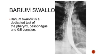
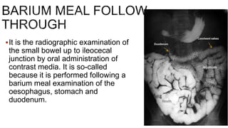
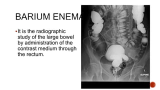
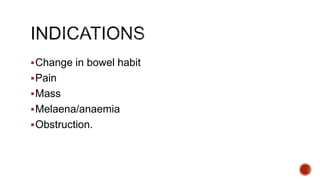
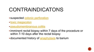
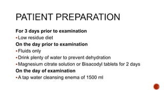
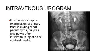
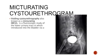
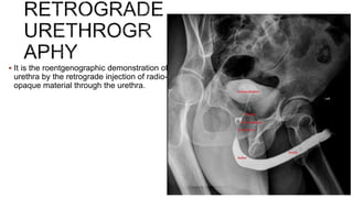
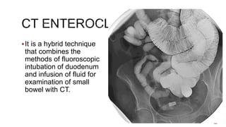
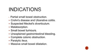
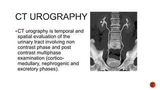
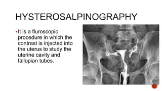
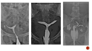
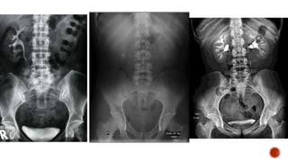
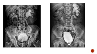
DR. Prithviraj Jadhav
Prof. Department of Radio-diagnosis,
DY Patil medical college, hospital & research institute Kolhapur
Barium swallow is a
dedicated test of
the pharynx, oesophagus
and GE Junction.
 Dysphagia and obstruction.
 Pain during swallowing.
 Assessment of mediastinal masses.
 Assessment of left atrial enlargement.
 Pre-op assessment of carcinoma bronchus and oesophagus.
 Motility disorders of oesophagus, E.g.: Achalasia and diffuse
oesophageal spasm, scleroderma.
 Assessment of site of perforation.
 Zenker's diverticulum and cricoid webs.
 Tracheo-oesophageal fistula.
 Perforation.
 Leakage of barium from an unsuspected perforation-granuloma
formation.
 Aspiration.
Nil orally for 6 h prior to the examination.
The patient is advised not to smoke on the day of the
examination, as it increases gastric motility.
It should be ensured that there are no contraindications to
the Preliminary film Technique or pharmacological agents
used.
Barium meal is the
radiological study of
stomach and duodenum.
It is done by oral
administration of contrast
media (Barium sulphate).
Dyspepsia
 Weight loss
Upper abdominal mass
Gastrointestinal haemorrhage (or unexplained iron-
deficiency anaemia)
 Partial obstruction
Assessment of site of perforation - it is essential that a
water- soluble contrast medium, e.g. Gastrografin or
LOCM, is used.
Complete large bowel obstruction.
Nil orally for 6 h prior to the examination.
The patient is advised not to smoke on the day of the
examination, as it increases gastric motility.
It should be ensured that there are no contraindications
to the pharmacological agents used.
It is the radiographic examination of
the small bowel up to ileocecal
junction by oral administration of
contrast media. It is so-called
because it is performed following a
barium meal examination of the
oesophagus, stomach and
duodenum.
 Patients who have low suspicion of small bowel disease
abdominal pain and diarrhoea.
 Patients with suspected complete (or) near complete small bowel
obstruction.
 Patients who are suspected of suffering from Crohn's disease.
 Patients who refuse placement of nasogastric tube/failed
intubation.
 Elderly patients with suspected jejunal diverticulosis who present
with malabsorption.
Colonic obstruction.
Suspected perforation.
Paralytic Ileus.
The colon should be cleaned by the administration of
a suitable purgative. (Purgative should be avoided in
patients with suspected obstruction, acute
exacerbation of Crohn's disease or an Ileostomy).
A low roughage diet and a high fluid intake is also
maintained for 48 hours prior to the investigation
No food or fluid should be taken for 12 hours before
the investigation. If the patient is taking tranquilizers,
antispasmodics and codeine, they should be stopped
for 24-48 hours before the examination.
It is the radiographic
study of the large bowel
by administration of the
contrast medium through
the rectum.
Change in bowel habit
Pain
Mass
Melaena/anaemia
Obstruction.
suspected colonic perforation
toxic megacolon
pseudomembranous colitis
imminent rectal biopsy within 7 days of the procedure or
within 7-10 days after the rectal biopsy
documented history of anaphylaxis to barium
For 3 days prior to examination
Low residue diet
On the day prior to examination
Fluids only
Drink plenty of water to prevent dehydration
Magnesium citrate solution or Bisacodyl tablets for 2 days
On the day of examination
A tap water cleansing enema of 1500 ml
It is the radiographic
examination of urinary
tract including renal
parenchyma, calyces
and pelvis after
intravenous injection of
contrast media.
1. Screening of the entire urinary tract especially in cases of haematuria or pyuria.
2. Diseases of the renal collecting system and renal pelvis
3. Differentiation of function of both kidneys.
4. Abnormalities of the ureter.
5. Obstructive uropathy- IVU is the gold standard.
6. TB of the urinary tract
7. Calculus disease.
8. Potential Renal Donors.
9. Prior to endo-urological procedures and surgery of urinary tract.
10. Suspected renal injury.
11. Renal colic or flank pain.
1. VATER anomalies: These patients have vertebral, anal, tracheo oesophageal, and
renal anomalies. Renal anomalies are seen in about 90% of patients.
2. Malformation of urinary tract, e.g., polycystic disease, PUJ obstruction etc.
3. Neurological disorders affecting urinary tract.
4. Malformation of genitalia like bilateral cryptorchidism, III degree hypospadias,
family history of urinary tract anomalies, urinary tract infection.
5. Enuresis in the presence of bacteriuria,abnormal urinary sediment, adolescents,
diurnal/nocturnal incontinence and history of recurrent urinary tract infection.
6. In girls with constant or intermittent dampness which suggests an ectopically
inserted ureter, IVU is mandatory.
7. Anorectal anomalies.
1. Iodine sensitivity.
2. Pregnancy.
3. Severe history of anaphylaxis previously carries 30% risk of similar reaction on a
subsequent occasion. The risk is lower with low osmolar contrast media.
4. Raised serum creatinine levels
• overnight fasting for 5 hours prior to the date of examination; a laxative
may not be necessary for bowel preparation as it does not improve
image quality
• on the day of the procedure take a scout/pilot film to check patient
preparation and also to look for radiopaque calculi
• check serum creatinine level to be within the normal range (as per
hospital guidelines)
• take a history of the patient for any known drug allergies followed by
written informed consent for the procedure
• emergency medications and emergency equipment must always be
available in case the patient has a reaction to contrast
 Voiding cystourethrography also
known as a micturating
(MCU), is a fluoroscopic study of
the lower urinary tract in which
introduced into the bladder via a
1.UTI-Usually done after some weeks after acute stage or may be done under
antibiotic coverage.
2.Voiding difficulties like dysuria, thin stream, dribbling, frequency, urgency.
3.Vesico ureteric reflux.
4.Other congenital anomalies : Meningomyelocele, Sacral agenesis,
5.Rectal anomalies.
6.Baseline study prior to lower UT surgery.
7.For post operative evaluation of ureteric abnormalities.
8.Pelvic Trauma.
Main indications
1. Trauma to urethra.
2. Urethral stricture.
3. Suspected urethral diverticula.
Other indications
1. UTI.
2. Reflux nephropathy prior to renal transplant of one/both kidneys.
3. Follow up of patients with spinal cord injury.
 The patient micturates prior to the examination.
 Acute urinary tract infection
 It is the roentgenographic demonstration of
urethra by the retrograde injection of radio-
opaque material through the urethra.
 Strictures
Urethral tears
Congenital abnormalities
Periurethral or prostatic abscess
Fistulae or false passages.
Bowel preparation is not required for mcu procedure
Acute urinary tract infection.
CT ENTEROCLYSIS
CT UROGRAPHY
It is a hybrid technique
that combines the
methods of fluoroscopic
intubation of duodenum
and infusion of fluid for
examination of small
bowel with CT.
 Partial small bowel obstruction.
 Crohn's disease and Ulcerative colitis.
 Suspected Meckel's diverticulum.
 Malabsorption.
 Small bowel tumours.
 Unexplained gastrointestinal bleeding.
 Complete colonic obstruction.
 Paralytic ileus.
 Massive small bowel dilatation.
Pregnancy
Gastric outlet obstruction
Low residue diet and good hydration.
Laxatives a day prior to the procedure and no oral dose on
the day of procedure.
Sedation can be used optionally if required.
CT urography is temporal and
spatial evaluation of the
urinary tract involving non
contrast phase and post
contrast multiphase
examination (cortico-
medullary, nephrogenic and
excretory phases).
 Urinary calculus disease
 Evaluation of Hematuria
 Suspected pelvis or ureteral obstruction
 Inflammatory conditions of the kidney and ureter
 Congenital anomalies of the kidney and ureter
 Urinary tract trauma
Allergy to contrast agents.
Asthmatic patients.
Patients with cardiac diseases.
Renal insufficiency.
Diabetic patients.
Pregnant patients.
Patients should be told to avoid food intake 6
hours before the examination. However, they
should maintain good hydration prior to the
examination.
It is a fluroscopic
procedure in which the
contrast is injected into
the uterus to study the
uterine cavity and
fallopian tubes.
1. Infertility:
• To demonstrate patency of the fallopian tubes and their communication
with the peritoneal cavity.
• Prior to artificial insemination.
2. Recurrent abortions: To demonstrate congenital abnormalities of the uterus
or incompetence of the internal os of the uterus.
3. Following tubal surgery: To monitor the effect of tubal surgery.
4. Migrated IUCD.
5. Uterine and tubal lesions like tuberculosis, submucous fibroids, polyps, and
synechiae.
The patient should be advised to abstain from
intercourse between booking the appointment and the
time of examination unless a reliable method of
contraception is used to avoid the possibility of
irradiating an early pregnancy.
The patient should be fasting 4 hours prior to the
procedure.
 Active Pelvic Sepsis.
 Sensitivity to contrast media.
 Recent dilatation and curettage.
 Pregnancy.
 The week prior to and the week following onset of
menstruation.
 Severe renal or cardiac disease.
 Cervicitis/purulent vaginal discharge.
radiological procedures presentation.pptx

radiological procedures presentation.pptx

  • 1.
    DR. Prithviraj Jadhav Prof.Department of Radio-diagnosis, DY Patil medical college, hospital & research institute Kolhapur
  • 2.
    Barium swallow isa dedicated test of the pharynx, oesophagus and GE Junction.
  • 3.
     Dysphagia andobstruction.  Pain during swallowing.  Assessment of mediastinal masses.  Assessment of left atrial enlargement.  Pre-op assessment of carcinoma bronchus and oesophagus.  Motility disorders of oesophagus, E.g.: Achalasia and diffuse oesophageal spasm, scleroderma.  Assessment of site of perforation.  Zenker's diverticulum and cricoid webs.
  • 4.
     Tracheo-oesophageal fistula. Perforation.  Leakage of barium from an unsuspected perforation-granuloma formation.  Aspiration.
  • 5.
    Nil orally for6 h prior to the examination. The patient is advised not to smoke on the day of the examination, as it increases gastric motility. It should be ensured that there are no contraindications to the Preliminary film Technique or pharmacological agents used.
  • 6.
    Barium meal isthe radiological study of stomach and duodenum. It is done by oral administration of contrast media (Barium sulphate).
  • 7.
    Dyspepsia  Weight loss Upperabdominal mass Gastrointestinal haemorrhage (or unexplained iron- deficiency anaemia)  Partial obstruction Assessment of site of perforation - it is essential that a water- soluble contrast medium, e.g. Gastrografin or LOCM, is used.
  • 8.
    Complete large bowelobstruction. Nil orally for 6 h prior to the examination. The patient is advised not to smoke on the day of the examination, as it increases gastric motility. It should be ensured that there are no contraindications to the pharmacological agents used.
  • 9.
    It is theradiographic examination of the small bowel up to ileocecal junction by oral administration of contrast media. It is so-called because it is performed following a barium meal examination of the oesophagus, stomach and duodenum.
  • 10.
     Patients whohave low suspicion of small bowel disease abdominal pain and diarrhoea.  Patients with suspected complete (or) near complete small bowel obstruction.  Patients who are suspected of suffering from Crohn's disease.  Patients who refuse placement of nasogastric tube/failed intubation.  Elderly patients with suspected jejunal diverticulosis who present with malabsorption.
  • 11.
  • 12.
    The colon shouldbe cleaned by the administration of a suitable purgative. (Purgative should be avoided in patients with suspected obstruction, acute exacerbation of Crohn's disease or an Ileostomy). A low roughage diet and a high fluid intake is also maintained for 48 hours prior to the investigation No food or fluid should be taken for 12 hours before the investigation. If the patient is taking tranquilizers, antispasmodics and codeine, they should be stopped for 24-48 hours before the examination.
  • 13.
    It is theradiographic study of the large bowel by administration of the contrast medium through the rectum.
  • 14.
    Change in bowelhabit Pain Mass Melaena/anaemia Obstruction.
  • 15.
    suspected colonic perforation toxicmegacolon pseudomembranous colitis imminent rectal biopsy within 7 days of the procedure or within 7-10 days after the rectal biopsy documented history of anaphylaxis to barium
  • 16.
    For 3 daysprior to examination Low residue diet On the day prior to examination Fluids only Drink plenty of water to prevent dehydration Magnesium citrate solution or Bisacodyl tablets for 2 days On the day of examination A tap water cleansing enema of 1500 ml
  • 17.
    It is theradiographic examination of urinary tract including renal parenchyma, calyces and pelvis after intravenous injection of contrast media.
  • 18.
    1. Screening ofthe entire urinary tract especially in cases of haematuria or pyuria. 2. Diseases of the renal collecting system and renal pelvis 3. Differentiation of function of both kidneys. 4. Abnormalities of the ureter. 5. Obstructive uropathy- IVU is the gold standard. 6. TB of the urinary tract 7. Calculus disease. 8. Potential Renal Donors. 9. Prior to endo-urological procedures and surgery of urinary tract. 10. Suspected renal injury. 11. Renal colic or flank pain.
  • 19.
    1. VATER anomalies:These patients have vertebral, anal, tracheo oesophageal, and renal anomalies. Renal anomalies are seen in about 90% of patients. 2. Malformation of urinary tract, e.g., polycystic disease, PUJ obstruction etc. 3. Neurological disorders affecting urinary tract. 4. Malformation of genitalia like bilateral cryptorchidism, III degree hypospadias, family history of urinary tract anomalies, urinary tract infection. 5. Enuresis in the presence of bacteriuria,abnormal urinary sediment, adolescents, diurnal/nocturnal incontinence and history of recurrent urinary tract infection. 6. In girls with constant or intermittent dampness which suggests an ectopically inserted ureter, IVU is mandatory. 7. Anorectal anomalies.
  • 20.
    1. Iodine sensitivity. 2.Pregnancy. 3. Severe history of anaphylaxis previously carries 30% risk of similar reaction on a subsequent occasion. The risk is lower with low osmolar contrast media. 4. Raised serum creatinine levels
  • 21.
    • overnight fastingfor 5 hours prior to the date of examination; a laxative may not be necessary for bowel preparation as it does not improve image quality • on the day of the procedure take a scout/pilot film to check patient preparation and also to look for radiopaque calculi • check serum creatinine level to be within the normal range (as per hospital guidelines) • take a history of the patient for any known drug allergies followed by written informed consent for the procedure • emergency medications and emergency equipment must always be available in case the patient has a reaction to contrast
  • 22.
     Voiding cystourethrographyalso known as a micturating (MCU), is a fluoroscopic study of the lower urinary tract in which introduced into the bladder via a
  • 23.
    1.UTI-Usually done aftersome weeks after acute stage or may be done under antibiotic coverage. 2.Voiding difficulties like dysuria, thin stream, dribbling, frequency, urgency. 3.Vesico ureteric reflux. 4.Other congenital anomalies : Meningomyelocele, Sacral agenesis, 5.Rectal anomalies. 6.Baseline study prior to lower UT surgery. 7.For post operative evaluation of ureteric abnormalities. 8.Pelvic Trauma.
  • 24.
    Main indications 1. Traumato urethra. 2. Urethral stricture. 3. Suspected urethral diverticula. Other indications 1. UTI. 2. Reflux nephropathy prior to renal transplant of one/both kidneys. 3. Follow up of patients with spinal cord injury.
  • 25.
     The patientmicturates prior to the examination.  Acute urinary tract infection
  • 26.
     It isthe roentgenographic demonstration of urethra by the retrograde injection of radio- opaque material through the urethra.
  • 27.
     Strictures Urethral tears Congenitalabnormalities Periurethral or prostatic abscess Fistulae or false passages.
  • 28.
    Bowel preparation isnot required for mcu procedure Acute urinary tract infection.
  • 29.
  • 30.
    It is ahybrid technique that combines the methods of fluoroscopic intubation of duodenum and infusion of fluid for examination of small bowel with CT.
  • 31.
     Partial smallbowel obstruction.  Crohn's disease and Ulcerative colitis.  Suspected Meckel's diverticulum.  Malabsorption.  Small bowel tumours.  Unexplained gastrointestinal bleeding.  Complete colonic obstruction.  Paralytic ileus.  Massive small bowel dilatation.
  • 32.
    Pregnancy Gastric outlet obstruction Lowresidue diet and good hydration. Laxatives a day prior to the procedure and no oral dose on the day of procedure. Sedation can be used optionally if required.
  • 33.
    CT urography istemporal and spatial evaluation of the urinary tract involving non contrast phase and post contrast multiphase examination (cortico- medullary, nephrogenic and excretory phases).
  • 34.
     Urinary calculusdisease  Evaluation of Hematuria  Suspected pelvis or ureteral obstruction  Inflammatory conditions of the kidney and ureter  Congenital anomalies of the kidney and ureter  Urinary tract trauma
  • 35.
    Allergy to contrastagents. Asthmatic patients. Patients with cardiac diseases. Renal insufficiency. Diabetic patients. Pregnant patients.
  • 36.
    Patients should betold to avoid food intake 6 hours before the examination. However, they should maintain good hydration prior to the examination.
  • 37.
    It is afluroscopic procedure in which the contrast is injected into the uterus to study the uterine cavity and fallopian tubes.
  • 38.
    1. Infertility: • Todemonstrate patency of the fallopian tubes and their communication with the peritoneal cavity. • Prior to artificial insemination. 2. Recurrent abortions: To demonstrate congenital abnormalities of the uterus or incompetence of the internal os of the uterus. 3. Following tubal surgery: To monitor the effect of tubal surgery. 4. Migrated IUCD. 5. Uterine and tubal lesions like tuberculosis, submucous fibroids, polyps, and synechiae.
  • 39.
    The patient shouldbe advised to abstain from intercourse between booking the appointment and the time of examination unless a reliable method of contraception is used to avoid the possibility of irradiating an early pregnancy. The patient should be fasting 4 hours prior to the procedure.
  • 40.
     Active PelvicSepsis.  Sensitivity to contrast media.  Recent dilatation and curettage.  Pregnancy.  The week prior to and the week following onset of menstruation.  Severe renal or cardiac disease.  Cervicitis/purulent vaginal discharge.