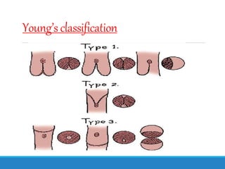
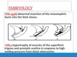
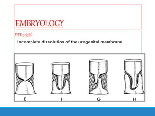
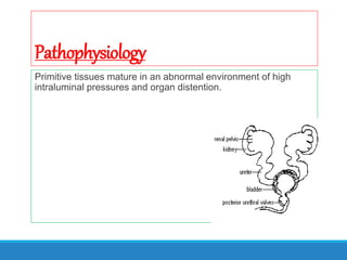
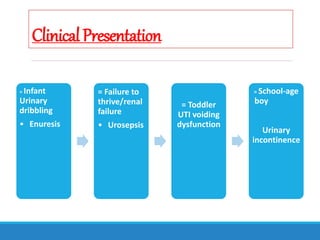
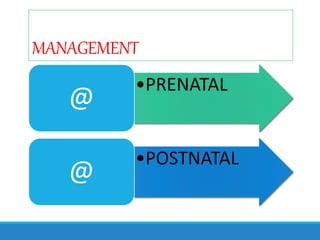
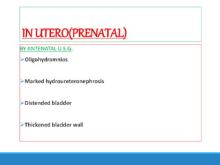
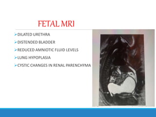
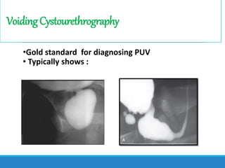
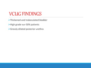
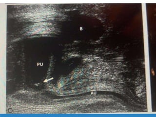
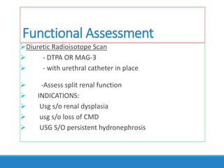
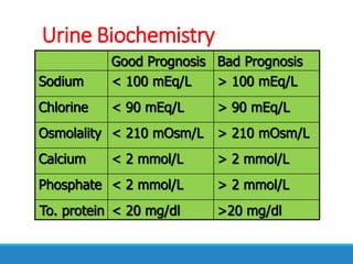
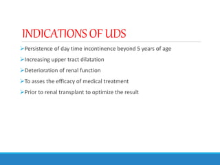
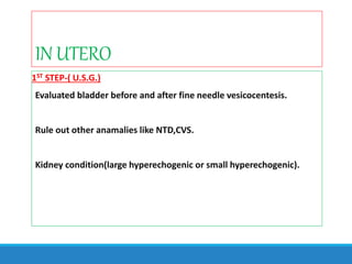
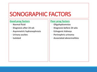
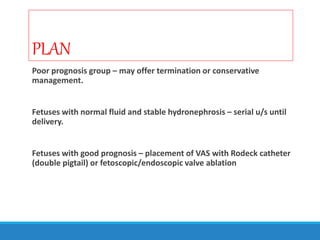
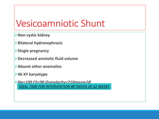
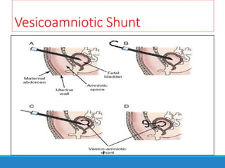
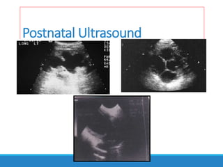
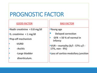
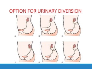
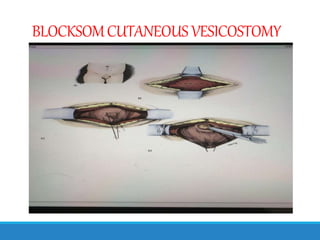
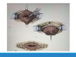
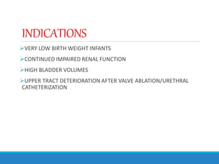
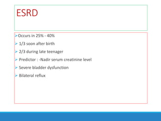
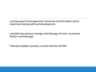
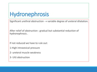
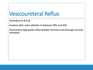
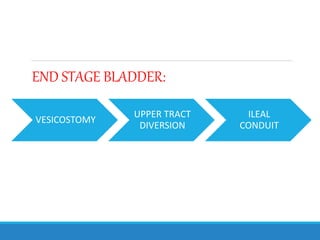
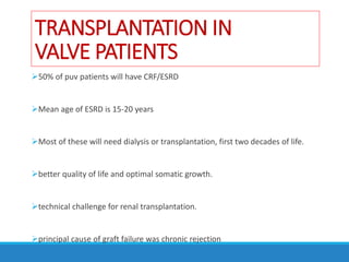
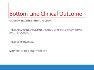
EVALUATIONAND MANAGEMENT
OFPOSTERIORURETHRALVALVE.
PRESENTED BY : DEVASHISH CHOUBEY
INTRODUCTION
Langenbeck : first reported congenital obstruction of the prostatic
urethra in 1802.
Hugh Hampton Young: define and name the condition as posterior
urethral valves(1919).
Incidence is 1.6 to 2.1 per 10000 births.
PUV M/C cause for LUTO followed by urethral atresia and prune belly
syndrome.
INTRODUCTION
1 in 1250 fetal ultrasound screenings.
Inheritance is poorly understood.
Affect siblings, twins, and successive generations.
They become obstructive during or after the eighth week of life.
When PU has developed
GENETIC BASIS
Short arm of chromosome 11
Inheritance may be autosomal recessive,x linked recessive
Young’s classification
EMBRYOLOGY
TYPE-1:(95%)abnormal insertion of the mesonephric
ducts into the fetal cloaca.
TYPE-2: hypertrophy of muscles of the superficial
trigone and prostatic urethra in response to high
voiding pressure from distal obstruction.
EMBRYOLOGY
TYPE-3 :(5%)
Incomplete dissolution of the urogenital membrane
Pathophysiology
Primitive tissues mature in an abnormal environment of high
intraluminal pressures and organ distention.
Pathophysiology
Bladder dysfunction is usually a lifelong problem resulting in
incontinence and poor emptying.
Persistent hydronephrosis is common and may be due to either ureteral
or bladder dysfunction.
Reflux usually improves and often resolves after valve ablation.
Pathophysiology
Posterior urethral valves damage the entire urinary tract proximal to the
valve.
Pulmonary hypoplasia is the most common cause of mortality in valve
patients.
Most renal damage occurs early in fetal life.
EFFECTONBLADDERDEVELOPMENT
With puv normal bladder
recycling fails and
function is altered
Initial period:near
complete bladder
emptying with high
voiding pressures
Compensated
phase:impairement in
bladder capacity and
compliance
Decompensated phase:
bladder capacity
compliance,contractility
EFFECT CONT….
DETRUSOR DECOMPENSATES FURTHER….
INEFFICIENT BLADDER PRESSURES
INCREASED PVR
TYPEIII INFILTRATES DETRUSOR MUSCLE
TYPEIII
TYPEI
ECM
EFFECTON RENAL/BLADDERFUNCTION
• 0-1 YEAR
HYPERTONIC
SMALL CAPACITY
DETRUSOR OVERACTIVITY
• 1-3 YEARS
HYPERREFLEXIC BLADDER
UNINHIBITED
CONTRACTIONS
• 4-12 YEARS
• INCREASED RESIDUAL URINE
MYOGENIC FAILURE
OVERFLOW
ClinicalPresentation
In utero
Older
Child
Neonatal
ClinicalPresentation
Variable
Age dependent
= Prenatally : 70%
of PUV by
ultrasound
= Newborn: -
Abdominal mass-
ascites-respiratory
distress-Urosepsis
ClinicalPresentation
= Infant
Urinary
dribbling
• Enuresis
= Failure to
thrive/renal
failure
• Urosepsis
= Toddler
UTI voiding
dysfunction
= School-age
boy
Urinary
incontinence
MANAGEMENT
•PRENATAL
@
•POSTNATAL
@
INUTERO(PRENATAL)
BY ANTENATAL U.S.G.
Oligohydramnios
Marked hydroureteronephrosis
Distended bladder
Thickened bladder wall
INUTERO
FETAL MRI
DILATED URETHRA
DISTENDED BLADDER
REDUCED AMNIOTIC FLUID LEVELS
LUNG HYPOPLASIA
CYSTIC CHANGES IN RENAL PARENCHYMA
VoidingCystourethrography
•Gold standard for diagnosing PUV
• Typically shows :
VCUGFINDINGS
Thickened and trabeculated bladder
High grade vur-50% patients
Grossly dilated posterior urethra
Functional Assessment
Diuretic Radioisotope Scan
 - DTPA OR MAG-3
 - with urethral catheter in place
 -Assess split renal function
 INDICATIONS:
 Usg s/o renal dysplasia
 usg s/o loss of CMD
 USG S/O persistent hydronephrosis
Urine Biochemistry
Good Prognosis Bad Prognosis
Sodium < 100 mEq/L > 100 mEq/L
Chlorine < 90 mEq/L > 90 mEq/L
Osmolality < 210 mOsm/L > 210 mOsm/L
Calcium < 2 mmol/L > 2 mmol/L
Phosphate < 2 mmol/L > 2 mmol/L
To. protein < 20 mg/dl >20 mg/dl
INDICATIONS OF UDS
Persistence of day time incontinence beyond 5 years of age
Increasing upper tract dilatation
Deterioration of renal function
To asses the efficacy of medical treatment
Prior to renal transplant to optimize the result
IN UTERO
1ST STEP-( U.S.G.)
Evaluated bladder before and after fine needle vesicocentesis.
Rule out other anamalies like NTD,CVS.
Kidney condition(large hyperechogenic or small hyperechogenic).
Needle Aspiration of Bladder
IN UTERO
2ND STEP:-
Prenatal evaluation for fetal karyotype.
Amniocentesis if fluid available.
FINAL STEP:-
Evaluation of fetal kidney function with sequential vesicocenteses
IN UTERO
Completely drain fetal bladder at 48-72 hr intervals at a minimum of
three occasions.
progressive hypotonicity and values that fall below threshold benefit
from in utero intervention i.e. shunt placement.
Prognosis
Outcome depends upon severity
Classified as good or poor
Poor prognostic factors :
- Diagnosis before 24 wks
- Oligohydramnios
- Renal dysplasia
- Marked hydronephrosis
SONOGRAPHIC FACTORS
Good prog factors
◦ Normal fluid
◦ Diagnosis after 24 wk
◦ Asymmetric hydronephrosis
◦ Urinary ascites
◦ Isolated
Poor prog factors
◦ Oligohydramnios
◦ Diagnosis before 24 wks
◦ Echogenic kidneys
◦ Perinephric urinoma
◦ Associated abnormalities
PLAN
Poor prognosis group – may offer termination or conservative
management.
Fetuses with normal fluid and stable hydronephrosis – serial u/s until
delivery.
Fetuses with good prognosis – placement of VAS with Rodeck catheter
(double pigtail) or fetoscopic/endoscopic valve ablation
Vesicoamniotic Shunt
Non cystic kidney
Bilateral hydronephrosis
Single pregnancy
Decreased amniotic fluid volume
Absent other anomalies
46 XY karyotype
Na<100 Ch<90 Osmolarity<210mosm/dl
IDEAL TIME FOR INTERVENTION BETWEEN 20-32 WEEKS
Vesicoamniotic Shunt
COMPLICATIONS
Premature labor
Fetal bowel injury
Fetal loss
Infection
PLUTO TRIAL
“PLUTO TRIAL comprehends the fact that
Vesicoamniotic shunting may have a survival
benefit for the infant in select cases,but there is no
clear benefit in the risk for renal failure.”
LIMITATIONS:
Poor enrolment
Pre mature termination of trial
PLAN
Consultation with pediatric urologist
Route of delivery – routine obstetric indications
Average age of delivery due to spontaneous rupture of membranes =
33-35 wks
Following delivery – sterile ostomy bag to abdomen until renal
function and anatomical evaluation by pediatric urologist.
InitialDiagnosticEvaluation
(POSTNATAL)
Ultrasound
Voiding Cystourethrography
Radionuclide Renal Scan(functional assesment)
Postnatal Ultrasound
To evaluate the effect of PUV on the urinary tract rather than to
diagnose PUV.
Typical finding: wide prostatic urethra,thick-walled bladder and upper
tract dilatation.
Assessment of renal parenchyma.
Postnatal Ultrasound
PROGNOSTIC FACTOR
GOOD FACTOR
Nadir creatinine < 0.8 mg/dl
S. creatinine < 1 mg/dl
Pop-off mechanism
 -VURD
 -Ascitis
 -Large bladder
 diverticulum.
BAD FACTOR
Young age
 Delayed correction
 GFR < 50 % of normal in
infancy
VUR – mortality (b/l - 57% u/l -
17%, non - 9%)
Loss of cortico medullary junction
Postnatal management
Initial management
- bladder drainage
- Tt life threatening condition by:-
*maximal ventilatory support
*extracorporeal membrane oxy-
genation.
*dialysis & parenteral nutrition
*control of hypertension
- Valve Ablation
Bladder Drainage
By infant feeding tube or catheter.
Monitor Creatinine, BUN, and electrolytes should be followed twice
daily for the first few days of life until they plateau.
Serum bicarbonate, sodium, and potassium are all critical factors to
monitor.
Valve Ablation
By hooks,balloon catheters, and valvulotomes.
A bugbee electrode or a pediatric resectoscope with a hook or cold
knife.
Incision prefer 12 or 4 & 8 o” clock position.
leave a catheter in place for 24 hrs.
BEFORE ABLATION AFTER ABLATION
Re-evaluation
By cystoscope and VCUG after 12wks.
VCUG debatable but use to see urethra, bladder capacity & reflux.
signs of successful relief of obstruction
- Decreased trabeculation
- Resolution of reflux
- Uniform urethral diameter
- PUD/AUD ratio 2.5 to 3
OPTION FOR URINARY DIVERSION
Vesicostomy
Distal ureterostomy
Proximal loop ureterostomy
Cutaneous pyeloplasty
Ring ureterostomy
Sober y ureterostomy
BLOCKSOMCUTANEOUS VESICOSTOMY
INDICATIONS
VERY LOW BIRTH WEIGHT INFANTS
CONTINUED IMPAIRED RENAL FUNCTION
HIGH BLADDER VOLUMES
UPPER TRACT DETERIORATION AFTER VALVE ABLATION/URETHRAL
CATHETERIZATION
COMPLICATION AND ITS
MANAGEMENT
I. RENAL DYSPLASIA
II. RENAL FUNCTION
III. RENAL TUBULAR FUNCTION
IV. HYDRONEPHROSIS
V. VUR
VI. VESICAL DYSFUNCTION
VII. VALVE BLADDER
Renal Dysplasia
Histological diagnosis
Cause 1- high pelvic pressure during nephrogenesis
2-primary embryologic abnormality from abnormal position of
uteteric bud .
Severity will determine ultimate renal function
Renal Function
Children with PUV may demonstrate gradual loss of renal function over time
Cause:
1- Renal parenchymal dysplasia
2- Incomplete relief of obstruction
3-parenchymal injury from :
* UTI
*HTN
*Progressive glomerulosclerosis from hyperfiltration
* Obstruction
Renal Tubular Function
50% of patients with PUV have impairment concentration ability
 Persistently high urinary flow rate regardless of fluid intake or state
of hydration
 severe dehydration and electrolyte imbalance
 ureteral dilatation and high resting vesical pressure
ESRD
Occurs in 25% - 40%
 1/3 soon after birth
 2/3 during late teenager
 Predictor : -Nadir serum creatinine level
 Severe bladder dysfunction
 Bilateral reflux
primary goal of management is preserve renal function and to
maximize renal growth and development.
provide low-pressure storage and drainage of urine to prevent
further renal damage.
Monitor bladder function, control infection & HTN.
Hydronephrosis
Significant urethral obstruction  variable degree of ureteral dilatation.
After relief of obstruction : gradual but substantial reduction of
hydronephrosis .
If not reduced we have to rule out:
1-High intravesical pressure
2- ureteral muscle weakness
3- UVJ obstruction
Vesicoureteral Reflux
Secondary to B.O.O.
resolves after valve ablation in between 20% and 32%
If persistent high-grade reflux bladder function and drainage must be
reviewed.
Vesicoureteral Reflux
50% VUR at time of diagnosis
Primary or Secondary
Vesical Dysfunction
Usually secondary to irreversible change in organization and function of
the smooth muscle from outlet obstruction
Present as as urinary incontinence (20%)
Bladder dysfunction persist in 75 % of cases after
valve ablation
VALVEBLADDER
Definition:
A CHRONIC CONDITION IN VALVE PATIENTS WHERE DESPITE
SUCCESFUL VALVE ABLATION,INTRINSIC BLADDER
DYSFUNCTION LEADS TO DETERIORATION OF THE UPPER
URINARY TRACTS AND INCONTINENCE.
VALVEBLADDER
CLINICAL FEATURES
Typically comfortable with large bladder volumes at high
pressure
Overflow incontinence
Incomplete void
Increased frequency
Nocturia
VALVE BLADDER
term coined by Mitchell.
HIGH
VOIDING
PRESSURES
INCREASED
URINE
PRODUCTI
ON
NEPHROGENIC
DIABETES
INSIPIDUS
NO PERIODS
OF
RELAXATION
DETRUSOR
FIBRES
INJURY
IMPARED
CONTRACTILIT
Y
TO SUMMARISE…..
POLYURIA BLADDERINSENSITIVITY
RESIDUAL URINE
VOLUME
VALVE BLADDER
Even after relief of obstruction a significant number of patient will
continue to have hypertonia and detrusor hyperreflexia and low
compliance
Physiological obstruction of the ureter associated with bladder filling
persistent hydronephrosis and/or urinary incontinence
MANAGEMENT OF BLADDER DYSFUNCTION.
Intensively individualized
Close monitoring by
-Urodynamics
- Urine volumes
- Renal function
-Infections
-Hydronephrosis
@
• anticholinergics
• α-blockers
@
• timed voiding
• double voiding
@
• intermittent catheterization
• Cont. nocturnal blad. drainage
@
• Augmentation bladder
• Appendicovesicostomy
INDICATIONS OF BLADDER
AUGMENTATION
Poor bladder compliance
Failed medical management/anticholinergics
Repeated UTI
Persisting VUR threatening kidney function( GFR)
BIOFEEDBACK AND PELVICFLOOR
EXERCISE
Provide significant and durable relief for persistent lower urinary tract
dysfunction
Consistent good response seen in 70% of patients
Alpha blockers/terazosin(0.25-2mg) can be added for bladder
empyting.
INDIAN JOURNAL OF UROLOGY OCT-DEC 2010 VOL 26 ISSUE 4
ROLE OF CIC
Increases GFR
Decreases UTI
Decrease upper tract deterioration
Increase continence
Increase compliance
END STAGE BLADDER:
VESICOSTOMY
UPPER TRACT
DIVERSION
ILEAL
CONDUIT
TRANSPLANTATION IN
VALVE PATIENTS
50% of puv patients will have CRF/ESRD
Mean age of ESRD is 15-20 years
Most of these will need dialysis or transplantation, first two decades of life.
better quality of life and optimal somatic growth.
technical challenge for renal transplantation.
principal cause of graft failure was chronic rejection
Bottom Line Clinical Outcome
MONITOR BLADDER & RENAL FUCTION.
TREAT ACCORDINGLY FOR PRESERVATION OF UPPER URINARY TRACT
AND ITS FUCTION.
TREAT COMPLICATION.
MAINTAIN BETTER QUALITY OF LIFE
Puv

Puv

  • 1.
  • 2.
    INTRODUCTION Langenbeck : firstreported congenital obstruction of the prostatic urethra in 1802. Hugh Hampton Young: define and name the condition as posterior urethral valves(1919). Incidence is 1.6 to 2.1 per 10000 births. PUV M/C cause for LUTO followed by urethral atresia and prune belly syndrome.
  • 3.
    INTRODUCTION 1 in 1250fetal ultrasound screenings. Inheritance is poorly understood. Affect siblings, twins, and successive generations. They become obstructive during or after the eighth week of life. When PU has developed
  • 4.
    GENETIC BASIS Short armof chromosome 11 Inheritance may be autosomal recessive,x linked recessive
  • 5.
  • 6.
    EMBRYOLOGY TYPE-1:(95%)abnormal insertion ofthe mesonephric ducts into the fetal cloaca. TYPE-2: hypertrophy of muscles of the superficial trigone and prostatic urethra in response to high voiding pressure from distal obstruction.
  • 7.
  • 8.
    Pathophysiology Primitive tissues maturein an abnormal environment of high intraluminal pressures and organ distention.
  • 9.
    Pathophysiology Bladder dysfunction isusually a lifelong problem resulting in incontinence and poor emptying. Persistent hydronephrosis is common and may be due to either ureteral or bladder dysfunction. Reflux usually improves and often resolves after valve ablation.
  • 10.
    Pathophysiology Posterior urethral valvesdamage the entire urinary tract proximal to the valve. Pulmonary hypoplasia is the most common cause of mortality in valve patients. Most renal damage occurs early in fetal life.
  • 12.
    EFFECTONBLADDERDEVELOPMENT With puv normalbladder recycling fails and function is altered Initial period:near complete bladder emptying with high voiding pressures Compensated phase:impairement in bladder capacity and compliance Decompensated phase: bladder capacity compliance,contractility
  • 13.
    EFFECT CONT…. DETRUSOR DECOMPENSATESFURTHER…. INEFFICIENT BLADDER PRESSURES INCREASED PVR TYPEIII INFILTRATES DETRUSOR MUSCLE TYPEIII TYPEI ECM
  • 14.
    EFFECTON RENAL/BLADDERFUNCTION • 0-1YEAR HYPERTONIC SMALL CAPACITY DETRUSOR OVERACTIVITY • 1-3 YEARS HYPERREFLEXIC BLADDER UNINHIBITED CONTRACTIONS • 4-12 YEARS • INCREASED RESIDUAL URINE MYOGENIC FAILURE OVERFLOW
  • 15.
  • 16.
    ClinicalPresentation Variable Age dependent = Prenatally: 70% of PUV by ultrasound = Newborn: - Abdominal mass- ascites-respiratory distress-Urosepsis
  • 17.
    ClinicalPresentation = Infant Urinary dribbling • Enuresis =Failure to thrive/renal failure • Urosepsis = Toddler UTI voiding dysfunction = School-age boy Urinary incontinence
  • 18.
  • 19.
    INUTERO(PRENATAL) BY ANTENATAL U.S.G. Oligohydramnios Markedhydroureteronephrosis Distended bladder Thickened bladder wall
  • 20.
  • 21.
    FETAL MRI DILATED URETHRA DISTENDEDBLADDER REDUCED AMNIOTIC FLUID LEVELS LUNG HYPOPLASIA CYSTIC CHANGES IN RENAL PARENCHYMA
  • 22.
    VoidingCystourethrography •Gold standard fordiagnosing PUV • Typically shows :
  • 23.
    VCUGFINDINGS Thickened and trabeculatedbladder High grade vur-50% patients Grossly dilated posterior urethra
  • 25.
    Functional Assessment Diuretic RadioisotopeScan  - DTPA OR MAG-3  - with urethral catheter in place  -Assess split renal function  INDICATIONS:  Usg s/o renal dysplasia  usg s/o loss of CMD  USG S/O persistent hydronephrosis
  • 26.
    Urine Biochemistry Good PrognosisBad Prognosis Sodium < 100 mEq/L > 100 mEq/L Chlorine < 90 mEq/L > 90 mEq/L Osmolality < 210 mOsm/L > 210 mOsm/L Calcium < 2 mmol/L > 2 mmol/L Phosphate < 2 mmol/L > 2 mmol/L To. protein < 20 mg/dl >20 mg/dl
  • 27.
    INDICATIONS OF UDS Persistenceof day time incontinence beyond 5 years of age Increasing upper tract dilatation Deterioration of renal function To asses the efficacy of medical treatment Prior to renal transplant to optimize the result
  • 28.
    IN UTERO 1ST STEP-(U.S.G.) Evaluated bladder before and after fine needle vesicocentesis. Rule out other anamalies like NTD,CVS. Kidney condition(large hyperechogenic or small hyperechogenic).
  • 29.
  • 30.
    IN UTERO 2ND STEP:- Prenatalevaluation for fetal karyotype. Amniocentesis if fluid available. FINAL STEP:- Evaluation of fetal kidney function with sequential vesicocenteses
  • 31.
    IN UTERO Completely drainfetal bladder at 48-72 hr intervals at a minimum of three occasions. progressive hypotonicity and values that fall below threshold benefit from in utero intervention i.e. shunt placement.
  • 32.
    Prognosis Outcome depends uponseverity Classified as good or poor Poor prognostic factors : - Diagnosis before 24 wks - Oligohydramnios - Renal dysplasia - Marked hydronephrosis
  • 33.
    SONOGRAPHIC FACTORS Good progfactors ◦ Normal fluid ◦ Diagnosis after 24 wk ◦ Asymmetric hydronephrosis ◦ Urinary ascites ◦ Isolated Poor prog factors ◦ Oligohydramnios ◦ Diagnosis before 24 wks ◦ Echogenic kidneys ◦ Perinephric urinoma ◦ Associated abnormalities
  • 34.
    PLAN Poor prognosis group– may offer termination or conservative management. Fetuses with normal fluid and stable hydronephrosis – serial u/s until delivery. Fetuses with good prognosis – placement of VAS with Rodeck catheter (double pigtail) or fetoscopic/endoscopic valve ablation
  • 35.
    Vesicoamniotic Shunt Non cystickidney Bilateral hydronephrosis Single pregnancy Decreased amniotic fluid volume Absent other anomalies 46 XY karyotype Na<100 Ch<90 Osmolarity<210mosm/dl IDEAL TIME FOR INTERVENTION BETWEEN 20-32 WEEKS
  • 36.
  • 37.
    COMPLICATIONS Premature labor Fetal bowelinjury Fetal loss Infection
  • 38.
    PLUTO TRIAL “PLUTO TRIALcomprehends the fact that Vesicoamniotic shunting may have a survival benefit for the infant in select cases,but there is no clear benefit in the risk for renal failure.” LIMITATIONS: Poor enrolment Pre mature termination of trial
  • 39.
    PLAN Consultation with pediatricurologist Route of delivery – routine obstetric indications Average age of delivery due to spontaneous rupture of membranes = 33-35 wks Following delivery – sterile ostomy bag to abdomen until renal function and anatomical evaluation by pediatric urologist.
  • 40.
  • 41.
    Postnatal Ultrasound To evaluatethe effect of PUV on the urinary tract rather than to diagnose PUV. Typical finding: wide prostatic urethra,thick-walled bladder and upper tract dilatation. Assessment of renal parenchyma.
  • 42.
  • 44.
    PROGNOSTIC FACTOR GOOD FACTOR Nadircreatinine < 0.8 mg/dl S. creatinine < 1 mg/dl Pop-off mechanism  -VURD  -Ascitis  -Large bladder  diverticulum. BAD FACTOR Young age  Delayed correction  GFR < 50 % of normal in infancy VUR – mortality (b/l - 57% u/l - 17%, non - 9%) Loss of cortico medullary junction
  • 45.
    Postnatal management Initial management -bladder drainage - Tt life threatening condition by:- *maximal ventilatory support *extracorporeal membrane oxy- genation. *dialysis & parenteral nutrition *control of hypertension - Valve Ablation
  • 46.
    Bladder Drainage By infantfeeding tube or catheter. Monitor Creatinine, BUN, and electrolytes should be followed twice daily for the first few days of life until they plateau. Serum bicarbonate, sodium, and potassium are all critical factors to monitor.
  • 47.
    Valve Ablation By hooks,ballooncatheters, and valvulotomes. A bugbee electrode or a pediatric resectoscope with a hook or cold knife. Incision prefer 12 or 4 & 8 o” clock position. leave a catheter in place for 24 hrs.
  • 48.
  • 49.
    Re-evaluation By cystoscope andVCUG after 12wks. VCUG debatable but use to see urethra, bladder capacity & reflux. signs of successful relief of obstruction - Decreased trabeculation - Resolution of reflux - Uniform urethral diameter - PUD/AUD ratio 2.5 to 3
  • 50.
  • 51.
    Vesicostomy Distal ureterostomy Proximal loopureterostomy Cutaneous pyeloplasty Ring ureterostomy Sober y ureterostomy
  • 52.
  • 54.
    INDICATIONS VERY LOW BIRTHWEIGHT INFANTS CONTINUED IMPAIRED RENAL FUNCTION HIGH BLADDER VOLUMES UPPER TRACT DETERIORATION AFTER VALVE ABLATION/URETHRAL CATHETERIZATION
  • 55.
    COMPLICATION AND ITS MANAGEMENT I.RENAL DYSPLASIA II. RENAL FUNCTION III. RENAL TUBULAR FUNCTION IV. HYDRONEPHROSIS V. VUR VI. VESICAL DYSFUNCTION VII. VALVE BLADDER
  • 56.
    Renal Dysplasia Histological diagnosis Cause1- high pelvic pressure during nephrogenesis 2-primary embryologic abnormality from abnormal position of uteteric bud . Severity will determine ultimate renal function
  • 57.
    Renal Function Children withPUV may demonstrate gradual loss of renal function over time Cause: 1- Renal parenchymal dysplasia 2- Incomplete relief of obstruction 3-parenchymal injury from : * UTI *HTN *Progressive glomerulosclerosis from hyperfiltration * Obstruction
  • 58.
    Renal Tubular Function 50%of patients with PUV have impairment concentration ability  Persistently high urinary flow rate regardless of fluid intake or state of hydration  severe dehydration and electrolyte imbalance  ureteral dilatation and high resting vesical pressure
  • 59.
    ESRD Occurs in 25%- 40%  1/3 soon after birth  2/3 during late teenager  Predictor : -Nadir serum creatinine level  Severe bladder dysfunction  Bilateral reflux
  • 60.
    primary goal ofmanagement is preserve renal function and to maximize renal growth and development. provide low-pressure storage and drainage of urine to prevent further renal damage. Monitor bladder function, control infection & HTN.
  • 61.
    Hydronephrosis Significant urethral obstruction variable degree of ureteral dilatation. After relief of obstruction : gradual but substantial reduction of hydronephrosis . If not reduced we have to rule out: 1-High intravesical pressure 2- ureteral muscle weakness 3- UVJ obstruction
  • 62.
    Vesicoureteral Reflux Secondary toB.O.O. resolves after valve ablation in between 20% and 32% If persistent high-grade reflux bladder function and drainage must be reviewed.
  • 63.
    Vesicoureteral Reflux 50% VURat time of diagnosis Primary or Secondary
  • 64.
    Vesical Dysfunction Usually secondaryto irreversible change in organization and function of the smooth muscle from outlet obstruction Present as as urinary incontinence (20%) Bladder dysfunction persist in 75 % of cases after valve ablation
  • 65.
    VALVEBLADDER Definition: A CHRONIC CONDITIONIN VALVE PATIENTS WHERE DESPITE SUCCESFUL VALVE ABLATION,INTRINSIC BLADDER DYSFUNCTION LEADS TO DETERIORATION OF THE UPPER URINARY TRACTS AND INCONTINENCE.
  • 66.
    VALVEBLADDER CLINICAL FEATURES Typically comfortablewith large bladder volumes at high pressure Overflow incontinence Incomplete void Increased frequency Nocturia
  • 67.
    VALVE BLADDER term coinedby Mitchell. HIGH VOIDING PRESSURES INCREASED URINE PRODUCTI ON NEPHROGENIC DIABETES INSIPIDUS NO PERIODS OF RELAXATION DETRUSOR FIBRES INJURY IMPARED CONTRACTILIT Y
  • 68.
  • 69.
    VALVE BLADDER Even afterrelief of obstruction a significant number of patient will continue to have hypertonia and detrusor hyperreflexia and low compliance Physiological obstruction of the ureter associated with bladder filling persistent hydronephrosis and/or urinary incontinence
  • 70.
    MANAGEMENT OF BLADDERDYSFUNCTION. Intensively individualized Close monitoring by -Urodynamics - Urine volumes - Renal function -Infections -Hydronephrosis
  • 71.
    @ • anticholinergics • α-blockers @ •timed voiding • double voiding @ • intermittent catheterization • Cont. nocturnal blad. drainage @ • Augmentation bladder • Appendicovesicostomy
  • 72.
    INDICATIONS OF BLADDER AUGMENTATION Poorbladder compliance Failed medical management/anticholinergics Repeated UTI Persisting VUR threatening kidney function( GFR)
  • 73.
    BIOFEEDBACK AND PELVICFLOOR EXERCISE Providesignificant and durable relief for persistent lower urinary tract dysfunction Consistent good response seen in 70% of patients Alpha blockers/terazosin(0.25-2mg) can be added for bladder empyting. INDIAN JOURNAL OF UROLOGY OCT-DEC 2010 VOL 26 ISSUE 4
  • 74.
    ROLE OF CIC IncreasesGFR Decreases UTI Decrease upper tract deterioration Increase continence Increase compliance
  • 75.
    END STAGE BLADDER: VESICOSTOMY UPPERTRACT DIVERSION ILEAL CONDUIT
  • 76.
    TRANSPLANTATION IN VALVE PATIENTS 50%of puv patients will have CRF/ESRD Mean age of ESRD is 15-20 years Most of these will need dialysis or transplantation, first two decades of life. better quality of life and optimal somatic growth. technical challenge for renal transplantation. principal cause of graft failure was chronic rejection
  • 77.
    Bottom Line ClinicalOutcome MONITOR BLADDER & RENAL FUCTION. TREAT ACCORDINGLY FOR PRESERVATION OF UPPER URINARY TRACT AND ITS FUCTION. TREAT COMPLICATION. MAINTAIN BETTER QUALITY OF LIFE