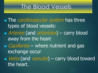
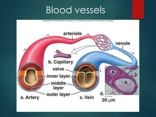
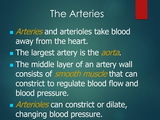
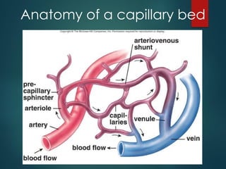
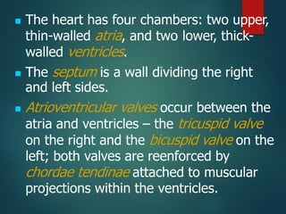
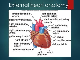
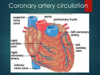
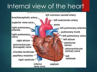
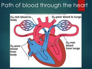
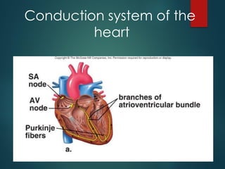
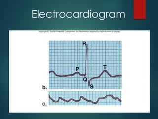
Introduction to CVS
• Components Of CVS
– Heart
– Blood Vessels
– Blood
• Two types of Circulation
– Systemic circulation
– Pulmonary circulation
The Blood Vessels
◼ The cardiovascular system has three
types of blood vessels:
◼ Arteries (and arterioles) – carry blood
away from the heart
◼ Capillaries – where nutrient and gas
exchange occur
◼ Veins (and venules) – carry blood toward
the heart.
Blood vessels
The Arteries
◼ Arteries and arterioles take blood
away from the heart.
◼ The largest artery is the aorta.
◼ The middle layer of an artery wall
consists of smooth muscle that can
constrict to regulate blood flow and
blood pressure.
◼ Arterioles can constrict or dilate,
changing blood pressure.
The Capillaries
◼ Capillaries have walls only one cell thick to
allow exchange of gases and nutrients
with tissue fluid.
◼ Capillary beds are present in all regions of
the body but not all capillary beds are
open at the same time.
◼ Contraction of a sphincter muscle closes
off a bed and blood can flow through an
arteriovenous shunt that bypasses the
capillary bed.
Anatomy of a capillary bed
The Veins
◼ Venules drain blood from capillaries,
then join to form veins that take blood
to the heart.
◼ Veins have much less smooth muscle
and connective tissue than arteries.
◼ Veins often have valves that prevent
the backward flow of blood when
closed.
◼ Veins carry about 70% of the body’s
blood and act as a reservoir during
hemorrhage.
◼ The heart is a cone-shaped, muscular
organ located between the lungs
behind the sternum.
◼ The heart muscle forms the
myocardium, with tightly interconnect
cells of cardiac muscle tissue.
◼ The pericardium is the outer
membranous sac with lubricating fluid.
The Heart
◼ The heart has four chambers: two upper,
thin-walled atria, and two lower, thick-
walled ventricles.
◼ The septum is a wall dividing the right
and left sides.
◼ Atrioventricular valves occur between the
atria and ventricles – the tricuspid valve
on the right and the bicuspid valve on the
left; both valves are reenforced by
chordae tendinae attached to muscular
projections within the ventricles.
External heart anatomy
Coronary artery circulation
Passage of Blood Through
the Heart
◼ Blood follows this sequence through the
heart: superior and inferior vena cava →
right atrium →tricuspid valve →right
ventricle →pulmonary semilunar valve →
pulmonary trunk and arteries to the lungs
→pulmonary veins leaving the lungs →left
atrium →bicuspid valve →left ventricle →
aortic semilunar valve →aorta →to the
body.
Internal view of the heart
◼ The pumping of the heart sends out
blood under pressure to the arteries.
◼ Blood pressure is greatest in the
aorta; the wall of the left ventricle is
thicker than that of the right ventricle
and pumps blood to the entire body.
◼ Blood pressure then decreases as the
cross-sectional area of arteries and
then arterioles increases.
Path of blood through the heart
The Heartbeat
◼ Each heartbeat is called a cardiac cycle.
◼ When the heart beats, the two atria
contract together, then the two
ventricles contract; then the whole heart
relaxes.
◼ Systole is the contraction of heart
chambers; diastole is their relaxation.
◼ The heart sounds, lub-dup, are due to
the closing of the atrioventricular valves,
followed by the closing of the semilunar
valves.
Intrinsic Control of Heartbeat
◼ The SA (sinoatrial) node, or pacemaker,
initiates the heartbeat and causes the
atria to contract on average every 0.85
seconds.
◼ The AV (atrioventricular) node conveys
the stimulus and initiates contraction of
the ventricles.
◼ The signal for the ventricles to contract
travels from the AV node through the
atrioventricular bundle to the smaller
Purkinje fibers.
Conduction system of the
heart
Extrinsic Control of Heartbeat
◼ A cardiac control center in the medulla
oblongata speeds up or slows down the
heart rate by way of the autonomic
nervous system branches:
parasympathetic system (slows heart
rate) and the sympathetic system
(increases heart rate).
◼ Hormones epinephrine and
norepinephrine from the adrenal
medulla also stimulate faster heart rate.
The Electrocardiogram
◼ An electrocardiogram (ECG) is a
recording of the electrical changes
that occur in the myocardium during a
cardiac cycle.
◼ Atrial depolarization creates the P
wave, ventricle depolarization creates
the QRS wave, and repolarization of
the ventricles produces the T wave.
Electrocardiogram

Cardiovascular system-1-pages-deleted.pdf

  • 1.
    Introduction to CVS •Components Of CVS – Heart – Blood Vessels – Blood • Two types of Circulation – Systemic circulation – Pulmonary circulation
  • 2.
    The Blood Vessels ◼The cardiovascular system has three types of blood vessels: ◼ Arteries (and arterioles) – carry blood away from the heart ◼ Capillaries – where nutrient and gas exchange occur ◼ Veins (and venules) – carry blood toward the heart.
  • 3.
  • 4.
    The Arteries ◼ Arteriesand arterioles take blood away from the heart. ◼ The largest artery is the aorta. ◼ The middle layer of an artery wall consists of smooth muscle that can constrict to regulate blood flow and blood pressure. ◼ Arterioles can constrict or dilate, changing blood pressure.
  • 5.
    The Capillaries ◼ Capillarieshave walls only one cell thick to allow exchange of gases and nutrients with tissue fluid. ◼ Capillary beds are present in all regions of the body but not all capillary beds are open at the same time. ◼ Contraction of a sphincter muscle closes off a bed and blood can flow through an arteriovenous shunt that bypasses the capillary bed.
  • 6.
    Anatomy of acapillary bed
  • 7.
    The Veins ◼ Venulesdrain blood from capillaries, then join to form veins that take blood to the heart. ◼ Veins have much less smooth muscle and connective tissue than arteries. ◼ Veins often have valves that prevent the backward flow of blood when closed. ◼ Veins carry about 70% of the body’s blood and act as a reservoir during hemorrhage.
  • 8.
    ◼ The heartis a cone-shaped, muscular organ located between the lungs behind the sternum. ◼ The heart muscle forms the myocardium, with tightly interconnect cells of cardiac muscle tissue. ◼ The pericardium is the outer membranous sac with lubricating fluid. The Heart
  • 9.
    ◼ The hearthas four chambers: two upper, thin-walled atria, and two lower, thick- walled ventricles. ◼ The septum is a wall dividing the right and left sides. ◼ Atrioventricular valves occur between the atria and ventricles – the tricuspid valve on the right and the bicuspid valve on the left; both valves are reenforced by chordae tendinae attached to muscular projections within the ventricles.
  • 10.
  • 11.
  • 12.
    Passage of BloodThrough the Heart ◼ Blood follows this sequence through the heart: superior and inferior vena cava → right atrium →tricuspid valve →right ventricle →pulmonary semilunar valve → pulmonary trunk and arteries to the lungs →pulmonary veins leaving the lungs →left atrium →bicuspid valve →left ventricle → aortic semilunar valve →aorta →to the body.
  • 13.
  • 14.
    ◼ The pumpingof the heart sends out blood under pressure to the arteries. ◼ Blood pressure is greatest in the aorta; the wall of the left ventricle is thicker than that of the right ventricle and pumps blood to the entire body. ◼ Blood pressure then decreases as the cross-sectional area of arteries and then arterioles increases.
  • 15.
    Path of bloodthrough the heart
  • 16.
    The Heartbeat ◼ Eachheartbeat is called a cardiac cycle. ◼ When the heart beats, the two atria contract together, then the two ventricles contract; then the whole heart relaxes. ◼ Systole is the contraction of heart chambers; diastole is their relaxation. ◼ The heart sounds, lub-dup, are due to the closing of the atrioventricular valves, followed by the closing of the semilunar valves.
  • 17.
    Intrinsic Control ofHeartbeat ◼ The SA (sinoatrial) node, or pacemaker, initiates the heartbeat and causes the atria to contract on average every 0.85 seconds. ◼ The AV (atrioventricular) node conveys the stimulus and initiates contraction of the ventricles. ◼ The signal for the ventricles to contract travels from the AV node through the atrioventricular bundle to the smaller Purkinje fibers.
  • 18.
  • 19.
    Extrinsic Control ofHeartbeat ◼ A cardiac control center in the medulla oblongata speeds up or slows down the heart rate by way of the autonomic nervous system branches: parasympathetic system (slows heart rate) and the sympathetic system (increases heart rate). ◼ Hormones epinephrine and norepinephrine from the adrenal medulla also stimulate faster heart rate.
  • 20.
    The Electrocardiogram ◼ Anelectrocardiogram (ECG) is a recording of the electrical changes that occur in the myocardium during a cardiac cycle. ◼ Atrial depolarization creates the P wave, ventricle depolarization creates the QRS wave, and repolarization of the ventricles produces the T wave.
  • 21.