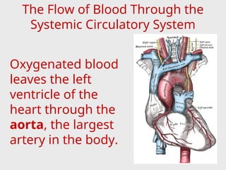
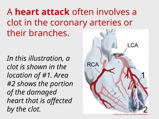
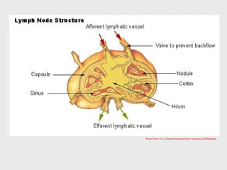
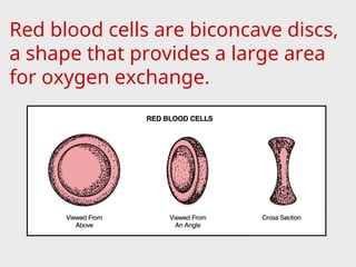
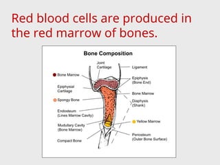
THE CIRCULATORY
SYSTEM
Agriscience 332
Animal Science
#8646-A
TEKS: (c)(2)(A) and (c)(2)(B)
Introduction
The circulatory system is
comprised of the heart, veins,
capillaries, arteries, lymph
vessels, and lymph glands,
which work together to supply
the body tissues with
nourishment and collect waste
materials.
Functions of the circulatory
system:
Distribute nutrients,
Transport and exchange
oxygen and carbon dioxide,
Remove waste materials,
Distribute secretions of
endocrine glands,
Prevent excessive bleeding,
Prevent infection, and
Regulate body temperature.
Anatomy and Physiology
of the Heart
The heart is a funnel-shaped,
hollow, muscular organ that is
responsible for pumping
blood to all parts of the body.
The heart is located near the
center of the thoracic cavity
between the lungs and is
contained in the pericardial sac.
The pericardial sac supports the
heart and contains some fluid
for lubrication.
The broad end, or base, of the
heart is also supported by large
arteries and veins.
The pointed end, or apex, of
the heart is directed toward the
abdomen.
The heart wall is made up of three
layers.
• Epicardium – outer layer of heart
wall, which is also the inner layer
of epicardial sac;
• Endocardium – inner layer that
consists of endothelial cells, which
line the heart, covers the heart
valves, and lines the blood vessels.
• Myocardium – middle layer
composed of cardiac muscle.
The cardiac muscle is an
involuntary, striated muscle
with fibers that intertwine.
In mammals and birds, the
heart is divided into a right and
left side and each side is divided
into an atrium and ventricle.
Therefore, the heart is said to
have four chambers (right
atrium, right ventricle, left atrium,
and left ventricle).
The atrioventricular valves
(AV valve) separate the atrium and
ventricle on each side of the heart.
The AV valves have flaps of tissues,
called leaflets or cusps, which open
and close to ensure that the blood
flows only in one direction and
does not backflow into the
atriums.
The AV valve on the right side of
the heart is called the tricuspid
valve because it has three leaflets
(cusps).
The AV valve on the left side of the
heart is called the bicuspid valve
(or mitral valve) because it has two
leaflets.
The pulmonary valve and the
aortic valve prevent blood from
back-flowing into their
respective ventricles.
The pulmonary valve is located
between the right ventricle and
the pulmonary artery.
The aortic valve is located
between the left ventricle and the
aortic artery.
Following the path that the blood
takes as it flows through the heart
and lungs is the best way to
understand the heart’s operation.
(This process will be discussed later in
the topic of pulmonary circulation.)
A group of cells called the
sinoatrial node (SA node) control
the beat of the heart by sending
out electrical signals to make the
heart pump.
Anatomy and Physiology
of the Vascular System
The vascular system is made up
of three types of blood vessels:
• Arteries,
• Capillaries, and
• Veins
Photo from U. S. Federal Government courtesy of Wikipedia.
Blood Vessels
Arteries are blood vessels that
carry blood, rich in oxygen, from
the heart to other parts of the
body.
The large arteries have thick walls
of elastic-like tissue that enables
them to withstand the blood
pressure created by the heart’s
beating.
As the arteries extend away
from the heart, they branch out
into smaller arteries called
arterioles.
The smaller arteries’ walls are
composed of large amounts of
smooth muscle instead of the
elastic tissue.
Arterioles branch into smaller
vessels called capillaries.
At this junction, the arterioles
have an especially thick layer of
smooth muscle in their walls that
carefully controls the amount of
blood each capillary receives.
Blood pressure for the entire
circulatory system is maintained by
the tension at the end of the
arterioles.
Shock is a serious condition that
occurs when the arterioles dilate
(relax) and allow a large volume of
blood into the capillary beds.
The reduced blood flow that occurs
with shock jeopardizes vital organs.
Capillaries are tiny, thin-walled
blood vessels that connect
arteries to veins and are located
in all body tissues.
Capillaries are so small in
diameter that blood cells pass
through in a single file.
The semi-permeable membrane
of capillary walls allows
nutrients, oxygen, and water to
diffuse from the blood to the
tissues.
Waste products, like carbon
dioxide, diffuse from the tissues
into the blood.
Capillary Bed Interaction of
molecules flowing in and out of blood at a capillary bed.
Larger tubular connectors, which
also connect arterioles to venules,
are located within the capillary
beds.
These tubules allow more blood to
flow through an area, help warm
tissues, and increase the return of
blood pressure to the heart.
Once blood passes through the
capillary beds, it begins its return
to the heart.
Veins are the blood vessels that
return blood to the heart from all
parts of the body.
Capillaries unite to form small
veins called venules.
The venules join together to
form larger veins, which have
thin walls and are collapsible.
For each artery, there is a much
larger vein counterpart.
Veins have valves that aid the
return flow of blood and prevent
the blood from reversing flow.
These valves allow for muscle
contractions and movement of
body parts.
The valves also assist the return
flow of blood to the heart when
blood pressure is low.
Parts of the
Circulatory System
The total circulatory system is
divided into two main parts:
• Pulmonary circulation, and
• Systemic circulation.
Pulmonary Circulation System
Red portion of heart and red blood vessels carry oxygen-rich blood.
Blue portion of heart and blue blood vessels carry oxygen-poor blood.
Pulmonary circulation is the part
of the circulatory system that
takes the blood from the heart to
the lungs, where it is oxygenated,
and returns it to the heart.
The main parts of the pulmonary
circulation system include the
heart, pulmonary arteries,
capillaries of the lungs, and
pulmonary veins.
Blood that is low in oxygen returns
to the heart through two large
veins called the superior (or
cranial) vena cava and the inferior
(or caudal) vena cava.
The un-oxygenated blood enters
the right atrium of the heart.
Flow of Blood in Pulmonary
Circulation
The blood then passes through
the right atrioventricular
(tricuspid) valve into the right
ventricle.
The right ventricle pumps the
blood through the pulmonary
valve into the pulmonary artery.
The pulmonary artery quickly
divides into two branches.
Each branch of the pulmonary
artery carries blood to a lung.
In the lungs the pulmonary
arteries branch into capillaries
that surround the alveoli.
Through diffusion, carbon
dioxide moves from the blood
into the alveoli and oxygen
moves from the alveoli into the
blood.
The oxygenated blood then
returns to the heart through the
pulmonary vein into the left
atrium.
From the left atrium, the blood
flows through the left
atrioventricular (bicuspid) valve
into the left ventricle.
The thick-walled left ventricle
pumps the blood through the
aortic valve into the aorta.
The amount of pressure that is
required for pulmonary circulation
is much less than what is required
for systemic circulation.
Therefore, the muscle mass
developed in the right ventricle is
much less that of the left ventricle.
Un-oxygenated blood is dark or
brownish red, while oxygenated
blood is bright red.
In the pulmonary system,
un-oxygenated blood is carried by
the pulmonary arteries and
oxygenated blood is carried by
pulmonary veins.
In the systemic system, arteries
carry oxygenated blood and veins
The Systemic Circulation System
The systemic circulation
includes the flow of oxygenated
blood from the heart to the
tissues in all parts of the body
and the return of un-
oxygenated blood back to the
heart.
The blood vessels, including the
arteries, capillaries, and veins,
are the main parts of systemic
Through systemic circulation,
oxygen and nutrients are
delivered to the body tissues via
the arteries.
Blood is filtered during systemic
circulation by the kidneys (most
of the waste) and liver (sugars).
The systemic circulatory system is
complex and its functions vary.
The systemic circulatory system is
divided into subsystems for
particular regions of the body.
The Flow of Blood Through the
Systemic Circulatory System
Oxygenated blood
leaves the left
ventricle of the
heart through the
aorta, the largest
artery in the body.
The left and right coronary
arteries immediately branch from
the aorta and carry fresh blood to
the heart muscle itself.
The coronary veins quickly return
that blood back to the heart.
A heart attack often involves a
clot in the coronary arteries or
their branches.
In this illustration, a
clot is shown in the
location of #1. Area
#2 shows the portion
of the damaged
heart that is affected
by the clot. Image by J. Heuser courtesy of Wikipedia.
The brachiocephalic trunk is the
next branch from the aorta.
The carotid arteries branch off
the brachiocephalic trunk and
carry oxygenated blood to the
neck and head region.
Blood from the neck and head
region returned by the jugular
veins.
The left and right brachial
arteries also branch from the
brachiocephalic trunk to supply
blood to the shoulders and
forelegs.
The thoracic aorta refers to the
portion of the aorta that goes
from the heart, through the
thoracic cavity to the diaphragm.
The portion of the aorta that
goes from the diaphragm,
through the abdominal region, to
the last lumbar vertebrae is
called the abdominal aorta.
Branches from the thoracic aorta
supply oxygenated blood to the
lungs (via bronchial arteries),
esophagus, ribs and diaphragm.
The celiac artery branches from
the aorta immediately past the
diaphragm and itself branches
into the gastric, splenic, and
hepatic arteries.
The gastric artery supplies blood
to the stomach.
The splenic artery supplies blood
to the spleen.
The hepatic artery supplies
blood to the liver.
The cranial and caudal
mesenteric arteries branch from
the abdominal aorta and carry
blood to the small and large
intestines.
The renal arteries are next to
branch from the abdominal aorta.
The renal arteries have two
important functions:
• supply blood to the kidneys,
and
• carry large volumes of blood to
the kidneys for filtration and
purification.
From the renal arteries arise
arteries that supply blood to the
testicles in males (internal
spermatic arteries) and parts of
the reproductive system in
females (uteroovarian arteries).
The abdominal aorta ends where
it branches into the internal and
external iliac arteries.
The internal iliac artery supplies
blood to the pelvic and hip region.
The external iliac artery
branches into the femoral
arteries.
The femoral arteries and their
branches supply oxygenated
blood to the hind legs.
Veins normally accompany
arteries and often have similar
names.
Veins are always larger than the
arteries and are sometimes more
visible than arteries because they
are closer to the skin surface.
Most veins eventually empty the
un-oxygenated blood into the
The cranial veins return the
blood from the head, neck,
forelegs, and part of the thoracic
cavity to the right atrium of the
heart via the superior vena cava.
These cranial veins include the
jugular vein, brachial veins,
internal thoracic veins, and the
vertebral veins.
The caudal veins return blood
from the iliac, lumbar, renal,
and adrenal veins to the right
atrium of the heart via the
inferior vena cava.
Before blood is returned to the
heart from the stomach,
pancreas, small intestine, and
spleen, it goes through the liver
for filtration.
This portion of the systemic
system is known as the hepatic
portal system.
The gastric vein (stomach),
splenic vein (spleen), pancreatic
vein (pancreas), and mesenteric
veins (small intestines) empty into
the portal vein that carries the
blood to the liver.
In the liver, the portal vein
branches into smaller venules and
finally into capillary beds.
In the capillary beds of the liver,
nutrients are exchanged for
storage and the blood is purified.
The capillaries then join into
venules that empty into the
hepatic vein, which carries blood
to the inferior (caudal) vena cava.
Liver of a sheep: (1) right lobe, (2) left lobe, (3) caudate lobe, (4) quadrate
lobe, (5) hepatic artery and portal vein, (6) hepatic lymph nodes, (7) gall
bladder.
Photo from Wikepedia.
Anatomy and Physiology of
the Lymphatic System
The lymphatic system is part of
the immune system and acts as a
secondary (accessory) circulatory
system.
Functions of the lymphatic system:
• remove excess fluids from
body tissues,
• absorb fatty acid and
transport fat to circulatory
system, and
• produce immune cells
(lymphocytes, monocytes, and
plasma cells).
Blood fluid escapes through the
thin-walled capillaries into
spaces between body tissue
cells.
Lymph vessels, which have very
thin walls, pick up these fluids
called lymph.
Flow of Blood & Lymph Within Tissue
The lymph vessels join to
form larger ducts that pass
through lymph nodes (or
glands).
Each lymph node has a
fibrous outer covering
(capsule), a cortex, and a
medulla.
Photo from U. S. Federal Government courtesy of Wikipedia.
Lymph nodes filter foreign
substances, such as bacteria and
cancer cells, from the lymph
before it is re-entered into the
blood system through the larger
veins.
Lymph nodes, which are
scattered among the lymph
vessels, act as the body’s first
defense against infection.
Lymph nodes produce the following
cells:
• Lymphocytes – a type of white
blood cell,
• Monocytes – a leukocyte that
protects against blood-borne
pathogens, and
• Plasma cells – produce
antibodies.
Each lymph node has its own blood
supply and venous drainage.
The lymph nodes usually have
names that are related to their
location in the body.
When a specific location gets
infected, the lymph nodes in
that area will enlarge to fight
the infection.
If the lymph node closest to an
infected area is unable to
eliminate the infection, other
lymph nodes in the system will
attempt to fight the infection.
This is particularly critical in the
case of cancer, which can be
spread from its point of origin
to all parts of the body through
the lymphatic system.
Anatomy and Physiology
of the Blood
Blood is an important
component of the circulatory
system.
Anatomically and functionally,
blood is a connective tissue.
The amount of blood that a
domestic animal has is expressed
in terms of percentage of body
weight.
Domestic Animal % of Body Weight
Cattle 7.7 %
Sheep 8.0%
Horses 9.7%
EXPECTED VOLUME OF BLOOD IN
DOMESTIC ANIMALS
Components of Blood
Plasma, which makes up 50 –
65% of the total volume of blood,
is a straw-colored liquid
containing water (90%) and
solids (10%).
The solids in plasma include
inorganic salts and organic
substances such as antibodies,
hormones, vitamins, enzymes,
proteins, and glucose (blood
The non-plasma, or cellular,
portion of blood is composed of
red blood cells, white blood cells,
and platelets.
Photo from U. S. Federal Government courtesy of Wikipedia.
From left to right:
Red blood cell
(erythrocyte);
Platelet
(thrombocyte);
White blood cell
(leukocyte).
Red blood cells, called
erythrocytes, are responsible for
carrying oxygen from the lungs to
various body tissues.
Red blood cells contain
hemoglobin, which gives them their
characteristic red color and helps
them carry the oxygen.
Red blood cells are biconcave discs,
a shape that provides a large area
for oxygen exchange.
Red blood cells are produced in
the red marrow of bones.
Most domestic animals have a
red blood cell count of seven
million cells per cubic millimeter
of blood.
Red blood cells will last from 90
to 120 days and are removed
from the blood by the spleen,
liver, bone marrow, or lymph
nodes when they are worn out.
Anemia is a condition caused by low
levels of red blood cells and
hemoglobin.
Anemia can be caused by the
following:
• Loss of blood due to injury,
• Infestations of blood-sucking
parasites, or
• Low levels of red cell production
due to poor nutrition.
Hemoconcentration is a
condition in which there is an
above normal level of red blood
cells.
Hemoconcentration is normally
caused by dehydration (loss of
body fluid), which can be the
result of vomiting, diarrhea, or
any chronic disease characterized
by high body temperatures.
Blood platelets, or thrombocytes,
are oval-shaped discs that are
formed in the bone marrow.
Blood platelets help prevent
blood loss from injuries to blood
vessels by forming clots (white
thrombus).
Platelets may secrete a
substance that causes the clot to
contract and solidify.
Platelets may also secrete a
substance that causes an injured
vessel to constrict at the injury.
White blood cells, or leukocytes,
are divided into two general
categories:
• Granulocytes, and
• Agranulocytes.
Granulocytes are the category of
leukocytes that contain granules
within the cytoplasm.
Granulocytes include:
• Neutrophils,
• Eosinophils, and
• Basophils.
Neutrophils – produced by bone
marrow, neutrophils fight disease by
migrating to the point of infection,
absorbing bacteria, and destroying
them.
Neutrophils dissolve
dead tissue resulting in
a semi-liquid material
called pus.
Abscess – a concentrated area of
Neutrophil (purple) migrating through tissue
to engulf bacteria through phagocytosis.
Courtesy of Wikipedia.
Eosinophils - a type of granulocyte
that plays a role in combating
infection by parasites, as well as,
impacting allergies and asthma.
They contain most of
the histamine
protein in the blood,
which is an
indication of allergic
reaction when elevated.
Images courtesy of Wikipedia.
Basophils – rare granulocytes
that are responsible for the
symptoms of allergies, including
inflammation.
Basophils
Image courtesy of Wikipedia.
Agranulocytes are the category
of leukocytes that contain very
little, if any, granules.
Agranulocytes are produced by
the lymph nodes, spleen,
thymus, and other lymphoid
tissue.
• Lymphocytes, and
• Monocytes.
There are two types of
agranulocytes:
Lymphocytes – agranulocytes
that produce and release
antibodies at site of infections to
fight disease.
Lymphocytes also
produce antibodies
that allow an animal
to build up immunities
to a particular disease.
Image from U. S. Federal Government courtesy of
Wikipedia.
Monocytes are agranulocytes that
absorb disease-producing
materials, such as bacteria that
cause tuberculosis, through
phagocytosis.
Unlike neutrophils,
monocytes do not
produce pus.
Monocytes join body tissue to form
larger, disease-absorbing masses
Image courtesy of Wikipedia.
In domestic animals,
approximately 85% to 90% of the
leukocytes in domestic mammals
are neutrophils and lymphocytes.
The total number of neutrophils
and lymphocytes are about equal,
but temporary stress increases
the ratio of neutrophils to
lymphocytes until that stress is
removed.
When bacterial infections occur,
the number of white blood cells
normally increases.
When viral infections occur, the
number of white blood cells
normally decreases.
Therefore, the concentration of
white blood cells can help
diagnose disease.
Blood clotting is called
coagulation and is important in
reducing blood loss caused by
injury and in healing the injury.
Fibrin is a thread-like mass
produced by fibrinogen
(fibrous protein in blood) and
thrombin.
Fibrin holds the red blood cells,
white blood cells, and platelets
together to form a blood clot.
White Cell Counts and Coagulation
Times for Domestic Animals
Species Normal White Cell Count
( Per Cubic Millimeter)
Coagulation Time
Cattle 9,000 6 ½ Minutes
Swine 15,000 3 ½ Minutes
Sheep 8,000 2 ½ Minutes
Horses 9,000 11 ½ Minutes
Vitamin K helps maintain
Antithromboplastin and
antithrombin, which are two
substances that prevent blood
from clotting within the
circulatory system.
Blood types are classified based
on certain antigens and
antibodies found on surface of
red blood cells.
For example, in humans there are a
total of 29 blood group systems based
on antigens on the surface of the red
blood cells, but the ABO and Rhesus
factor (positive or negative) are the
commonly used groups to determine
Human ABO Blood Types
Image courtesy of Wikipedia.
Young animals can receive
certain antibodies from their
mothers.
These antibodies must be
passed on to the young animal
through the colostrum milk
because the placental
membrane is fairly
impermeable.
When two different blood types,
an antigen and its antibody,
combine as a result of mating,
the reaction would cause
agglutination or the clumping
together of red blood cells.
This may cause some deaths
during the early embryonic
development in animals.
Many blood types and groups
have been identified in domestic
animals.
• Cattle have 9 recognized blood
groups;
• Horses have 8 recognized blood
groups; and
• Canine have 13 described
groups, but only 8 recognized
groups.
Some blood types can cause
disease in the offspring of
animals.
Individual animals and their
parents can be identified using
blood-typing.
Bulls used for commercial artificial
insemination must be blood-
typed.
Certain blood types may be
connected to superior
production and/or performance
in animals.
For example, egg production and
hatchability can be improved in
chickens and Pork Stress
Syndrome (PSS) can be identified
in swine.
ALL RIGHTS RESERVED
Reproduction or redistribution of all, or
part, of this presentation without
written permission is prohibited.
Instructional Materials Service
Texas A&M University
2588 TAMUS
College Station, Texas 77843-2588
http://www-ims.tamu.edu
2007

The circulatoy system: a comparative estudy

  • 1.
    THE CIRCULATORY SYSTEM Agriscience 332 AnimalScience #8646-A TEKS: (c)(2)(A) and (c)(2)(B)
  • 2.
    Introduction The circulatory systemis comprised of the heart, veins, capillaries, arteries, lymph vessels, and lymph glands, which work together to supply the body tissues with nourishment and collect waste materials.
  • 3.
    Functions of thecirculatory system: Distribute nutrients, Transport and exchange oxygen and carbon dioxide, Remove waste materials, Distribute secretions of endocrine glands,
  • 4.
    Prevent excessive bleeding, Preventinfection, and Regulate body temperature.
  • 5.
    Anatomy and Physiology ofthe Heart The heart is a funnel-shaped, hollow, muscular organ that is responsible for pumping blood to all parts of the body.
  • 6.
    The heart islocated near the center of the thoracic cavity between the lungs and is contained in the pericardial sac. The pericardial sac supports the heart and contains some fluid for lubrication.
  • 7.
    The broad end,or base, of the heart is also supported by large arteries and veins. The pointed end, or apex, of the heart is directed toward the abdomen.
  • 8.
    The heart wallis made up of three layers. • Epicardium – outer layer of heart wall, which is also the inner layer of epicardial sac; • Endocardium – inner layer that consists of endothelial cells, which line the heart, covers the heart valves, and lines the blood vessels.
  • 9.
    • Myocardium –middle layer composed of cardiac muscle. The cardiac muscle is an involuntary, striated muscle with fibers that intertwine.
  • 10.
    In mammals andbirds, the heart is divided into a right and left side and each side is divided into an atrium and ventricle. Therefore, the heart is said to have four chambers (right atrium, right ventricle, left atrium, and left ventricle).
  • 12.
    The atrioventricular valves (AVvalve) separate the atrium and ventricle on each side of the heart. The AV valves have flaps of tissues, called leaflets or cusps, which open and close to ensure that the blood flows only in one direction and does not backflow into the atriums.
  • 13.
    The AV valveon the right side of the heart is called the tricuspid valve because it has three leaflets (cusps). The AV valve on the left side of the heart is called the bicuspid valve (or mitral valve) because it has two leaflets.
  • 14.
    The pulmonary valveand the aortic valve prevent blood from back-flowing into their respective ventricles.
  • 15.
    The pulmonary valveis located between the right ventricle and the pulmonary artery. The aortic valve is located between the left ventricle and the aortic artery.
  • 16.
    Following the paththat the blood takes as it flows through the heart and lungs is the best way to understand the heart’s operation. (This process will be discussed later in the topic of pulmonary circulation.)
  • 17.
    A group ofcells called the sinoatrial node (SA node) control the beat of the heart by sending out electrical signals to make the heart pump.
  • 18.
    Anatomy and Physiology ofthe Vascular System The vascular system is made up of three types of blood vessels: • Arteries, • Capillaries, and • Veins
  • 19.
    Photo from U.S. Federal Government courtesy of Wikipedia. Blood Vessels
  • 20.
    Arteries are bloodvessels that carry blood, rich in oxygen, from the heart to other parts of the body. The large arteries have thick walls of elastic-like tissue that enables them to withstand the blood pressure created by the heart’s beating.
  • 21.
    As the arteriesextend away from the heart, they branch out into smaller arteries called arterioles. The smaller arteries’ walls are composed of large amounts of smooth muscle instead of the elastic tissue.
  • 22.
    Arterioles branch intosmaller vessels called capillaries. At this junction, the arterioles have an especially thick layer of smooth muscle in their walls that carefully controls the amount of blood each capillary receives.
  • 23.
    Blood pressure forthe entire circulatory system is maintained by the tension at the end of the arterioles. Shock is a serious condition that occurs when the arterioles dilate (relax) and allow a large volume of blood into the capillary beds. The reduced blood flow that occurs with shock jeopardizes vital organs.
  • 24.
    Capillaries are tiny,thin-walled blood vessels that connect arteries to veins and are located in all body tissues. Capillaries are so small in diameter that blood cells pass through in a single file.
  • 25.
    The semi-permeable membrane ofcapillary walls allows nutrients, oxygen, and water to diffuse from the blood to the tissues. Waste products, like carbon dioxide, diffuse from the tissues into the blood.
  • 26.
    Capillary Bed Interactionof molecules flowing in and out of blood at a capillary bed.
  • 27.
    Larger tubular connectors,which also connect arterioles to venules, are located within the capillary beds. These tubules allow more blood to flow through an area, help warm tissues, and increase the return of blood pressure to the heart.
  • 28.
    Once blood passesthrough the capillary beds, it begins its return to the heart. Veins are the blood vessels that return blood to the heart from all parts of the body.
  • 29.
    Capillaries unite toform small veins called venules. The venules join together to form larger veins, which have thin walls and are collapsible. For each artery, there is a much larger vein counterpart.
  • 30.
    Veins have valvesthat aid the return flow of blood and prevent the blood from reversing flow. These valves allow for muscle contractions and movement of body parts. The valves also assist the return flow of blood to the heart when blood pressure is low.
  • 31.
    Parts of the CirculatorySystem The total circulatory system is divided into two main parts: • Pulmonary circulation, and • Systemic circulation.
  • 32.
    Pulmonary Circulation System Redportion of heart and red blood vessels carry oxygen-rich blood. Blue portion of heart and blue blood vessels carry oxygen-poor blood.
  • 33.
    Pulmonary circulation isthe part of the circulatory system that takes the blood from the heart to the lungs, where it is oxygenated, and returns it to the heart. The main parts of the pulmonary circulation system include the heart, pulmonary arteries, capillaries of the lungs, and pulmonary veins.
  • 34.
    Blood that islow in oxygen returns to the heart through two large veins called the superior (or cranial) vena cava and the inferior (or caudal) vena cava. The un-oxygenated blood enters the right atrium of the heart. Flow of Blood in Pulmonary Circulation
  • 35.
    The blood thenpasses through the right atrioventricular (tricuspid) valve into the right ventricle. The right ventricle pumps the blood through the pulmonary valve into the pulmonary artery.
  • 36.
    The pulmonary arteryquickly divides into two branches. Each branch of the pulmonary artery carries blood to a lung. In the lungs the pulmonary arteries branch into capillaries that surround the alveoli.
  • 37.
    Through diffusion, carbon dioxidemoves from the blood into the alveoli and oxygen moves from the alveoli into the blood. The oxygenated blood then returns to the heart through the pulmonary vein into the left atrium.
  • 38.
    From the leftatrium, the blood flows through the left atrioventricular (bicuspid) valve into the left ventricle.
  • 39.
    The thick-walled leftventricle pumps the blood through the aortic valve into the aorta. The amount of pressure that is required for pulmonary circulation is much less than what is required for systemic circulation. Therefore, the muscle mass developed in the right ventricle is much less that of the left ventricle.
  • 40.
    Un-oxygenated blood isdark or brownish red, while oxygenated blood is bright red. In the pulmonary system, un-oxygenated blood is carried by the pulmonary arteries and oxygenated blood is carried by pulmonary veins. In the systemic system, arteries carry oxygenated blood and veins
  • 41.
  • 42.
    The systemic circulation includesthe flow of oxygenated blood from the heart to the tissues in all parts of the body and the return of un- oxygenated blood back to the heart. The blood vessels, including the arteries, capillaries, and veins, are the main parts of systemic
  • 43.
    Through systemic circulation, oxygenand nutrients are delivered to the body tissues via the arteries. Blood is filtered during systemic circulation by the kidneys (most of the waste) and liver (sugars).
  • 44.
    The systemic circulatorysystem is complex and its functions vary. The systemic circulatory system is divided into subsystems for particular regions of the body.
  • 45.
    The Flow ofBlood Through the Systemic Circulatory System Oxygenated blood leaves the left ventricle of the heart through the aorta, the largest artery in the body.
  • 46.
    The left andright coronary arteries immediately branch from the aorta and carry fresh blood to the heart muscle itself. The coronary veins quickly return that blood back to the heart.
  • 47.
    A heart attackoften involves a clot in the coronary arteries or their branches. In this illustration, a clot is shown in the location of #1. Area #2 shows the portion of the damaged heart that is affected by the clot. Image by J. Heuser courtesy of Wikipedia.
  • 48.
    The brachiocephalic trunkis the next branch from the aorta. The carotid arteries branch off the brachiocephalic trunk and carry oxygenated blood to the neck and head region. Blood from the neck and head region returned by the jugular veins.
  • 49.
    The left andright brachial arteries also branch from the brachiocephalic trunk to supply blood to the shoulders and forelegs.
  • 50.
    The thoracic aortarefers to the portion of the aorta that goes from the heart, through the thoracic cavity to the diaphragm. The portion of the aorta that goes from the diaphragm, through the abdominal region, to the last lumbar vertebrae is called the abdominal aorta.
  • 51.
    Branches from thethoracic aorta supply oxygenated blood to the lungs (via bronchial arteries), esophagus, ribs and diaphragm. The celiac artery branches from the aorta immediately past the diaphragm and itself branches into the gastric, splenic, and hepatic arteries.
  • 52.
    The gastric arterysupplies blood to the stomach. The splenic artery supplies blood to the spleen. The hepatic artery supplies blood to the liver.
  • 53.
    The cranial andcaudal mesenteric arteries branch from the abdominal aorta and carry blood to the small and large intestines. The renal arteries are next to branch from the abdominal aorta.
  • 54.
    The renal arterieshave two important functions: • supply blood to the kidneys, and • carry large volumes of blood to the kidneys for filtration and purification.
  • 55.
    From the renalarteries arise arteries that supply blood to the testicles in males (internal spermatic arteries) and parts of the reproductive system in females (uteroovarian arteries).
  • 56.
    The abdominal aortaends where it branches into the internal and external iliac arteries. The internal iliac artery supplies blood to the pelvic and hip region. The external iliac artery branches into the femoral arteries.
  • 57.
    The femoral arteriesand their branches supply oxygenated blood to the hind legs.
  • 58.
    Veins normally accompany arteriesand often have similar names. Veins are always larger than the arteries and are sometimes more visible than arteries because they are closer to the skin surface. Most veins eventually empty the un-oxygenated blood into the
  • 59.
    The cranial veinsreturn the blood from the head, neck, forelegs, and part of the thoracic cavity to the right atrium of the heart via the superior vena cava. These cranial veins include the jugular vein, brachial veins, internal thoracic veins, and the vertebral veins.
  • 60.
    The caudal veinsreturn blood from the iliac, lumbar, renal, and adrenal veins to the right atrium of the heart via the inferior vena cava. Before blood is returned to the heart from the stomach, pancreas, small intestine, and spleen, it goes through the liver for filtration.
  • 61.
    This portion ofthe systemic system is known as the hepatic portal system. The gastric vein (stomach), splenic vein (spleen), pancreatic vein (pancreas), and mesenteric veins (small intestines) empty into the portal vein that carries the blood to the liver.
  • 62.
    In the liver,the portal vein branches into smaller venules and finally into capillary beds. In the capillary beds of the liver, nutrients are exchanged for storage and the blood is purified. The capillaries then join into venules that empty into the hepatic vein, which carries blood to the inferior (caudal) vena cava.
  • 63.
    Liver of asheep: (1) right lobe, (2) left lobe, (3) caudate lobe, (4) quadrate lobe, (5) hepatic artery and portal vein, (6) hepatic lymph nodes, (7) gall bladder. Photo from Wikepedia.
  • 64.
    Anatomy and Physiologyof the Lymphatic System The lymphatic system is part of the immune system and acts as a secondary (accessory) circulatory system.
  • 65.
    Functions of thelymphatic system: • remove excess fluids from body tissues, • absorb fatty acid and transport fat to circulatory system, and • produce immune cells (lymphocytes, monocytes, and plasma cells).
  • 66.
    Blood fluid escapesthrough the thin-walled capillaries into spaces between body tissue cells. Lymph vessels, which have very thin walls, pick up these fluids called lymph.
  • 67.
    Flow of Blood& Lymph Within Tissue
  • 68.
    The lymph vesselsjoin to form larger ducts that pass through lymph nodes (or glands). Each lymph node has a fibrous outer covering (capsule), a cortex, and a medulla.
  • 69.
    Photo from U.S. Federal Government courtesy of Wikipedia.
  • 70.
    Lymph nodes filterforeign substances, such as bacteria and cancer cells, from the lymph before it is re-entered into the blood system through the larger veins. Lymph nodes, which are scattered among the lymph vessels, act as the body’s first defense against infection.
  • 71.
    Lymph nodes producethe following cells: • Lymphocytes – a type of white blood cell, • Monocytes – a leukocyte that protects against blood-borne pathogens, and • Plasma cells – produce antibodies.
  • 72.
    Each lymph nodehas its own blood supply and venous drainage. The lymph nodes usually have names that are related to their location in the body.
  • 73.
    When a specificlocation gets infected, the lymph nodes in that area will enlarge to fight the infection. If the lymph node closest to an infected area is unable to eliminate the infection, other lymph nodes in the system will attempt to fight the infection.
  • 74.
    This is particularlycritical in the case of cancer, which can be spread from its point of origin to all parts of the body through the lymphatic system.
  • 75.
    Anatomy and Physiology ofthe Blood Blood is an important component of the circulatory system. Anatomically and functionally, blood is a connective tissue.
  • 76.
    The amount ofblood that a domestic animal has is expressed in terms of percentage of body weight. Domestic Animal % of Body Weight Cattle 7.7 % Sheep 8.0% Horses 9.7% EXPECTED VOLUME OF BLOOD IN DOMESTIC ANIMALS
  • 77.
  • 78.
    Plasma, which makesup 50 – 65% of the total volume of blood, is a straw-colored liquid containing water (90%) and solids (10%). The solids in plasma include inorganic salts and organic substances such as antibodies, hormones, vitamins, enzymes, proteins, and glucose (blood
  • 79.
    The non-plasma, orcellular, portion of blood is composed of red blood cells, white blood cells, and platelets. Photo from U. S. Federal Government courtesy of Wikipedia. From left to right: Red blood cell (erythrocyte); Platelet (thrombocyte); White blood cell (leukocyte).
  • 80.
    Red blood cells,called erythrocytes, are responsible for carrying oxygen from the lungs to various body tissues. Red blood cells contain hemoglobin, which gives them their characteristic red color and helps them carry the oxygen.
  • 81.
    Red blood cellsare biconcave discs, a shape that provides a large area for oxygen exchange.
  • 82.
    Red blood cellsare produced in the red marrow of bones.
  • 83.
    Most domestic animalshave a red blood cell count of seven million cells per cubic millimeter of blood. Red blood cells will last from 90 to 120 days and are removed from the blood by the spleen, liver, bone marrow, or lymph nodes when they are worn out.
  • 84.
    Anemia is acondition caused by low levels of red blood cells and hemoglobin. Anemia can be caused by the following: • Loss of blood due to injury, • Infestations of blood-sucking parasites, or • Low levels of red cell production due to poor nutrition.
  • 85.
    Hemoconcentration is a conditionin which there is an above normal level of red blood cells. Hemoconcentration is normally caused by dehydration (loss of body fluid), which can be the result of vomiting, diarrhea, or any chronic disease characterized by high body temperatures.
  • 86.
    Blood platelets, orthrombocytes, are oval-shaped discs that are formed in the bone marrow. Blood platelets help prevent blood loss from injuries to blood vessels by forming clots (white thrombus).
  • 87.
    Platelets may secretea substance that causes the clot to contract and solidify. Platelets may also secrete a substance that causes an injured vessel to constrict at the injury.
  • 88.
    White blood cells,or leukocytes, are divided into two general categories: • Granulocytes, and • Agranulocytes.
  • 89.
    Granulocytes are thecategory of leukocytes that contain granules within the cytoplasm. Granulocytes include: • Neutrophils, • Eosinophils, and • Basophils.
  • 90.
    Neutrophils – producedby bone marrow, neutrophils fight disease by migrating to the point of infection, absorbing bacteria, and destroying them. Neutrophils dissolve dead tissue resulting in a semi-liquid material called pus. Abscess – a concentrated area of Neutrophil (purple) migrating through tissue to engulf bacteria through phagocytosis. Courtesy of Wikipedia.
  • 91.
    Eosinophils - atype of granulocyte that plays a role in combating infection by parasites, as well as, impacting allergies and asthma. They contain most of the histamine protein in the blood, which is an indication of allergic reaction when elevated. Images courtesy of Wikipedia.
  • 92.
    Basophils – raregranulocytes that are responsible for the symptoms of allergies, including inflammation. Basophils Image courtesy of Wikipedia.
  • 93.
    Agranulocytes are thecategory of leukocytes that contain very little, if any, granules. Agranulocytes are produced by the lymph nodes, spleen, thymus, and other lymphoid tissue.
  • 94.
    • Lymphocytes, and •Monocytes. There are two types of agranulocytes:
  • 95.
    Lymphocytes – agranulocytes thatproduce and release antibodies at site of infections to fight disease. Lymphocytes also produce antibodies that allow an animal to build up immunities to a particular disease. Image from U. S. Federal Government courtesy of Wikipedia.
  • 96.
    Monocytes are agranulocytesthat absorb disease-producing materials, such as bacteria that cause tuberculosis, through phagocytosis. Unlike neutrophils, monocytes do not produce pus. Monocytes join body tissue to form larger, disease-absorbing masses Image courtesy of Wikipedia.
  • 98.
    In domestic animals, approximately85% to 90% of the leukocytes in domestic mammals are neutrophils and lymphocytes. The total number of neutrophils and lymphocytes are about equal, but temporary stress increases the ratio of neutrophils to lymphocytes until that stress is removed.
  • 99.
    When bacterial infectionsoccur, the number of white blood cells normally increases. When viral infections occur, the number of white blood cells normally decreases. Therefore, the concentration of white blood cells can help diagnose disease.
  • 100.
    Blood clotting iscalled coagulation and is important in reducing blood loss caused by injury and in healing the injury.
  • 101.
    Fibrin is athread-like mass produced by fibrinogen (fibrous protein in blood) and thrombin. Fibrin holds the red blood cells, white blood cells, and platelets together to form a blood clot.
  • 102.
    White Cell Countsand Coagulation Times for Domestic Animals Species Normal White Cell Count ( Per Cubic Millimeter) Coagulation Time Cattle 9,000 6 ½ Minutes Swine 15,000 3 ½ Minutes Sheep 8,000 2 ½ Minutes Horses 9,000 11 ½ Minutes
  • 103.
    Vitamin K helpsmaintain Antithromboplastin and antithrombin, which are two substances that prevent blood from clotting within the circulatory system.
  • 104.
    Blood types areclassified based on certain antigens and antibodies found on surface of red blood cells. For example, in humans there are a total of 29 blood group systems based on antigens on the surface of the red blood cells, but the ABO and Rhesus factor (positive or negative) are the commonly used groups to determine
  • 105.
    Human ABO BloodTypes Image courtesy of Wikipedia.
  • 106.
    Young animals canreceive certain antibodies from their mothers. These antibodies must be passed on to the young animal through the colostrum milk because the placental membrane is fairly impermeable.
  • 107.
    When two differentblood types, an antigen and its antibody, combine as a result of mating, the reaction would cause agglutination or the clumping together of red blood cells. This may cause some deaths during the early embryonic development in animals.
  • 108.
    Many blood typesand groups have been identified in domestic animals. • Cattle have 9 recognized blood groups; • Horses have 8 recognized blood groups; and • Canine have 13 described groups, but only 8 recognized groups.
  • 109.
    Some blood typescan cause disease in the offspring of animals. Individual animals and their parents can be identified using blood-typing. Bulls used for commercial artificial insemination must be blood- typed.
  • 110.
    Certain blood typesmay be connected to superior production and/or performance in animals. For example, egg production and hatchability can be improved in chickens and Pork Stress Syndrome (PSS) can be identified in swine.
  • 111.
    ALL RIGHTS RESERVED Reproductionor redistribution of all, or part, of this presentation without written permission is prohibited. Instructional Materials Service Texas A&M University 2588 TAMUS College Station, Texas 77843-2588 http://www-ims.tamu.edu 2007