The document provides an overview of the anatomy of the heart, including:
1. The heart is a cone-shaped four-chambered muscular organ located in the mediastinum between the lungs.
2. It has an external epicardium layer, thick middle myocardium layer, and inner endocardium layer.
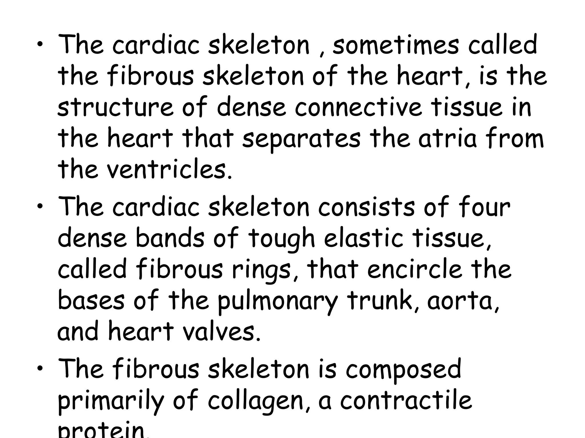
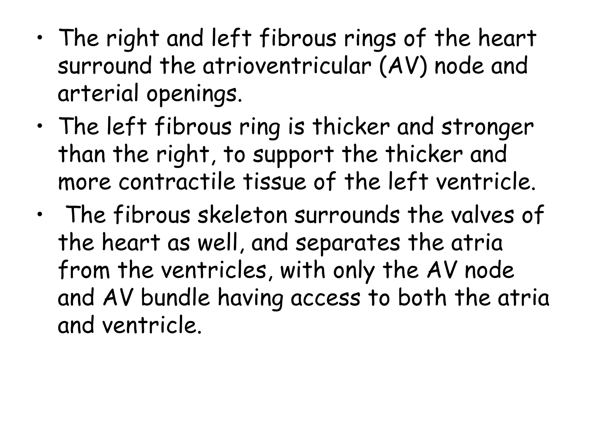
3. The fibrous skeleton surrounds the heart valves and separates the atria from the ventricles, allowing electrical signals to pass through only at the atrioventricular node.
4. The heart has four chambers - two superior atria that receive blood and two inferior ventricles that pump blood, with the left ventricle having a thicker muscular wall.
Title: Sense of Smell
Presenter: Dr. Faiza, Assistant Professor of Physiology
Qualifications:
MBBS (Best Graduate, AIMC Lahore)
FCPS Physiology
ICMT, CHPE, DHPE (STMU)
MPH (GC University, Faisalabad)
MBA (Virtual University of Pakistan)
Learning Objectives:
Describe the primary categories of smells and the concept of odor blindness.
Explain the structure and location of the olfactory membrane and mucosa, including the types and roles of cells involved in olfaction.
Describe the pathway and mechanisms of olfactory signal transmission from the olfactory receptors to the brain.
Illustrate the biochemical cascade triggered by odorant binding to olfactory receptors, including the role of G-proteins and second messengers in generating an action potential.
Identify different types of olfactory disorders such as anosmia, hyposmia, hyperosmia, and dysosmia, including their potential causes.
Key Topics:
Olfactory Genes:
3% of the human genome accounts for olfactory genes.
400 genes for odorant receptors.
Olfactory Membrane:
Located in the superior part of the nasal cavity.
Medially: Folds downward along the superior septum.
Laterally: Folds over the superior turbinate and upper surface of the middle turbinate.
Total surface area: 5-10 square centimeters.
Olfactory Mucosa:
Olfactory Cells: Bipolar nerve cells derived from the CNS (100 million), with 4-25 olfactory cilia per cell.
Sustentacular Cells: Produce mucus and maintain ionic and molecular environment.
Basal Cells: Replace worn-out olfactory cells with an average lifespan of 1-2 months.
Bowman’s Gland: Secretes mucus.
Stimulation of Olfactory Cells:
Odorant dissolves in mucus and attaches to receptors on olfactory cilia.
Involves a cascade effect through G-proteins and second messengers, leading to depolarization and action potential generation in the olfactory nerve.
Quality of a Good Odorant:
Small (3-20 Carbon atoms), volatile, water-soluble, and lipid-soluble.
Facilitated by odorant-binding proteins in mucus.
Membrane Potential and Action Potential:
Resting membrane potential: -55mV.
Action potential frequency in the olfactory nerve increases with odorant strength.
Adaptation Towards the Sense of Smell:
Rapid adaptation within the first second, with further slow adaptation.
Psychological adaptation greater than receptor adaptation, involving feedback inhibition from the central nervous system.
Primary Sensations of Smell:
Camphoraceous, Musky, Floral, Pepperminty, Ethereal, Pungent, Putrid.
Odor Detection Threshold:
Examples: Hydrogen sulfide (0.0005 ppm), Methyl-mercaptan (0.002 ppm).
Some toxic substances are odorless at lethal concentrations.
Characteristics of Smell:
Odor blindness for single substances due to lack of appropriate receptor protein.
Behavioral and emotional influences of smell.
Transmission of Olfactory Signals:
From olfactory cells to glomeruli in the olfactory bulb, involving lateral inhibition.
Primitive, less old, and new olfactory systems with different path
Ozempic: Preoperative Management of Patients on GLP-1 Receptor Agonists Saeid Safari
Preoperative Management of Patients on GLP-1 Receptor Agonists like Ozempic and Semiglutide
ASA GUIDELINE
NYSORA Guideline
2 Case Reports of Gastric Ultrasound
Anti ulcer drugs and their Advance pharmacology ||
Anti-ulcer drugs are medications used to prevent and treat ulcers in the stomach and upper part of the small intestine (duodenal ulcers). These ulcers are often caused by an imbalance between stomach acid and the mucosal lining, which protects the stomach lining.
||Scope: Overview of various classes of anti-ulcer drugs, their mechanisms of action, indications, side effects, and clinical considerations.
- Video recording of this lecture in English language: https://youtu.be/lK81BzxMqdo
- Video recording of this lecture in Arabic language: https://youtu.be/Ve4P0COk9OI
- Link to download the book free: https://nephrotube.blogspot.com/p/nephrotube-nephrology-books.html
- Link to NephroTube website: www.NephroTube.com
- Link to NephroTube social media accounts: https://nephrotube.blogspot.com/p/join-nephrotube-on-social-media.html
These lecture slides, by Dr Sidra Arshad, offer a quick overview of physiological basis of a normal electrocardiogram.
Learning objectives:
1. Define an electrocardiogram (ECG) and electrocardiography
2. Describe how dipoles generated by the heart produce the waveforms of the ECG
3. Describe the components of a normal electrocardiogram of a typical bipolar leads (limb II)
4. Differentiate between intervals and segments
5. Enlist some common indications for obtaining an ECG
Study Resources:
1. Chapter 11, Guyton and Hall Textbook of Medical Physiology, 14th edition
2. Chapter 9, Human Physiology - From Cells to Systems, Lauralee Sherwood, 9th edition
3. Chapter 29, Ganong’s Review of Medical Physiology, 26th edition
4. Electrocardiogram, StatPearls - https://www.ncbi.nlm.nih.gov/books/NBK549803/
5. ECG in Medical Practice by ABM Abdullah, 4th edition
6. ECG Basics, http://www.nataliescasebook.com/tag/e-c-g-basics
Explore natural remedies for syphilis treatment in Singapore. Discover alternative therapies, herbal remedies, and lifestyle changes that may complement conventional treatments. Learn about holistic approaches to managing syphilis symptoms and supporting overall health.
Ethanol (CH3CH2OH), or beverage alcohol, is a two-carbon alcohol
that is rapidly distributed in the body and brain. Ethanol alters many
neurochemical systems and has rewarding and addictive properties. It
is the oldest recreational drug and likely contributes to more morbidity,
mortality, and public health costs than all illicit drugs combined. The
5th edition of the Diagnostic and Statistical Manual of Mental Disorders
(DSM-5) integrates alcohol abuse and alcohol dependence into a single
disorder called alcohol use disorder (AUD), with mild, moderate,
and severe subclassifications (American Psychiatric Association, 2013).
In the DSM-5, all types of substance abuse and dependence have been
combined into a single substance use disorder (SUD) on a continuum
from mild to severe. A diagnosis of AUD requires that at least two of
the 11 DSM-5 behaviors be present within a 12-month period (mild
AUD: 2–3 criteria; moderate AUD: 4–5 criteria; severe AUD: 6–11 criteria).
The four main behavioral effects of AUD are impaired control over
drinking, negative social consequences, risky use, and altered physiological
effects (tolerance, withdrawal). This chapter presents an overview
of the prevalence and harmful consequences of AUD in the U.S.,
the systemic nature of the disease, neurocircuitry and stages of AUD,
comorbidities, fetal alcohol spectrum disorders, genetic risk factors, and
pharmacotherapies for AUD.
Couples presenting to the infertility clinic- Do they really have infertility...Sujoy Dasgupta
Dr Sujoy Dasgupta presented the study on "Couples presenting to the infertility clinic- Do they really have infertility? – The unexplored stories of non-consummation" in the 13th Congress of the Asia Pacific Initiative on Reproduction (ASPIRE 2024) at Manila on 24 May, 2024.
Title: Sense of Taste
Presenter: Dr. Faiza, Assistant Professor of Physiology
Qualifications:
MBBS (Best Graduate, AIMC Lahore)
FCPS Physiology
ICMT, CHPE, DHPE (STMU)
MPH (GC University, Faisalabad)
MBA (Virtual University of Pakistan)
Learning Objectives:
Describe the structure and function of taste buds.
Describe the relationship between the taste threshold and taste index of common substances.
Explain the chemical basis and signal transduction of taste perception for each type of primary taste sensation.
Recognize different abnormalities of taste perception and their causes.
Key Topics:
Significance of Taste Sensation:
Differentiation between pleasant and harmful food
Influence on behavior
Selection of food based on metabolic needs
Receptors of Taste:
Taste buds on the tongue
Influence of sense of smell, texture of food, and pain stimulation (e.g., by pepper)
Primary and Secondary Taste Sensations:
Primary taste sensations: Sweet, Sour, Salty, Bitter, Umami
Chemical basis and signal transduction mechanisms for each taste
Taste Threshold and Index:
Taste threshold values for Sweet (sucrose), Salty (NaCl), Sour (HCl), and Bitter (Quinine)
Taste index relationship: Inversely proportional to taste threshold
Taste Blindness:
Inability to taste certain substances, particularly thiourea compounds
Example: Phenylthiocarbamide
Structure and Function of Taste Buds:
Composition: Epithelial cells, Sustentacular/Supporting cells, Taste cells, Basal cells
Features: Taste pores, Taste hairs/microvilli, and Taste nerve fibers
Location of Taste Buds:
Found in papillae of the tongue (Fungiform, Circumvallate, Foliate)
Also present on the palate, tonsillar pillars, epiglottis, and proximal esophagus
Mechanism of Taste Stimulation:
Interaction of taste substances with receptors on microvilli
Signal transduction pathways for Umami, Sweet, Bitter, Sour, and Salty tastes
Taste Sensitivity and Adaptation:
Decrease in sensitivity with age
Rapid adaptation of taste sensation
Role of Saliva in Taste:
Dissolution of tastants to reach receptors
Washing away the stimulus
Taste Preferences and Aversions:
Mechanisms behind taste preference and aversion
Influence of receptors and neural pathways
Impact of Sensory Nerve Damage:
Degeneration of taste buds if the sensory nerve fiber is cut
Abnormalities of Taste Detection:
Conditions: Ageusia, Hypogeusia, Dysgeusia (parageusia)
Causes: Nerve damage, neurological disorders, infections, poor oral hygiene, adverse drug effects, deficiencies, aging, tobacco use, altered neurotransmitter levels
Neurotransmitters and Taste Threshold:
Effects of serotonin (5-HT) and norepinephrine (NE) on taste sensitivity
Supertasters:
25% of the population with heightened sensitivity to taste, especially bitterness
Increased number of fungiform papillae
Lung Cancer: Artificial Intelligence, Synergetics, Complex System Analysis, S...Oleg Kshivets
RESULTS: Overall life span (LS) was 2252.1±1742.5 days and cumulative 5-year survival (5YS) reached 73.2%, 10 years – 64.8%, 20 years – 42.5%. 513 LCP lived more than 5 years (LS=3124.6±1525.6 days), 148 LCP – more than 10 years (LS=5054.4±1504.1 days).199 LCP died because of LC (LS=562.7±374.5 days). 5YS of LCP after bi/lobectomies was significantly superior in comparison with LCP after pneumonectomies (78.1% vs.63.7%, P=0.00001 by log-rank test). AT significantly improved 5YS (66.3% vs. 34.8%) (P=0.00000 by log-rank test) only for LCP with N1-2. Cox modeling displayed that 5YS of LCP significantly depended on: phase transition (PT) early-invasive LC in terms of synergetics, PT N0—N12, cell ratio factors (ratio between cancer cells- CC and blood cells subpopulations), G1-3, histology, glucose, AT, blood cell circuit, prothrombin index, heparin tolerance, recalcification time (P=0.000-0.038). Neural networks, genetic algorithm selection and bootstrap simulation revealed relationships between 5YS and PT early-invasive LC (rank=1), PT N0—N12 (rank=2), thrombocytes/CC (3), erythrocytes/CC (4), eosinophils/CC (5), healthy cells/CC (6), lymphocytes/CC (7), segmented neutrophils/CC (8), stick neutrophils/CC (9), monocytes/CC (10); leucocytes/CC (11). Correct prediction of 5YS was 100% by neural networks computing (area under ROC curve=1.0; error=0.0).
CONCLUSIONS: 5YS of LCP after radical procedures significantly depended on: 1) PT early-invasive cancer; 2) PT N0--N12; 3) cell ratio factors; 4) blood cell circuit; 5) biochemical factors; 6) hemostasis system; 7) AT; 8) LC characteristics; 9) LC cell dynamics; 10) surgery type: lobectomy/pneumonectomy; 11) anthropometric data. Optimal diagnosis and treatment strategies for LC are: 1) screening and early detection of LC; 2) availability of experienced thoracic surgeons because of complexity of radical procedures; 3) aggressive en block surgery and adequate lymph node dissection for completeness; 4) precise prediction; 5) adjuvant chemoimmunoradiotherapy for LCP with unfavorable prognosis.
Maxilla, Mandible & Hyoid Bone & Clinical Correlations by Dr. RIG.pptx
Anatomy of heart presenter dr.smita valani
1. Anatomy of heart
by
Dr.Smita Valani
Moderater: Dr. Anupama H
Dept of cardiology
M S Ramaih medical college
Bangalore
2. Heart
• Cone shaped four chamber muscular
organ
• Located in the mediastinum
– Behind sternum
– Between 2nd
and 6th
ribs
– Between T5-T8
• Lies on its side on the diaphragm
• Base : widest part of heart,directed
upwards and towards right shoulder
• Apex points to left & downwards,in 5th
ICS,1 cm medial to mid clavicular line.
6. Coverings of the Heart
• Pericardium – membranous sac surrounding the heart
• Base : directed downwards,fused partly with tendon
of diaphragm
• Apex : fused with great blood vessels
– Fibrous pericardium – tough, loose-fitting,
inelastic
– Serous pericardium
• Parietal layer: lines the inside of the fibrous
pericardium
• Visceral layer: adheres to outside of the
heart;also called as epicardium.
– Pericardial space: between parietal and
visceral layer
• Filled with 30-50mL of pericardial fluid
• Decreases friction
7. Walls of the Heart
• Epicardium – outer layer
– Epicardium = serous pericardium
• Myocardium – thick, contractile layer composed
of cardiac muscle cells
– Framework of collagenous fibres with blood vessels
that supply the myocardium and nerve fibres that
regulate the heart.
– Contraction of myocardium pumps blood from the heart
into the major arteries.
– Arrangement of muscle pattern is elegant and complex,
muscles cells swirl and spiral round the chambers of
heart.
– Figure of 8 around the atria and base of the great
vessels
8. Contd
-Deeper ventricular muscle also form figure of 8 around
both the ventricles and proceed towards the apex.
-More superficial layers of ventricular muscle wraps
around both the ventricles.
This swirling pattern helps pimp blood more effectively.
• Endocardium – interior of cardiac wall
– Endothelial tissue(simple squamous epithelium)
– Covers projections of myocardial tissue called
trabeculae
– Joins myocardium with a thin layer of connective
tissue.
10. Fibrous Skeleton
• Surrounds all four
valves
– Composed of dense
connective tissue
• Functions
– Anchors valve cusps
– Prevents overdilation
of valve openings
– Main point of insertion
for cardiac muscle
– Blocks direct spread
of electrical impulses
11. • The cardiac skeleton , sometimes called
the fibrous skeleton of the heart, is the
structure of dense connective tissue in
the heart that separates the atria from
the ventricles.
• The cardiac skeleton consists of four
dense bands of tough elastic tissue,
called fibrous rings, that encircle the
bases of the pulmonary trunk, aorta,
and heart valves.
• The fibrous skeleton is composed
primarily of collagen, a contractile
12. • The right and left fibrous rings of the heart
surround the atrioventricular (AV) node and
arterial openings.
• The left fibrous ring is thicker and stronger
than the right, to support the thicker and
more contractile tissue of the left ventricle.
• The fibrous skeleton surrounds the valves of
the heart as well, and separates the atria
from the ventricles, with only the AV node
and AV bundle having access to both the atria
and ventricle.
13. • The left atrioventricular ring is closely
connected, by its right margin, with the aortic
arterial ring; between these and the right
atrioventricular ring is a triangular mass of
fibrous tissue, the Fibrous trigone(central
fibrous body), which represents the os
cordis seen in the heart of some of the larger
animals, as the ox and elephant.[2]
• Lastly, there is the tendinous band, the
posterior surface of the conus arteriosus.[
• 2]
The fibrous rings surrounding the arterial
orifices serve for the attachment of the great
vessels and semilunar valves, they are known
as The aortic annulus.[
• ]
14. • 2
Each ring receives, by its ventricular
margin, the attachment of some of the
muscular fibers of the ventricles; its
opposite margin presents three deep
semicircular notches, to which the
middle coat of the artery is firmly
fixed
• From the margins of the semicircular
notches the fibrous structure of the
ring is continued into the segments of
the valves.[2]
15. • The tendon of Todaro is a collagenous band
within the subendocardium that constitutes
part of the fibrous skeleton of the heart. It
originates from the central fibrous body. It is
palpable in the right atrium running from the
interatrial septum inferiorly and to the right
to the inferomedial part of the inferior vena
caval valve.
16.
17. Chambers of the Heart
• Atria – two superior chambers
– “Receiving chambers”
– Blood from veins enters atria
• Ventricles – two inferior chambers
– “pumping chambers”
– Thick muscular walls to increase force of
pumping action
• Left > right
– Separated by interventricular septum
19. Contd
• Consist of rough anterior part,the atrium
proper,sinus venerium
• Superior vena cava opens in superior and
posterior part.
• Inferior vena cava opens in inferior part
close to inter atrial septum.it is bounded by
a fold called ‘the VALVE OF IVC’.
• Coronary sinus opens just left of valve of
IVC
• Vene cordis minimae : openings of small
veins of heart which opens besides opening
of coronary sinus.
20. Interior of heart
• Crista terminalis: sinus venarum &atrium
proper meet at a muscular ridge called
Crista terminalis,corresponds to sulcus
terminalis externally.
• Musculi pectinati: small muscular ridges
found in atrium proper
• IA septum : separates the two atria;shows a
oval depression which forms FOSSA
OVALIS in inferior part and a curved ridge
in upper part called the LIMBUS FOSSA
OVALIS.
• RA opens into RV through AV valve.
21. • he triangular area of the right atrium is
bounded by the:
• base of the septal leaflet of the tricuspid
valve inferiorly
• anterior margin of the coronary sinus
orifice
• tendon of Todaro anterosuperiorly
• Usually, the septal part of the
atrioventricular node lies within this region.
23. Contd
• Thin walled cavity with smooth wall.
• Musculi pectinati are present in auricle only.
• Pulmonary vein orifices lie on the
posterolateral (left pulmonary veins) and
posteromedial (right pulmonary veins) aspects
of the left atrial cavity. The left and right
upper pulmonary veins are directed
anterosuperiorly, whereas the lower veins
enter the left atrium nearly perpendicular to
the posterior atrial wall
• Left atrial appendage arises anterolaterally
and lies in left AV groove, It is smaller, more
tortous and less pyramidal than its right
counterpart.
24. Interior of Right ventricle
• a right-anterior structure
• comprised of an inlet and trabecular and
outflow segments
• inlet component extends from the tricuspid
annulus to the insertions of the papillary
muscles.
• An apical trabecular zone extends inferiorly
beyond the attachments of the papillary
muscles toward the ventricular apex and
about halfway along the anterior wall.
• The outflow portion, also known as the conus
or infundibulum, is a smooth-walled muscular
subpulmonary channel
25. • A prominent arch-shaped muscular ridge
known as the crista supraventricularis
separates the tricuspid and pulmonary valves.
• It is made up of three components ie,
parietal band, infundibular septum, and septal
band
• The parietal band is a free-wall structure
• the adjacent infundibular septum is
intracardiac and separates the two
ventricular outflow tracts beneath the right
and left cusps of both semilunar valves
26. • The septal band forms a Y-shaped muscle, the
two upper limbs of which cradle the
infundibular septum. From this branching
point of the septal band emanates the medial
tricuspid papillary muscle
• The moderator band forms an intracavitary
muscle that connects the septal band with the
anterior tricuspid papillary muscle
• the right ventricular chamber has a
crescentic appearance
28. Interior of left ventricle
• The left ventricle, like the right
ventricle, is made of an inlet portion
comprised of the mitral valve apparatus,
a subaortic outflow portion, and a finely
trabeculated apical zone.
• The LV free wall is normally thickest
toward the base and thinnest toward
the apex, where it averages only 1 to 2
mm in thickness, even in hypertrophied
hearts.
29. • The left ventricular free-wall and septal
thicknesses are three times the thickness of
the right ventricular free wall.
• The mitral and aortic valves share fibrous
continuity
• the mitral valve has an elliptical orifice and
no septal attachments
• the left ventricular chamber appears circular
in cross section,
30. • Left ventricular false tendons, also
referred to as pseudotendons or bands,
are discrete, thin, cordlike
fibromuscular structures that connect
two walls, the two papillary muscles, or a
papillary muscle to a wall, usually the
ventricular septum
32. Contd
• IV septum : separates RV & LV
- Forms anterior wall & some part of wall on
right side of LV
- comprised of four parts: (1) inlet, (2)
trabecular, (3) membranous, and (4)
infundibular.
- Can also be divided into two parts :
MEMBRANOUS AND MUSCULAR
- Muscular part is thick & forms major part
- Membranous part is thin upper part of the
septum
- Third part of septum can be considered atrio
ventricular part due to its position above
septal cusp of tricuspid valve between atrium
33. Contd
• Inflow part begins just infront of AV
orifice,runs forwards and left towards the
apex of the heart.
- has rough inner surface due to
TRABECULAE CARNAE(bundles of
muscle fibres)
- Papillary muscles : finger like processes
belongs functionally to AV valves.
- There are 3 pappilary muscles in RV
:anterior,posterior & septal
- There are 2 pappillary muscles in
LV:anterior & posterior.
34. Atrioventricular orifices
• Right & left AV orifices are oval apertures
• Directed forwards,downwards & towards
left
• Guarded by AV valves
• Right orifice is larger than left
35. Valves of the Heart
• Permit blood flow in one direction during
circulation
• Each cusp consist of double fold of
endocardium within which there is some
fibrous tissue.
• Has two surfaces ,atrial & ventricular
• Has a base attached to the ring of fibrous
tissue around AV orifice
• It has apex & free margins which give
attachment to CHORDA TENDINAE
36. Contd
• At the other end chordae are attached
to pappillary muscles.
• As a result adjoining margins of cusps
are drawn together when papillary
muscle contracts.
37. • Atrioventricular valves (AV valves)
– Also cuspid valves
– Between atria and ventricles
• Semilunar (SL valves)
– Between R ventricle and pulmonary arteries
and L ventricle and aorta
38. Atrioventricular Valves
• Tricuspid valve
– Btwn R atrium and ventricle
– 5 components: annulus, leaflets,
commissures, chordae and papillary muscles.
– 3 flaps of endocardium called cusps
viz.,anterior ,posterior and septal cusp.
– The septal and the anterior leaflets are
larger;anterior cusp separates inflow part
ofRV from infundibulum and is most mobile
• The septal leaflet is in immediate proximity of
the membranous ventricular septum, and its
extension provides a basis for spontaneous
closure of the perimembranous ventricular
septal defect. It is least mobile becoz has
39. • The anterior leaflet is attached to the
anterolateral margin of the annulus
– Cusps are connected to ventricular papillary
muscle via chordae tendinae
– The tricuspid valve is the most apically (or
caudally) placed valve with the largest orifice
among the four valves.
– Chordae tendinae are attached to cusp by ant
papillary or posterior papillary muscle or
directly from interventricular septum or from
small papillary muscle attached to septum
– Chordae from ant papillary muscle attached to
ant and post cusps, from post papillar muscle to
posterior and septal, and those from septal to
septal and anterior cusps.
40. Contd
-base of each cusp is attached to fibrous ring
that surrounds the AV orifice called annulus
-The tricuspid annulus is oval-shaped and when
dilated becomes more circular.
-20% larger than MV annulus .
-Normal TV annulus= 3.0 3.5 cm
-papillary muscles as described above
• Bicuspid valve
-also called as mitral valve
– Btwn L atrium and ventricle
41.
42.
43. Anatomy of mitral valve
Mitral valve
apparatus :-
Left Atrial Wall
Mitral valve Annulus.
Mitral leaflets with
commissures.
Chordae tendinae.
Papillary muscles.
Supporting LV Wall.
Altogether called as
mitral valve complex.
Resembles the Bishops
“mitre” .
44. Mitral valve Annulus
Annulus :fibroelastic
ring that connects
the valve leaflets.
Annulus is elliptical in
shape in systole &
circular in diastole.
D shaped. Straight
border posterior to
aortic valve.
45. • Thin, pliable, soft translucent
• Each has atrial and ventricular surface
• Free edge has indentations
• Two indentation as anterolateral and
postero-medial commissures divide
leaflets into anterior leaflet and post
leaflet.
Mitral leaflets & commissures
46. Mitral leaflets & commissures
AML :-
Semicircular, free
edge no indentation
In continuity of
aortic annulus, post
to aortic root.
Encircles on 1/3rd
of
annulus, but covers
2/3rd
of valve orifice
area.
2 zones on anterior
leaflets:
PML :-
Quadrangular in shape.
Occupies 2/3rd
of the
annulus, but covers
only 1/3rd
of the valve
area.
Free edge has 2
identations –3 scallops.
3 zones: Basal, clear
and rough
47. Contd
• Rough and clear zone
depending on
attachment of chordae
tendinae insertion.
• 2 zones separated by
prominent ridge on
atrial surface –line of
leaflet closure, located
approx. 1 cm from free
edge of ant.leaflet.
• Distal to ridge-
cresentric shape-rough
zone. Is thick, has
chordae attachment on
ventricular surface
• Rough zone: distal to
ridge of line of closure
of leaflets. Broadest at
distal part and tapers
towards identation.
• Clear zone: no
attachments
• Basal zone: Only in
middle scallop.
--Located between
annulus and clear
zone
--Attachment of basal
chorda tendinae.
48. Chorae Tendinae
-Thin fibrous strings arising from apical portion of
papillary muscle or directly from ventricular wall &
insert into valve leaflets.
-2 types: True or false chordae
A)Commissural chordae: Chordae that insert into
interleaflet or commisural area at junction of
anterios and posterior leaflets.
--Anterolateral and posteromedial commissural
chordae
B) Leaflet Chordae: That insert directly into ant or
posterior leaflets.
49. Contd
• Anterior Leaflet: 2 types
1)Rough zone chordae: insert into distal part of
rough zone of anterior leaflets.
2)Strut Chordae: They branch before inserting to
ant leaflet. (not present in post leaflet)
• Posterior Leaflet: 3 types
1)Rough zone chordae
2)Basal Chordae
3)Cleft Chordae: Insert into clefts of posterior leaflet
dividing it into 3 scallops.
51. Papillary Muscles
Located at the
junction of the
apical (lower) third &
middle third of the
left ventricle.
2 in number.
APM :- antero-
lateral wall of LV.
PPM :- postero-
medial wall of LV.
APM :- has dual
blood supply.
OM of CX.
D1 of LAD.
PPM:- has single
blood supply.
Last OM/ RCA.
52.
53. • Free edge of each cusp curves upward from the
commissure and forms a slight thickening at
the tip or midpoint, called the Arantius nodule
– When the valve closes, the three nodes meet
in the center, allowing coaptation to occur
along three lines that radiate out from this
center point
• Overlap of valve tissue along the lines of
closure produces a tight seal and prevents
backflow during diastole
54. Semilunar Valves
• Aorta semilunar valve
• Btwn L ventricle and aorta
• The area of a normal aortic valve is 3 to 4 cm2
• Normal opening generally produces 2 cm of
leaflet separation
• Aortic valve is composed of three cusps of
equal size, each of which is surrounded by a
sinus
• Cusps are separated by three commissures and
supported by a fibrous anulus
• Each cusp is crescent shaped and capable of
opening fully to allow unimpeded forward flow,
then closing tightly to prevent regurgitation
55. Contd• Free edge of each cusp curves upward from the
commissure and forms a slight thickening at the
tip or midpoint, called the Arantius nodule
– When the valve closes, the three nodes meet in
the center, allowing coaptation to occur along
three lines that radiate out from this center
point
– Overlap of valve tissue along the lines of
closure produces a tight seal and prevents
backflow during diastole
• Behind each cusp is its associated Valsalva sinus
• Sinuses represent outpunching in the aortic root
directly behind each cusp
• Function to support the cusps during systole and
provide a reservoir of blood to augment coronary
artery flow during diastole
56. • Pulmonary semilunar valve
– Btwn R ventricle and pulmonary trunk
– virtually identical in design to the aortic
valve
– the pulmonary and tricuspid valves are
separated by infundibular muscle
– The pulmonic valve is typically cephalad to
the aortic valve such that the supravalvular
portion of the aortic valve lies in immediate
proximity to portions of the pulmonary
valve. This relationship is important for the
electrophysiologist
59. Arterial Supply of the Heart
• The arterial supply of
the heart is provided
by the right and left
coronary arteries,
which arise from the
ascending aorta
immediately above
the aortic valve.
60. • Left and right coronary arteries arise
from the left and right sinuses,
respectively, and are associated with
the left and right aortic cusps
61. Coronary Arteries
The origins of the coronary
arteries are as follows:
• The left coronary artery
arises from the aortic
sinus immediately above
the left posterior cusp of
the aortic valve .
• The right coronary artery
arises from the aortic
sinus immediately above
the anterior cusp of the
aortic valve.
62. Right coronary artery
Branches
– Right marginal arteries
(acute marginal artery)
– Posterior
interventricular artery.
(in post. IV sulcus)
– Sinoatrial nodal artery.
– Atrioventricular nodal
artery.
63. Left coronary artery
Branches
– Left anterior
descending (LAD) or
anterior
interventricular
artery. (lies in
anterior IV sulcus)
• Septal branches
• Diagonal branches
– Left marginal artery.
(Obtuse marginal
artery)
– Left circumflex
artery.
66. Venous Drainage of the Heart
• The venous drainage
of the heart is by
three means:
– Coronary sinus.
– Anterior cardiac veins
– Venae Cordis
minimae.
67. CORONARY SINUS
•This is the largest of vein of heart situated in
the left posterior coronary sulcus. It is about 3
cm long and ends by opening into the posterior
wall of the right atrium.
•Its tributaries are:
−Great cardiac vein: It enters the left end
of the coronary sinus.
−Middle cardiac vein: It accompanies the
posterior interventricular artery and joins
the right end of the coronary sinus.
−Small cardiac vein: It accompanies the
right coronary artery and joins the right
68. − Posterior vein of left ventricle: It runs on the
diaphragmatic surface of the left ventricle and ends in
the middle of the coronary sinus.
− Oblique vein of left atrium ( of Marshall): It runs on the
posterior surface of the left atrium, joins the left end of
coronary sinus and develops from the left common
cardinal vein.
− The right marginal vein: It accompanies the marginal
branch of the right coronary artery.
69. ANTERIOR CARDIAC VEIN
3 to 4 small veins run on the anterior wall of
the right ventricle, open directly into the right atrium.
VENAE CORDIS MINIMAE
(also called smallest cardiac veins, venae cardiacae minimae, or
Thebesian veins)
•Numerous small veins present in all 4 chambers of
heart which open directly into the cavities.
•The Thebesian venous network is considered an
alternative (secondary) pathway of venous drainage
of the myocardium.
72. Lymphatics of heart
• Lymphatics of the heart accompany the
coronary arteries and form 2 trunks.
• Right trunk ends in brachiocephalic
nodes and the left trunk into the
tracheobronchial lymph nodes at the
bifurcation of the trachea.
73. Conduction System of the Heart
• Four structures composed of modified
cardiac muscle
• Sinoatrial Node (SA Node)
– Pacemaker of the heart
– 100s of cells in the R atrium near the
opening of the superior vena cava
• Atrioventricular Node (AV Node)
– Small mass of cardiac muscle tissue
– Left lower border of R atrium
74. Conduction System of the Heart
• Atrioventricular Bundle
– Also Bundle of His
– Bundle of specialized cardiac muscle fibers
originating in the AV node
– Branches into R and L branches eventually
becoming Purkinje fibers
– Extend into the walls of the ventricles and
papillary muscles
75. Nerve Supply of the Heart
• The heart is innervated by sympathetic
and parasympathetic fibers of the
autonomic nervous system via the cardiac
plexuses situated below the arch of the
aorta.
• The sympathetic supply arises from the
cervical and upper thoracic portions of the
sympathetic trunks, and the
parasympathetic supply comes from the
vagus nerves.