Download to read offline


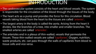
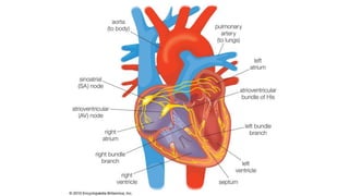
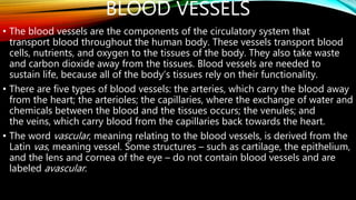
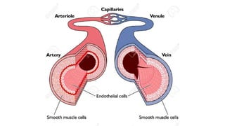
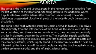
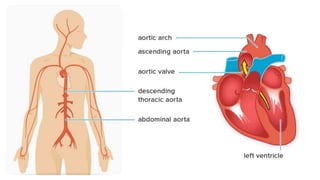
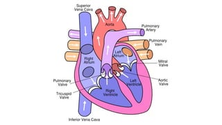
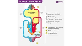


The document discusses the structure and function of the circulatory system. It describes the heart as a hollow muscular organ that pumps blood through arteries, capillaries, and veins. The heart has four chambers and a conduction system that generates electrical signals to coordinate contractions. Blood vessels branch throughout the body to deliver oxygen, nutrients and remove waste. Arteries carry blood away from the heart, veins carry blood back to the heart, and capillaries facilitate exchange of materials between blood and tissues.