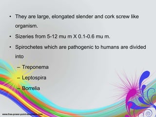
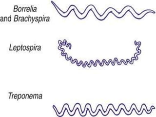
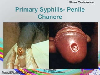
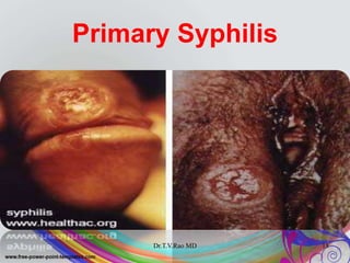
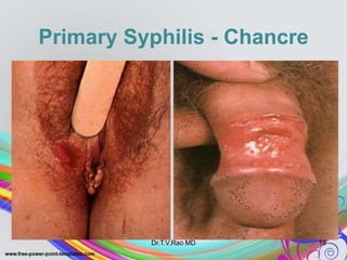
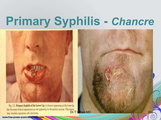
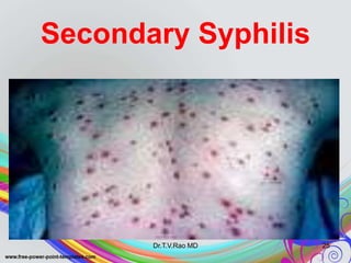
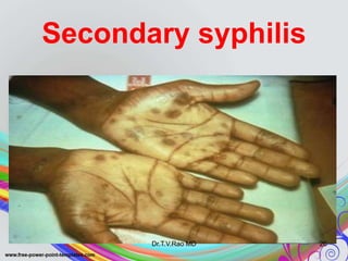
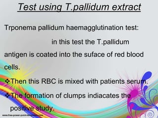
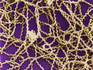
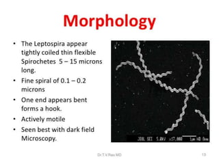
SPIROCHETES
RATHEESH R.L
• They are large, elongated slender and cork screw like
organism.
• Sizeries from 5-12 mu m X 0.1-0.6 mu m.
• Spirochetes which are pathogenic to humans are divided
into
– Treponema
– Leptospira
– Borrelia
TREPONEMA
• The important pathogenic species the
genus is
TREPONEMA PALLIDUM
MORPHOLOGY
• They are thin ,long 6-14 mu m in length
and 0.2 mu m in width.
• The body is coiled with 6-14 evenly
spaced coils.
• Actively motile.
CULTURAL
CHARACTERISTICS
• Treponema pallidum has not been
cultured in any artificial medias.
PATHOGENESIS
• Syphilis is a chronic infectious disease
caused by the spirochaete Treponema
pallidum.
• The disease is transmitted during sexual
contact or can be transmitted congenitally.
• So that it is divided into,
1. ACQUIRED SYPHILLIS
2. CONGENITAL SYPHILLIS
ACQUIRED SYPHILIS
• It is mainly transmitted because of unprotected
sexual contacts.
• The organism penetrates the mucous surface and
multiplies at the site of entry.
• Incubation period is 2-4 weeks and it ranges from
10-100 days.
• During the incubation period the organism
invades the regional lymph nodes(inguinal
in males & pelvic in females) and leads to
lymphadenitis
• From the lymph nodes the organism
spreads to the body parts through the
blood.
• Clinically syphilis is divided into 4stages,
• 1. primary syphilis or primary stage
• 2. secondary syphilis or secondary stage
• 3. latent syphilis or latent stage
• 4. tertiary syphilis or tertiary stage
PRIMARY SYPHILIS OR
PRIMARY STAGE
• In this stage the papule appears in the
genital area that ulcerates and forming a
primary lesion called “chancre” or “hard
chancre”.
• It is characterized by red, flat, painless and
indurated ulcer with 1-2 cm in diameter.
• This lesion is also called as hunterian
chancre, which is most frequently appears
in the external genitalia(penis,labia,vagina)
• Occasionally the chancre may appear on
mouth, lips, cheeks and nipples.
Primary Syphilis
Dr.T.V.Rao MD 16
Primary Syphilis- Penile
Chancre
17
Clinical Manifestations
Source: CDC/ NCHSTP/ Division of STD Prevention, STD Clinical Slides
Dr.T.V.Rao MD
Primary Syphilis
Dr.T.V.Rao MD 18
Primary Syphilis - Chancre
Dr.T.V.Rao MD 19
Primary Syphilis - Chancre
Dr.T.V.Rao MD 20
SECONDARY SYPHILIS OR
SECONDARY STAGE
• The secondary stage is characterized by
appearance of widespread papular skin
rashus particularlly on trunk and extrimities
and mucous patches in the oropharynx.
• There may be enlargement of lymph
nodes will occur in the second stage.
• Other features are retinitis, arthritis,
periostitis and meningitis.
Secondary Syphilis
Dr.T.V.Rao MD 23
Secondary Syphilis
Dr.T.V.Rao MD 24
Secondary Syphilis
Dr.T.V.Rao MD 25
Secondary syphilis
Dr.T.V.Rao MD 26
LATENT SYPHILIS OR
LATENT STAGE
• After disappearance of the secondary
lesions 30% of cases will get cured without
any treatment.
• Remaining 30% will continue without
producing any symptoms and that can
lead to tertiary syphilis.
LATENT SYPHILIS OR
LATENT STAGE
• This stage is slowly progressive and the
lesions may be formed anywhere in the
body and may affect any organ.
• The granulomatous nodules will be formed
in the skin, mucous membrane, bones,
tongue, liver and myocardium.
CONGENITAL SYPHILLIS
• The organism is capable of crossing the
placenta and leading to congenital
syphilis.
• Transmission can be occur from the 10th
week until the late in pregnancy.
• The infected feces may die and resulting
in miscarriage.
LABORATORY DIAGNOSIS
• Hematological investigations
• Bacteriological investigations
– Direct demonstration of treponema
• Microscopic studies
• Direct fluorescent antibody staining for T.Pallidum
• Tissue biopsy
– Serological diagnosis
• Non treponemal or standard test
• Treponemal tests
Hematological investigations
• In acute cases the leucocytes will be
increased and in chronic cases the
lymphocytes will be elevated.
Bacteriological investigations
• Microscopic studies
When the specimen is observed
under a dark ground microscope after
covering the specimen by a cover slip, the
organism appears as spiral, slender with
flexion and extension movements.
Direct fluorescent antibody
staining for T.Pallidum(DFA-TP)
• The smears made from the exudates can
be directly stained with fluorescein labelled
antitrponemal antibody.
• This is also called as fluoroscein labelled
antibody test.
Tissue biopsy
• The organism can be demonstrated in
tissues by silver staining or by
immunofluorescence.
SEROLOGICAL TESTS
• Serological tests are the most effective
diagnostic method for syphilis.
• Two types of antibodies are produced in this
disease, specific anti-treponemal antibody &
non-specific reagin antibody.
• Therefore two types of antigen are used in the
serological studies which include treponemal
antigen and non-treponemal antigen.
• Thus depending upon the antigen used,
serological test for syphilis can be two
types,
• Non treponemal or standard test
• Treponemal tests
NON TREPONEMAL OR STANDARD TEST
• 1. flocculation test
VDRL test
RPR test
kahn test
• 2. Compliment fixation test
TREPONEMAL TEST
• Test using cultivable treponemes as
antigen.
• Tests using pathogenic treponemes as
antigen.
– Test using live T.pallidum
– Test using killed T.palludum
– Test using T.pallidum extract
VDRL TEST
• It is a most widely used screening test
which is rapid and economical.
• In VDRL test, 0.05 ml of serum which is
heated at 56degree C for 30 min is taken.
• One drop of freshly prepared cardiolipin
antigen is added over that.
• The slide is rotated at 180 rpm for 4
minutes.
• Positive: formation of clumps
• Negative: formation of uniformly disturbed
crystals.
RAPID PLASMA REAGIN
(RPR)
• The test is almost similar to VDRL test.
• One drop of patients serum is mixed with
one drop of modified VDRL antigen on a
disposable plastic card with a disposable
stick.
• The card is gently rocked to and fro for 8
minutes and observed under microscope.
• Positive: aggregation of carbon particles
occurs in the periphery of the specimen.
• Negative: absence of black aggregation.
Kahn test
• Also known as khan tube test
• In this test 0.15ml of serum which is
heated at 56degree C for 30 minutes is
added to three test tubes containing
0.05,0.025, and 0.0125 ml of freshly
prepared cardiolipin antigen.
• Shake the tube for 3-4 minutes.
• Presence of floccules indicates a positive
test.
• Absence of floccules indicates a negative
test.
Compliment fixation test
• Otherwise known as wassermann
reaction.
• In this study serum which is heated at 56
degree C for 30 minutes is mixed with the
cardiolipin antigen.
• Then add 2 units of serum from guinea pig
to the mixture.
• The mixture is intubated at 37 degree C
for one hour.
• The absence of hemolysis indicates a
positive test.
• The presence of hemolysis indicates a
negative test.
TREPONEMAL TEST
• Test using cultivable treponemes as
antigen.
Test using reiter’s treponeme:
in this test the antigen used
is derived from reiters strain of T.pallidum
and most probably derived from the
endoflagella of the organism.
• The antigen is protein in nature hence the
test is also called as Reiter protein
compliment fixation test.
• The procedure and the results are same
as that of compliment fixation test.
Tests using pathogenic
treponemes as antigen.
 Test using live T.pallidum:
1. Treponema pallidum immobilization test:
In this study the suspected
serum is incubated anaerobically. if antibodies are
present, the immobilized organism can be found
under dark ground microscope.
• REACTIVE: more than 50% are
immobilized
• NON-REACTIVE: less than 20% are
immobilized
• DOUBTFUL TEST: 20-50% are
immobilized
Test using killed T.pallidum:
1. Treponama pallidum immune adherence test:
 in this test the serum is mixed with
inactivated treponemes.
 Fresh heparinized whole blood of a normal
individual is added to the above mixture.
 The mixture is incubated.
 If antibodies are present in the patients serum,
the treponemes will adhere to the erythrocytes.
 If no antibodies are present, no such adherence
will occur.
2. Treponema pallidum agglutination test:
In this method the serum is mixed with
T.pallidum and the mixture is intubated.
Under dark ground microscope, the
organism is found agglutinated only if the
serum contains specific antibodies.
3. Fluorescent treponemal antibody test:
In this test smears of T.pallidum are made
on slides and fixed with acetone.
The patients serum is added to the mixture
and incubated for the presence of
antibodies that will react with treponemes
in the smear.
The excess serum is washed off and the
antibodies that binds to the organisms in
smear are detected by treating the smear
with fluorescein labelled antihuman
immunoglobin.
The slide is again incubated and washed
and examined under microscope.
The organism shows fluorescence in
positive cases.
4. Fluorescent treponemal antibody adsorption
test:
In the test the patients serum is first absorbed
by non-pathogenic treponemal antigen(Reiter’s
strain) to remove group specific treponemal
antibody present in the serum.
The test can detect both IgG & IgM antibodies
present in patients serum.
Test using T.pallidum extract
Trponema pallidum haemagglutination test:
in this test the T.pallidum
antigen is coated into the suface of red blood
cells.
Then this RBC is mixed with patients serum.
The formation of clumps indiacates the
positive study.
TREATMENT
• Early stages
benzathine benzyl penicillin-single dose
doxycycline- twice for 5 days
• Late stages
benzathine benzyl penicillin- once in a
week for 3 weeks.
• THE IMPORTANT SPECIES COMING
UNDER THIS GENUS IS……
Leptospira icterohemorrhagica
CULTURAL
CHARACTERISTICS
• They are aerobic and grows well in the
temparature of 25-30 degree C at the pH
of 7.2-7.5.
• The organism can be cultivated in the
enrichment media which contains rabbit
serum.
PATHOGENESIS
The disease caused by the micro organism
is known as leptospirosis (weils disease/
infectious jaundice)
Leptospires are believed to enter the host
through the following:
• Abrasions in healthy skin
• Animal and rodent bites
• Mucous membranes or conjunctiva
• The placenta during pregnancy
• Wild rats are the reservoir of the micro
organism.
• The organism localize in the kidneys and
colonizing in the convoluted tubules of the
rats.
• Humans acquires infection when the
ingestion of contaminated food or water
occur which is infected with the rats urine.
• Incubation period is 1-8 weeks.
• After that the organism is spreading to
whole body through the circulating blood
and causes severe leptospirosis.
• The symptoms include,
– Fever
– Conjunctivitis
– Albuminuria
– Hemorrhage
– jaundice
LABORATORY DIAGNOSIS
• Hematological studies
– Increased number of leucocytes, ESR and
polymorphous cells.
– Blood urea and serum bilirubin level
increased.
• Bacteriological investigations
– Microscopy:
under dark ground microsopy can
observe coiled, motile spirochetes.
– Culture studies:
the specimen is placed in stewart or
korthof’s medium. Then the bottles are
incubated at 30 degree C for 2days- 2weeks.
Under microscope can observe the micro
organism.
• Serological investigations
it includes microscopic
agglutination test and indirect fluorescent
antibody test.
TREATMENT
• Tetracycline and penicillin are effective
against the micro organism
• The important pathogenic species is
BORRELIA BURGDOEFERI
MORPHOLOGY
• They are gram negative
• Helical and flexible in structure
• Size varies from 4-5mu m X 0.2-0.25mu m
CULTURAL
CHARACTERISTICS
• The organism can be cultivated in tissue
culture and chorioallantoic membrane of
chick embryo.
• They grow best at 34 drgree C
PATHOGENESIS
• The organism is primarily a parasite of small
mammals like rats and rodents.
• Then it is transmitted to man the bite of ixodid
tick which carries these spirochetes.
• Incubation period is 2-3 weeks and the
disease is known as lyme disease or lyme
arthritis.
• The features are fever, head ache, stiff
neck, myalgia, arthralgia,
lymphadenopathy and rashus or lesions.
• These rashus/lesions is known as
erythema chronium migrans.
LABORATORY DIAGNOSIS
• The diagnosting methods are,
-ELISA
- Immunofluorescence studies
TREATMENT
• Tetracycline and penicillin are the drug of
choice for this lyme disease.
17. spirochetes

17. spirochetes

  • 1.
  • 3.
    • They arelarge, elongated slender and cork screw like organism. • Sizeries from 5-12 mu m X 0.1-0.6 mu m. • Spirochetes which are pathogenic to humans are divided into – Treponema – Leptospira – Borrelia
  • 5.
    TREPONEMA • The importantpathogenic species the genus is TREPONEMA PALLIDUM
  • 6.
    MORPHOLOGY • They arethin ,long 6-14 mu m in length and 0.2 mu m in width. • The body is coiled with 6-14 evenly spaced coils. • Actively motile.
  • 8.
    CULTURAL CHARACTERISTICS • Treponema pallidumhas not been cultured in any artificial medias.
  • 9.
    PATHOGENESIS • Syphilis isa chronic infectious disease caused by the spirochaete Treponema pallidum. • The disease is transmitted during sexual contact or can be transmitted congenitally.
  • 10.
    • So thatit is divided into, 1. ACQUIRED SYPHILLIS 2. CONGENITAL SYPHILLIS
  • 11.
    ACQUIRED SYPHILIS • Itis mainly transmitted because of unprotected sexual contacts. • The organism penetrates the mucous surface and multiplies at the site of entry. • Incubation period is 2-4 weeks and it ranges from 10-100 days.
  • 12.
    • During theincubation period the organism invades the regional lymph nodes(inguinal in males & pelvic in females) and leads to lymphadenitis • From the lymph nodes the organism spreads to the body parts through the blood.
  • 13.
    • Clinically syphilisis divided into 4stages, • 1. primary syphilis or primary stage • 2. secondary syphilis or secondary stage • 3. latent syphilis or latent stage • 4. tertiary syphilis or tertiary stage
  • 14.
    PRIMARY SYPHILIS OR PRIMARYSTAGE • In this stage the papule appears in the genital area that ulcerates and forming a primary lesion called “chancre” or “hard chancre”. • It is characterized by red, flat, painless and indurated ulcer with 1-2 cm in diameter.
  • 15.
    • This lesionis also called as hunterian chancre, which is most frequently appears in the external genitalia(penis,labia,vagina) • Occasionally the chancre may appear on mouth, lips, cheeks and nipples.
  • 16.
  • 17.
    Primary Syphilis- Penile Chancre 17 ClinicalManifestations Source: CDC/ NCHSTP/ Division of STD Prevention, STD Clinical Slides Dr.T.V.Rao MD
  • 18.
  • 19.
    Primary Syphilis -Chancre Dr.T.V.Rao MD 19
  • 20.
    Primary Syphilis -Chancre Dr.T.V.Rao MD 20
  • 21.
    SECONDARY SYPHILIS OR SECONDARYSTAGE • The secondary stage is characterized by appearance of widespread papular skin rashus particularlly on trunk and extrimities and mucous patches in the oropharynx.
  • 22.
    • There maybe enlargement of lymph nodes will occur in the second stage. • Other features are retinitis, arthritis, periostitis and meningitis.
  • 23.
  • 24.
  • 25.
  • 26.
  • 27.
    LATENT SYPHILIS OR LATENTSTAGE • After disappearance of the secondary lesions 30% of cases will get cured without any treatment. • Remaining 30% will continue without producing any symptoms and that can lead to tertiary syphilis.
  • 28.
    LATENT SYPHILIS OR LATENTSTAGE • This stage is slowly progressive and the lesions may be formed anywhere in the body and may affect any organ. • The granulomatous nodules will be formed in the skin, mucous membrane, bones, tongue, liver and myocardium.
  • 29.
    CONGENITAL SYPHILLIS • Theorganism is capable of crossing the placenta and leading to congenital syphilis. • Transmission can be occur from the 10th week until the late in pregnancy. • The infected feces may die and resulting in miscarriage.
  • 30.
    LABORATORY DIAGNOSIS • Hematologicalinvestigations • Bacteriological investigations – Direct demonstration of treponema • Microscopic studies • Direct fluorescent antibody staining for T.Pallidum • Tissue biopsy – Serological diagnosis • Non treponemal or standard test • Treponemal tests
  • 31.
    Hematological investigations • Inacute cases the leucocytes will be increased and in chronic cases the lymphocytes will be elevated.
  • 32.
    Bacteriological investigations • Microscopicstudies When the specimen is observed under a dark ground microscope after covering the specimen by a cover slip, the organism appears as spiral, slender with flexion and extension movements.
  • 33.
    Direct fluorescent antibody stainingfor T.Pallidum(DFA-TP) • The smears made from the exudates can be directly stained with fluorescein labelled antitrponemal antibody. • This is also called as fluoroscein labelled antibody test.
  • 34.
    Tissue biopsy • Theorganism can be demonstrated in tissues by silver staining or by immunofluorescence.
  • 35.
    SEROLOGICAL TESTS • Serologicaltests are the most effective diagnostic method for syphilis. • Two types of antibodies are produced in this disease, specific anti-treponemal antibody & non-specific reagin antibody. • Therefore two types of antigen are used in the serological studies which include treponemal antigen and non-treponemal antigen.
  • 36.
    • Thus dependingupon the antigen used, serological test for syphilis can be two types, • Non treponemal or standard test • Treponemal tests
  • 37.
    NON TREPONEMAL ORSTANDARD TEST • 1. flocculation test VDRL test RPR test kahn test • 2. Compliment fixation test
  • 38.
    TREPONEMAL TEST • Testusing cultivable treponemes as antigen. • Tests using pathogenic treponemes as antigen. – Test using live T.pallidum – Test using killed T.palludum – Test using T.pallidum extract
  • 39.
    VDRL TEST • Itis a most widely used screening test which is rapid and economical. • In VDRL test, 0.05 ml of serum which is heated at 56degree C for 30 min is taken. • One drop of freshly prepared cardiolipin antigen is added over that. • The slide is rotated at 180 rpm for 4 minutes.
  • 40.
    • Positive: formationof clumps • Negative: formation of uniformly disturbed crystals.
  • 41.
    RAPID PLASMA REAGIN (RPR) •The test is almost similar to VDRL test. • One drop of patients serum is mixed with one drop of modified VDRL antigen on a disposable plastic card with a disposable stick. • The card is gently rocked to and fro for 8 minutes and observed under microscope.
  • 42.
    • Positive: aggregationof carbon particles occurs in the periphery of the specimen. • Negative: absence of black aggregation.
  • 43.
    Kahn test • Alsoknown as khan tube test • In this test 0.15ml of serum which is heated at 56degree C for 30 minutes is added to three test tubes containing 0.05,0.025, and 0.0125 ml of freshly prepared cardiolipin antigen. • Shake the tube for 3-4 minutes.
  • 44.
    • Presence offloccules indicates a positive test. • Absence of floccules indicates a negative test.
  • 45.
    Compliment fixation test •Otherwise known as wassermann reaction. • In this study serum which is heated at 56 degree C for 30 minutes is mixed with the cardiolipin antigen. • Then add 2 units of serum from guinea pig to the mixture. • The mixture is intubated at 37 degree C for one hour.
  • 46.
    • The absenceof hemolysis indicates a positive test. • The presence of hemolysis indicates a negative test.
  • 47.
    TREPONEMAL TEST • Testusing cultivable treponemes as antigen. Test using reiter’s treponeme: in this test the antigen used is derived from reiters strain of T.pallidum and most probably derived from the endoflagella of the organism.
  • 48.
    • The antigenis protein in nature hence the test is also called as Reiter protein compliment fixation test. • The procedure and the results are same as that of compliment fixation test.
  • 49.
    Tests using pathogenic treponemesas antigen.  Test using live T.pallidum: 1. Treponema pallidum immobilization test: In this study the suspected serum is incubated anaerobically. if antibodies are present, the immobilized organism can be found under dark ground microscope.
  • 50.
    • REACTIVE: morethan 50% are immobilized • NON-REACTIVE: less than 20% are immobilized • DOUBTFUL TEST: 20-50% are immobilized
  • 51.
    Test using killedT.pallidum: 1. Treponama pallidum immune adherence test:  in this test the serum is mixed with inactivated treponemes.  Fresh heparinized whole blood of a normal individual is added to the above mixture.  The mixture is incubated.  If antibodies are present in the patients serum, the treponemes will adhere to the erythrocytes.  If no antibodies are present, no such adherence will occur.
  • 52.
    2. Treponema pallidumagglutination test: In this method the serum is mixed with T.pallidum and the mixture is intubated. Under dark ground microscope, the organism is found agglutinated only if the serum contains specific antibodies.
  • 53.
    3. Fluorescent treponemalantibody test: In this test smears of T.pallidum are made on slides and fixed with acetone. The patients serum is added to the mixture and incubated for the presence of antibodies that will react with treponemes in the smear. The excess serum is washed off and the antibodies that binds to the organisms in smear are detected by treating the smear with fluorescein labelled antihuman immunoglobin.
  • 54.
    The slide isagain incubated and washed and examined under microscope. The organism shows fluorescence in positive cases.
  • 55.
    4. Fluorescent treponemalantibody adsorption test: In the test the patients serum is first absorbed by non-pathogenic treponemal antigen(Reiter’s strain) to remove group specific treponemal antibody present in the serum. The test can detect both IgG & IgM antibodies present in patients serum.
  • 56.
    Test using T.pallidumextract Trponema pallidum haemagglutination test: in this test the T.pallidum antigen is coated into the suface of red blood cells. Then this RBC is mixed with patients serum. The formation of clumps indiacates the positive study.
  • 57.
    TREATMENT • Early stages benzathinebenzyl penicillin-single dose doxycycline- twice for 5 days • Late stages benzathine benzyl penicillin- once in a week for 3 weeks.
  • 59.
    • THE IMPORTANTSPECIES COMING UNDER THIS GENUS IS…… Leptospira icterohemorrhagica
  • 62.
    CULTURAL CHARACTERISTICS • They areaerobic and grows well in the temparature of 25-30 degree C at the pH of 7.2-7.5. • The organism can be cultivated in the enrichment media which contains rabbit serum.
  • 63.
    PATHOGENESIS The disease causedby the micro organism is known as leptospirosis (weils disease/ infectious jaundice) Leptospires are believed to enter the host through the following: • Abrasions in healthy skin • Animal and rodent bites • Mucous membranes or conjunctiva • The placenta during pregnancy
  • 64.
    • Wild ratsare the reservoir of the micro organism. • The organism localize in the kidneys and colonizing in the convoluted tubules of the rats. • Humans acquires infection when the ingestion of contaminated food or water occur which is infected with the rats urine.
  • 65.
    • Incubation periodis 1-8 weeks. • After that the organism is spreading to whole body through the circulating blood and causes severe leptospirosis.
  • 66.
    • The symptomsinclude, – Fever – Conjunctivitis – Albuminuria – Hemorrhage – jaundice
  • 67.
    LABORATORY DIAGNOSIS • Hematologicalstudies – Increased number of leucocytes, ESR and polymorphous cells. – Blood urea and serum bilirubin level increased.
  • 68.
    • Bacteriological investigations –Microscopy: under dark ground microsopy can observe coiled, motile spirochetes. – Culture studies: the specimen is placed in stewart or korthof’s medium. Then the bottles are incubated at 30 degree C for 2days- 2weeks. Under microscope can observe the micro organism.
  • 69.
    • Serological investigations itincludes microscopic agglutination test and indirect fluorescent antibody test.
  • 70.
    TREATMENT • Tetracycline andpenicillin are effective against the micro organism
  • 72.
    • The importantpathogenic species is BORRELIA BURGDOEFERI
  • 74.
    MORPHOLOGY • They aregram negative • Helical and flexible in structure • Size varies from 4-5mu m X 0.2-0.25mu m
  • 75.
    CULTURAL CHARACTERISTICS • The organismcan be cultivated in tissue culture and chorioallantoic membrane of chick embryo. • They grow best at 34 drgree C
  • 76.
    PATHOGENESIS • The organismis primarily a parasite of small mammals like rats and rodents. • Then it is transmitted to man the bite of ixodid tick which carries these spirochetes.
  • 77.
    • Incubation periodis 2-3 weeks and the disease is known as lyme disease or lyme arthritis. • The features are fever, head ache, stiff neck, myalgia, arthralgia, lymphadenopathy and rashus or lesions. • These rashus/lesions is known as erythema chronium migrans.
  • 78.
    LABORATORY DIAGNOSIS • Thediagnosting methods are, -ELISA - Immunofluorescence studies
  • 79.
    TREATMENT • Tetracycline andpenicillin are the drug of choice for this lyme disease.