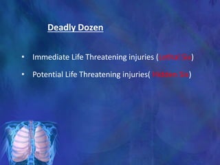
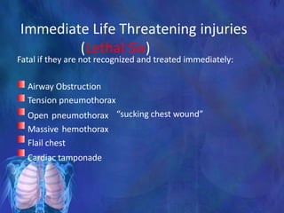
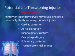
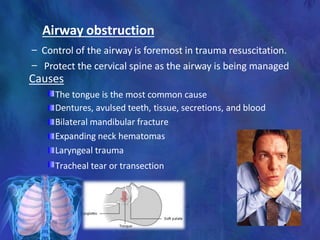
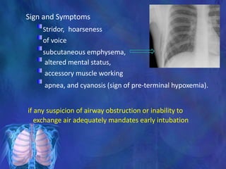
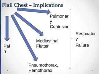
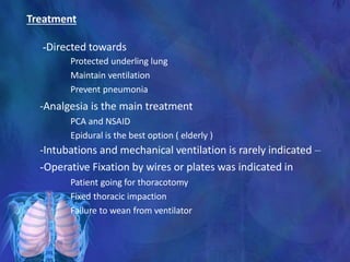
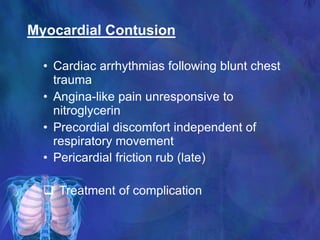
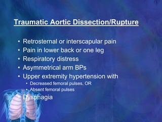
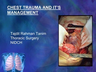
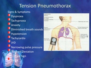
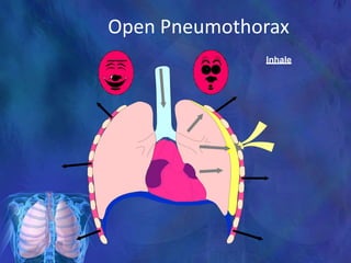
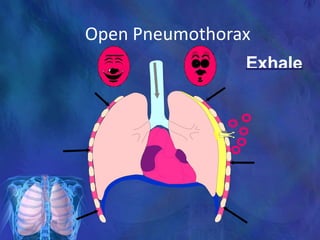
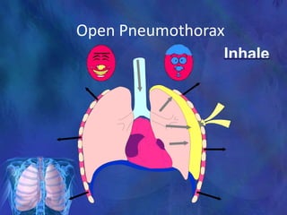
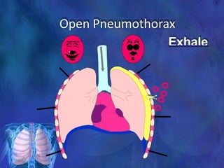
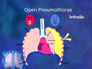
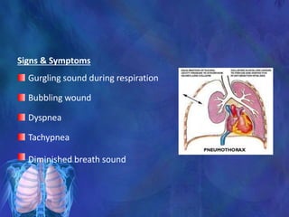
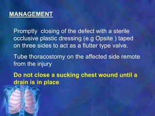
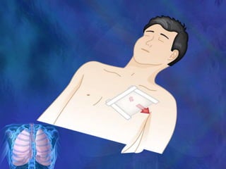
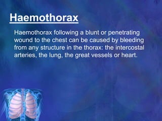
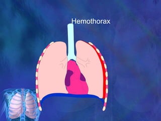
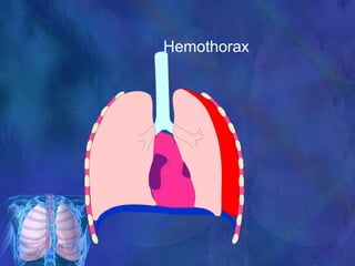
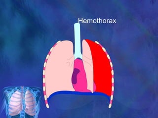
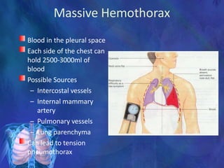
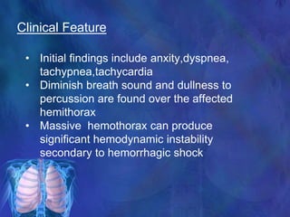
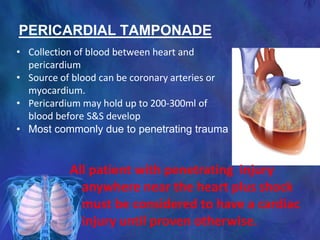
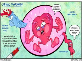
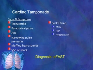
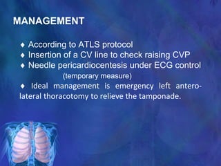
SlideShare a Scribd company logo
CHEST TRAUMA AND IT’S
MANAGEMENT
Tajdit Rahman Tanim
Thoracic Surgery
NIDCH
Introduction
• Chest trauma is often sudden and
dramatic
• Accounts for 25% of all trauma deaths
• 2/3 of deaths occur after reaching hospital
• Second leading cause of trauma deaths
after head injury (in USA)
• RTA is common etiology 65-70 %
• Abdominal injuries are common with chest
trauma.
Epidemiology
• The fact that it has become possible in recent
decades for millions of people to travel at high speed
had led to a phenomenal increase in blunt injury to
the chest - a most lethal type of injury.
Types of Chest injury
• Blunt thoracic injuries
 Forces distributed over large
area
• Penetrating thoracic injuries
 Forces distributed over small
area
 Organs injured usually those
that lie along path of
penetrating object
CAUSES OF CHEST INJURIES
BLUNT
TRAUMA
• Motor vehicle
accidents
• Auto vs.
pedestrian
• Falls
• Blast injuries
PENETRATI
NG TRAUMA
• Gunshot
wounds
• Stab wounds
• Shrapnel
wounds
Pathophysiology in Chest Injury
Flail chest
Contusion
Pneumothorax
Haemothorax
Heart & vessel
Associated injury
Decreased
alveolar
ventilation
Shock
Rt to Lt Shunting
Decreased CO
Hypoxia
Hypotension
Respiratory Acidosis
Metabolic Acidosis
Death
Initial assessment and
management
• Primary survey by ATLS guideline
• Resuscitation of vital functions
• Detailed secondary survey
• Definitive care
The first step is to make a rough estimate of
the status of the circulatory and respiratory
systems. This provides the first diagnostic clues and
often determines which therapeutic action is to be
taken. Specific questions are then posed pertaining
to individual injuries or their consequences.
Assessment of patient with
Thoracic injury
• The evaluation of thoracic injuries is only
one aspect of the total assessment of
severely injured patients.
• Both diagnosis and therapy go hand in
hand.
• The basic principle of elective surgery -
“First investigate and make the diagnosis,
then treat the illness” - is a dangerous
illusion.
Assessment of patient with
Thoracic injury
Immediately life-
threatening; diagnosis
and therapy before
taking other steps
TEN QUESTIONS to be asked in the initial
assessment of severe blunt thoracic injuries
1. Hypovolemia?
2. Respiratory insufficiency?
3. Tension pneumothorax?
4. Cardiac tamponade?
1. Multiple rib fractures? (Paradoxical respiration?)
2. Pneumothorax ? (subcutaneous emphysema?
mediastinal emphysema?)
3. Hemothorax?
4. Diaphragmatic rupture?
5. Aortic rupture?
6. Cardiac contusion?
TEN QUESTIONS to be asked in the initial
assessment of severe blunt thoracic injuries
Monitoring and evaluating the
patient with Thoracic trauma
• Radiograph of the thorax
(Chest wall i.e. ribs, sternum,
vertebral, clavicles).
• Mediastinum (wide or normal)
shifted or not.
• Lung parenchyma (Contusion).
• The heart (cardiac tamponade).
• Diaphragm.
• Pneumothorax, hemothorax.



ECG
CVP
Arterial blood
gases.
Urine output.
Lab. Investigations.
Others.



• The treatment of polytraumatized patient must follow
a certain protocol which includes.
• Adequate oxygenation.
• Fluid replacement.
• Surgical intervention.
• Treatment of septic complications.
• Adequate caloric and substrate supplementation.
• Prevention of stress bleeding.
• Finally, be alert of possible complication (CNS,
ARDS, hepatic, renal, coagulation disorders,
sepsis.
Management of patients with
Thoracic Trauma
Deadly Dozen
• Immediate Life Threatening injuries (Lethal Six)
• Potential Life Threatening injuries( Hidden Six)
Immediate Life Threatening injuries
(Lethal Six)
Fatal if they are not recognized and treated immediately:
Airway Obstruction
Tension pneumothorax
Open pneumothorax
Massive hemothorax
Flail chest
Cardiac tamponade
“sucking chest wound”
Potential Life Threatening injuries
( Hidden Six)
Primary or secondary survey may reveal one of six
potentially life-threatening thoracic injuries
• Cardiac contusion
• Aortic disruption
• Diaphragmatic rupture
• Oesophageal injury
• Pulmonary contusion
• Tracheo-bronchial injuries
Airway obstruction
– Control of the airway is foremost in trauma resuscitation.
– Protect the cervical spine as the airway is being managed
Causes
The tongue is the most common cause
Dentures, avulsed teeth, tissue, secretions, and blood
Bilateral mandibular fracture
Expanding neck hematomas
Laryngeal trauma
Tracheal tear or transection
Sign and Symptoms
Stridor, hoarseness
of voice
subcutaneous emphysema,
altered mental status,
accessory muscle working
apnea, and cyanosis (sign of pre-terminal hypoxemia).
if any suspicion of airway obstruction or inability to
exchange air adequately mandates early intubation
Management
Intubate using a controlled rapid sequence when in
doubt of obstruction
Provide inline cervical spine immobilization during
intubation.
Early intubate in cases of neck hematoma or possible
airway edema
Emergency cricothyroidotomy should perform if
endotracheal intubation fails.
Tension Pneumothorax
Air enters pleural space and becomes trapped leads to
pressure increase
Increased pressure which collapses lung and shifts
mediastinum to unaffected side
Increased dyspnoea and compressed heart and great vessels
leads to decreased cardiac output.
Leads to Cardiogenic Shock
Each time we inhale,
the lung collapses further. There
is no place for the air to
escape..
Tension Pneumothorax
Each time we inhale,
the lung collapses further. There
is no place for the air to
escape..
Tension Pneumothorax
Heart is being
compressed
The trachea is
pushed to
the opposite side
Tension Pneumothorax
Signs & Symptoms
Dyspnoea
Tachypnoea
Anxiety
Diminished breath sounds
Hypotension
Tachycardia
JVD
Narrowing pulse pressure
Tracheal Deviation
– Late Sign
Shock
Inferior vena cava
Treatment
Needle Thoracostomy ( 12 or 14 gauge IV catheter in 2nd ICS )
Tube Thoracostomy
Open Pneumothorax
(Sucking Chest Wound)
Open chest wall injury
Stab wounds usually self-sealing
Extensive damage
Air passes through opening into
the pleural space and remains
outside of lung
Large open defect in chest wall
(>3 cm diameter) with
equilibration between
intrathoracic and atmospheric
pressure
MECHANISM
Opening of chest wall exceeds two third of the
diameter of the trachea
with each inspiratory cycle air is drawn through
the defect rather than through the trachea
Accumulation of air in hemithorax rather than in the
lung
Leading to profound hypoventilation on the affected
side and hypoxia
If there is valvular effect increasing air
accumulation in the pleural cavity results in a
tension pneumothorax
Open Pneumothorax
Open Pneumothorax
Inhale
Open Pneumothorax
Exhale
Open Pneumothorax
Inhale
Open Pneumothorax
Exhale
Open Pneumothorax
Inhale
Open Pnuemothorax
Inhale
Signs & Symptoms
Gurgling sound during respiration
Bubbling wound
Dyspnea
Tachypnea
Diminished breath sound
MANAGEMENT
Promptly closing of the defect with a sterile
occlusive plastic dressing (e.g Opsite ) taped
on three sides to act as a flutter type valve.
Tube thoracostomy on the affected side remote
from the injury
Do not close a sucking chest wound until a
drain is in place
Haemothorax
Haemothorax following a blunt or penetrating
wound to the chest can be caused by bleeding
from any structure in the thorax: the intercostal
arteries, the lung, the great vessels or heart.
Hemothorax
Hemothorax
Hemothorax
Hemothorax
May put pressure on the heart
Lots of blood vessels
Where does the blood come
from.
Massive Hemothorax
Blood in the pleural space
Each side of the chest can
hold 2500-3000ml of
blood
Possible Sources
– Intercostal vessels
– Internal mammary
artery
– Pulmonary vessels
– Lung parenchyma
Can lead to tension
pneumothorax
Clinical Feature
• Initial findings include anxity,dyspnea,
tachypnea,tachycardia
• Diminish breath sound and dullness to
percussion are found over the affected
hemithorax
• Massive hemothorax can produce
significant hemodynamic instability
secondary to hemorrhagic shock
Diagnosis
By physical
examination and
CXR
Indication of Thoracaotomy
• Persistent shock
• Suspect or proven cardiac injury
• Massive hemothorax(>1500ml on chest tube
insertion)
• Persistent hemothorax(>500ml/hr initial hour of
drainage or >200ml/hr for several hours)
• Retained hemothorax
PERICARDIAL TAMPONADE
All patient with penetrating injury
anywhere near the heart plus shock
must be considered to have a cardiac
injury until proven otherwise.
• Collection of blood between heart and
pericardium
• Source of blood can be coronary arteries or
myocardium.
• Pericardium may hold up to 200-300ml of
blood before S&S develop
• Most commonly due to penetrating trauma
Cardiac Tamponade
Diagnosis- eFAST
Signs & Symptoms
Tachycardia
Paradoxical pulse
JVD
Narrowing pulse
pressures
Muffled heart sounds
S&S of shock
Beck’s Triad
MHS
JVD
Hypotension
MANAGEMENT
 According to ATLS protocol
 Insertion of a CV line to check raising CVP
 Needle pericardiocentesis under ECG control
(temporary measure)
 Ideal management is emergency left antero-
lateral thoracotomy to relieve the tamponade.
Three or more ribs
fractured in two or
more places
Flail Chest
• Blunt Force
• The blunt force typically also produces an
underlying pulmonary contusion.
• Dx is made clinically in pts who are not
ventilated, not radiography.
• Paradoxical respiration.
Flailed Chest
Treatment
-Directed towards
Protected underling lung
Maintain ventilation
Prevent pneumonia
-Analgesia is the main treatment
PCA and NSAID
Epidural is the best option ( elderly )
-Intubations and mechanical ventilation is rarely indicated –
-Operative Fixation by wires or plates was indicated in
Patient going for thoracotomy
Fixed thoracic impaction
Failure to wean from ventilator
Myocardial Contusion
• Cardiac arrhythmias following blunt chest
trauma
• Angina-like pain unresponsive to
nitroglycerin
• Precordial discomfort independent of
respiratory movement
• Pericardial friction rub (late)
 Treatment of complication
Traumatic Aortic Dissection/Rupture
• Retrosternal or interscapular pain
• Pain in lower back or one leg
• Respiratory distress
• Asymmetrical arm BPs
• Upper extremity hypertension with
• Decreased femoral pulses, OR
• Absent femoral pulses
• Dysphagia
-Widening of mediastinum
-Loss of aortic contour
-Tracheal shift to right
-Nasogastric tube shift
-Left sided haemothorax
MANAGEMENT
1. Control of systolic blood pressure (
less than 100 mm of Hg)
2. Placing of an endo vascular intra
aortic stent
3. Surgical repair / Aortic resection
with grafting-
surgical removal of some
portion of diseased aorta is called
aortic resection. A polyester
graft(Dacron) is used to replace
the diseased aortic tissue.
Esophageal injury
• Results from penetrating trauma; blunt injury is rare
Patient can present with odynophagia,
subcutaneous or mediastinal emphysema,
pleural effusion, air in the retro-oesophageal space an
unexplained fever within 24 hours of injury
• Combination of oesophagogram and oesophagoscopy confirm
diagnosis
• CT can be done
• Treatment is operative repair and drainage
• Mid-oesophageal injury => Right thoracotomy
• Distal oesophageal injury => Left thoracotomy
Diaphragmatic Rupture
• A tear in the Diaphragm that allows the abdominal
organs enter the chest cavity
• Any penetrating injury to or below 5th intercostal
space can cause diaphragmatic penetration &
abdominal injury
• Blunt injury to the diaphragm is usually caused by a
compressive force applied to the pelvis & abdomen.
• More common on Left side due to liver helps protect
the right side of diaphragm
• Associated with multiple injury patients
Diaphragm Rupture
S/S of Diaphragmatic Rupture
• Abdominal Pain
• Shortness of Air
• Decreased Breath Sounds on side of rupture
• Bowel Sounds heard in chest cavity
Diagnosis
• Chest radiography after placement of a
nasogastric tube, Contrast studies of the
upper or lower gastrointestinal tract, CT
scan & diagnostic peritoneal lavage.
• Most accurate evaluation is by video-assisted
thoracoscopy (VATS) or laparoscopy.
Treatment of Diaphragmatic Rupture
 Operative Repair.
Penetrating diaphragmatic injury must be
repaired via the abdomen and not the chest,
to rule out penetrating hollow viscus injury.
Laproscopy can be done.
Pulmonary Contusion
 Crushing and bruising of the lung parenchyma.
 Sudden blow or blunt injury to the chest => compression
of thoracic cavity and lung followed by an equally sudden
decompression. Concussive and compressive force is
most important cause.
 The natural progression of pulmonary contusion is
worsening hypoxemia for the first 24 to 48 hours.
 X-ray findings not significant initially.
 CT with contrast is confirmatory.
Contusion of left upper & lower lobe with
aspirated blood around
S/S of Pul. Contusion
• Hemoptysis
• Dyspnea
• Cough
• Chest wall abrasion
• Echymosis.
Tx. Of Pul. contusion
• Oxygen administration
• Pul. Toilet
• Mechanical ventilation => in severe case
Tracheo-bronchial Injuries
• Blunt and penetrating trauma
• Presented as hoarseness, SCE.
• Dyspnea , Pneumothorax , hemoptysis ,
Mediastinal shift
• Intercostal retractions, Respiratory distress ,
Stridor.
• Chest drain will reveal a large air leak and the
collapsed lung may fail to re-expand.
• Diagnosis => Bronchoscopy
Managment
• Priority is to stabilize AIRWAY.
• Intubation of the unaffected bronchus and
operative repair.
THORACOTOMY & TUBE
THORACOSTOMY
INDICATIONS FOR THORACOTOMY
• Post-traumatic cardiovascular collapse
• Pericardial tamponade
• Vascular injury to the thoracic outlet
• Control of Hge from lung injury
• Massive Air leak
• Proved tracheobronchial injury
• Internal cardiac massage
Types
• Emergency / Resuscitative Thoracotomy
For bleeding control
• Planned Thoracotomy
For repair of specific injury
Approaches
• Median Sternotomy
• Anterolateral & Posterolateral
Approach
131
Median Sternotomy
• Anterior aspect of the heart
• Anterior mediastinum
• Ascending and arch of aorta
• Pul. Arteries
• Trachea.. Carina
Lt A/L Thoracotomy
• Lt lung and hilum
• Thoracic Aorta
• Origin of Lt Sub clavian artery
• Lt side of the heart
• Lower esophagus
Rt A/L Thoracotomy
• Rt lung and hilum
• Azygous vein
• SVC
• IVC
• Upper esophagus
• Thoracic trachea
134
Tube Thoracostomy
Triangle Of Safety
Indications
• Pneumothorax
• Malignant pleural effusion
• Empyema and complicated parapneumonic
pleural effusion
• Traumatic haemopneumothorax
• Postoperative—for example, thoracotomy,
oesophagectomy, cardiac surgery.
Contra- Indications
• Aďsolute…. NO
• Relatives are
Bleeding Diathesis
Anti-coagulation
Adhesions
Loculations
Pulmonary bullae
69
Complications of tube thoracostomy
• Hemorrhage
• Infection
• Trauma to the Liver, spleen, Diaphragm,
Aorta, Heart.
• Minor complications like,
Subcut hematoma, Cough, dyspnea,
• Improper placement
Take home message
• Chest Injuries are common and life
threatening
• Early identification and prompt action
needed
• Follow ATLS protocol
• Airway maintenance is essential
• Aggressive treatment may be needed
THANKS A LOT

More Related Content

What's hot

Chest trauma
Chest trauma Chest trauma
Chest trauma
jyotiraj2001
 
Chest trauma by dr.damodhar.m.v
Chest trauma by dr.damodhar.m.vChest trauma by dr.damodhar.m.v
Chest trauma by dr.damodhar.m.v
Dr.Damodhar.M.V MBBS,CSSGB,MBA,CPHQ
 
Chest trauma
Chest trauma Chest trauma
Chest trauma
Ela Maran
 
Thoracic Trauma
Thoracic TraumaThoracic Trauma
Thoracic Trauma
Narenthorn EMS Center
 
Chest trauma presentation
Chest trauma  presentationChest trauma  presentation
Chest trauma presentation
OM VERMA
 
Thoracic trauma
Thoracic traumaThoracic trauma
Thoracic trauma
VASS Yukon
 
Surgery 6th year, Tutorial (Dr. Aram Baram)
Surgery 6th year, Tutorial (Dr. Aram Baram)Surgery 6th year, Tutorial (Dr. Aram Baram)
Surgery 6th year, Tutorial (Dr. Aram Baram)
College of Medicine, Sulaymaniyah
 
Approach to a trauma patient - Advanced Trauma Life Support
Approach to a trauma patient - Advanced Trauma Life SupportApproach to a trauma patient - Advanced Trauma Life Support
Approach to a trauma patient - Advanced Trauma Life Support
Parthasarathi Ghosh
 
Chest trauma
Chest traumaChest trauma
Chest trauma
Kaung Myat
 
Pulmonary contusion
Pulmonary contusionPulmonary contusion
Pulmonary contusion
Hasnein Mohamedali MD
 
Penetrating chest injury
Penetrating chest injuryPenetrating chest injury
Penetrating chest injuryNote Noteenote
 
Thoracic injury
Thoracic injury Thoracic injury
Thoracic injury
Gokul Nachiketh
 
General management of trauma
General management of traumaGeneral management of trauma
General management of trauma
Ahmad Sulong
 
Thoracic Trauma
Thoracic TraumaThoracic Trauma
Thoracic Trauma
Michail Papoulas
 
INITIAL ASSESSMENT OF TRAUMA PATIENTS....(INSPIRED FROM CTLS AND ATLS GUIDELI...
INITIAL ASSESSMENT OF TRAUMA PATIENTS....(INSPIRED FROM CTLS AND ATLS GUIDELI...INITIAL ASSESSMENT OF TRAUMA PATIENTS....(INSPIRED FROM CTLS AND ATLS GUIDELI...
INITIAL ASSESSMENT OF TRAUMA PATIENTS....(INSPIRED FROM CTLS AND ATLS GUIDELI...
Prerna Biswal
 
Neck Trauma
Neck TraumaNeck Trauma
Neck Trauma
kk 555888
 
thoracic trauma
thoracic traumathoracic trauma
thoracic trauma
Kamal hafiz
 
ATLS initial assessment 2019
ATLS initial assessment 2019ATLS initial assessment 2019
ATLS initial assessment 2019
Dr Abd Elaal Elbahnasy
 
Polytrauma
PolytraumaPolytrauma
Polytrauma
Akshay Shah
 

What's hot (20)

Chest trauma
Chest trauma Chest trauma
Chest trauma
 
Chest trauma by dr.damodhar.m.v
Chest trauma by dr.damodhar.m.vChest trauma by dr.damodhar.m.v
Chest trauma by dr.damodhar.m.v
 
Chest trauma
Chest trauma Chest trauma
Chest trauma
 
Thoracic Trauma
Thoracic TraumaThoracic Trauma
Thoracic Trauma
 
12100053 pulmonary contusion
12100053 pulmonary contusion12100053 pulmonary contusion
12100053 pulmonary contusion
 
Chest trauma presentation
Chest trauma  presentationChest trauma  presentation
Chest trauma presentation
 
Thoracic trauma
Thoracic traumaThoracic trauma
Thoracic trauma
 
Surgery 6th year, Tutorial (Dr. Aram Baram)
Surgery 6th year, Tutorial (Dr. Aram Baram)Surgery 6th year, Tutorial (Dr. Aram Baram)
Surgery 6th year, Tutorial (Dr. Aram Baram)
 
Approach to a trauma patient - Advanced Trauma Life Support
Approach to a trauma patient - Advanced Trauma Life SupportApproach to a trauma patient - Advanced Trauma Life Support
Approach to a trauma patient - Advanced Trauma Life Support
 
Chest trauma
Chest traumaChest trauma
Chest trauma
 
Pulmonary contusion
Pulmonary contusionPulmonary contusion
Pulmonary contusion
 
Penetrating chest injury
Penetrating chest injuryPenetrating chest injury
Penetrating chest injury
 
Thoracic injury
Thoracic injury Thoracic injury
Thoracic injury
 
General management of trauma
General management of traumaGeneral management of trauma
General management of trauma
 
Thoracic Trauma
Thoracic TraumaThoracic Trauma
Thoracic Trauma
 
INITIAL ASSESSMENT OF TRAUMA PATIENTS....(INSPIRED FROM CTLS AND ATLS GUIDELI...
INITIAL ASSESSMENT OF TRAUMA PATIENTS....(INSPIRED FROM CTLS AND ATLS GUIDELI...INITIAL ASSESSMENT OF TRAUMA PATIENTS....(INSPIRED FROM CTLS AND ATLS GUIDELI...
INITIAL ASSESSMENT OF TRAUMA PATIENTS....(INSPIRED FROM CTLS AND ATLS GUIDELI...
 
Neck Trauma
Neck TraumaNeck Trauma
Neck Trauma
 
thoracic trauma
thoracic traumathoracic trauma
thoracic trauma
 
ATLS initial assessment 2019
ATLS initial assessment 2019ATLS initial assessment 2019
ATLS initial assessment 2019
 
Polytrauma
PolytraumaPolytrauma
Polytrauma
 

Similar to Chest Trauma

Thoracic surgical emergencies
Thoracic surgical emergenciesThoracic surgical emergencies
Thoracic surgical emergencies
Nabarun Biswas
 
chest trauma
chest traumachest trauma
chest trauma
KIST Surgery
 
Chest trauma .pptx
Chest trauma .pptxChest trauma .pptx
Chest trauma .pptx
Donia45
 
4._management_of_chest_injury_2015/23.pptx
4._management_of_chest_injury_2015/23.pptx4._management_of_chest_injury_2015/23.pptx
4._management_of_chest_injury_2015/23.pptx
DagnachewAychiluhem
 
Chest 12. Chest Trauma.pptx
Chest 12. Chest Trauma.pptxChest 12. Chest Trauma.pptx
Chest 12. Chest Trauma.pptx
TsegayeChebo
 
Chest trauma (Emergency Medicine)
Chest trauma (Emergency Medicine)Chest trauma (Emergency Medicine)
Chest trauma (Emergency Medicine)
kalyan ram
 
Thoracic Injuries 03.ppt
Thoracic Injuries 03.pptThoracic Injuries 03.ppt
Thoracic Injuries 03.ppt
AnanthaKrishnan968945
 
Presentation1.pptx
Presentation1.pptxPresentation1.pptx
Presentation1.pptx
arunvishwakarma47
 
M1_Kamis_Thoracic Trauma_File EMAS.pptx
M1_Kamis_Thoracic Trauma_File EMAS.pptxM1_Kamis_Thoracic Trauma_File EMAS.pptx
M1_Kamis_Thoracic Trauma_File EMAS.pptx
residenbedahudayana
 
thoracic and abd.trauma.pptx
thoracic and abd.trauma.pptxthoracic and abd.trauma.pptx
thoracic and abd.trauma.pptx
bizuisrael648
 
Thoracic trauma
Thoracic traumaThoracic trauma
Thoracic trauma
Pium Pisey
 
Chest trauma
Chest traumaChest trauma
Chest trauma
Dr Mubashir Bashir
 
Chest injuries and related medical conditions.pptx
Chest injuries and related medical conditions.pptxChest injuries and related medical conditions.pptx
Chest injuries and related medical conditions.pptx
colmanny
 
Penetrating chest trauma.pptx
Penetrating chest  trauma.pptxPenetrating chest  trauma.pptx
Penetrating chest trauma.pptx
Tsholanang2
 
Chest trauma Doc1 course 2014-2015 by Dr BAYISENGA Justin
Chest trauma Doc1 course 2014-2015 by Dr BAYISENGA JustinChest trauma Doc1 course 2014-2015 by Dr BAYISENGA Justin
Chest trauma Doc1 course 2014-2015 by Dr BAYISENGA Justin
MWIZERWA JEAN-LUC
 
Chest trauma and indication for thoracotomy.ppt
Chest trauma and indication for thoracotomy.pptChest trauma and indication for thoracotomy.ppt
Chest trauma and indication for thoracotomy.ppt
MadhuSM4
 
CHEST TRAUMA and Thoracic outlet syndrome- AMRAN UN.pptx
CHEST TRAUMA and Thoracic outlet syndrome- AMRAN UN.pptxCHEST TRAUMA and Thoracic outlet syndrome- AMRAN UN.pptx
CHEST TRAUMA and Thoracic outlet syndrome- AMRAN UN.pptx
draliBarat1
 
CHEST TRAUMA
CHEST TRAUMACHEST TRAUMA
CHEST TRAUMA
owshidha
 
chest injury.pptx, hemothorax# pneumothorax # management
chest injury.pptx, hemothorax# pneumothorax # managementchest injury.pptx, hemothorax# pneumothorax # management
chest injury.pptx, hemothorax# pneumothorax # management
khadeejakhurshid
 
Chest injuries
Chest injuriesChest injuries
Chest injuries
Aaron Mascarenhas
 

Similar to Chest Trauma (20)

Thoracic surgical emergencies
Thoracic surgical emergenciesThoracic surgical emergencies
Thoracic surgical emergencies
 
chest trauma
chest traumachest trauma
chest trauma
 
Chest trauma .pptx
Chest trauma .pptxChest trauma .pptx
Chest trauma .pptx
 
4._management_of_chest_injury_2015/23.pptx
4._management_of_chest_injury_2015/23.pptx4._management_of_chest_injury_2015/23.pptx
4._management_of_chest_injury_2015/23.pptx
 
Chest 12. Chest Trauma.pptx
Chest 12. Chest Trauma.pptxChest 12. Chest Trauma.pptx
Chest 12. Chest Trauma.pptx
 
Chest trauma (Emergency Medicine)
Chest trauma (Emergency Medicine)Chest trauma (Emergency Medicine)
Chest trauma (Emergency Medicine)
 
Thoracic Injuries 03.ppt
Thoracic Injuries 03.pptThoracic Injuries 03.ppt
Thoracic Injuries 03.ppt
 
Presentation1.pptx
Presentation1.pptxPresentation1.pptx
Presentation1.pptx
 
M1_Kamis_Thoracic Trauma_File EMAS.pptx
M1_Kamis_Thoracic Trauma_File EMAS.pptxM1_Kamis_Thoracic Trauma_File EMAS.pptx
M1_Kamis_Thoracic Trauma_File EMAS.pptx
 
thoracic and abd.trauma.pptx
thoracic and abd.trauma.pptxthoracic and abd.trauma.pptx
thoracic and abd.trauma.pptx
 
Thoracic trauma
Thoracic traumaThoracic trauma
Thoracic trauma
 
Chest trauma
Chest traumaChest trauma
Chest trauma
 
Chest injuries and related medical conditions.pptx
Chest injuries and related medical conditions.pptxChest injuries and related medical conditions.pptx
Chest injuries and related medical conditions.pptx
 
Penetrating chest trauma.pptx
Penetrating chest  trauma.pptxPenetrating chest  trauma.pptx
Penetrating chest trauma.pptx
 
Chest trauma Doc1 course 2014-2015 by Dr BAYISENGA Justin
Chest trauma Doc1 course 2014-2015 by Dr BAYISENGA JustinChest trauma Doc1 course 2014-2015 by Dr BAYISENGA Justin
Chest trauma Doc1 course 2014-2015 by Dr BAYISENGA Justin
 
Chest trauma and indication for thoracotomy.ppt
Chest trauma and indication for thoracotomy.pptChest trauma and indication for thoracotomy.ppt
Chest trauma and indication for thoracotomy.ppt
 
CHEST TRAUMA and Thoracic outlet syndrome- AMRAN UN.pptx
CHEST TRAUMA and Thoracic outlet syndrome- AMRAN UN.pptxCHEST TRAUMA and Thoracic outlet syndrome- AMRAN UN.pptx
CHEST TRAUMA and Thoracic outlet syndrome- AMRAN UN.pptx
 
CHEST TRAUMA
CHEST TRAUMACHEST TRAUMA
CHEST TRAUMA
 
chest injury.pptx, hemothorax# pneumothorax # management
chest injury.pptx, hemothorax# pneumothorax # managementchest injury.pptx, hemothorax# pneumothorax # management
chest injury.pptx, hemothorax# pneumothorax # management
 
Chest injuries
Chest injuriesChest injuries
Chest injuries
 

Recently uploaded

heat stroke and heat exhaustion in children
heat stroke and heat exhaustion in childrenheat stroke and heat exhaustion in children
heat stroke and heat exhaustion in children
SumeraAhmad5
 
Triangles of Neck and Clinical Correlation by Dr. RIG.pptx
Triangles of Neck and Clinical Correlation by Dr. RIG.pptxTriangles of Neck and Clinical Correlation by Dr. RIG.pptx
Triangles of Neck and Clinical Correlation by Dr. RIG.pptx
Dr. Rabia Inam Gandapore
 
Cervical & Brachial Plexus By Dr. RIG.pptx
Cervical & Brachial Plexus By Dr. RIG.pptxCervical & Brachial Plexus By Dr. RIG.pptx
Cervical & Brachial Plexus By Dr. RIG.pptx
Dr. Rabia Inam Gandapore
 
Ozempic: Preoperative Management of Patients on GLP-1 Receptor Agonists
Ozempic: Preoperative Management of Patients on GLP-1 Receptor Agonists  Ozempic: Preoperative Management of Patients on GLP-1 Receptor Agonists
Ozempic: Preoperative Management of Patients on GLP-1 Receptor Agonists
Saeid Safari
 
POST OPERATIVE OLIGURIA and its management
POST OPERATIVE OLIGURIA and its managementPOST OPERATIVE OLIGURIA and its management
POST OPERATIVE OLIGURIA and its management
touseefaziz1
 
For Better Surat #ℂall #Girl Service ❤85270-49040❤ Surat #ℂall #Girls
For Better Surat #ℂall #Girl Service ❤85270-49040❤ Surat #ℂall #GirlsFor Better Surat #ℂall #Girl Service ❤85270-49040❤ Surat #ℂall #Girls
For Better Surat #ℂall #Girl Service ❤85270-49040❤ Surat #ℂall #Girls
Savita Shen $i11
 
Flu Vaccine Alert in Bangalore Karnataka
Flu Vaccine Alert in Bangalore KarnatakaFlu Vaccine Alert in Bangalore Karnataka
Flu Vaccine Alert in Bangalore Karnataka
addon Scans
 
Hemodialysis: Chapter 3, Dialysis Water Unit - Dr.Gawad
Hemodialysis: Chapter 3, Dialysis Water Unit - Dr.GawadHemodialysis: Chapter 3, Dialysis Water Unit - Dr.Gawad
Hemodialysis: Chapter 3, Dialysis Water Unit - Dr.Gawad
NephroTube - Dr.Gawad
 
Charaka Samhita Sutra sthana Chapter 15 Upakalpaniyaadhyaya
Charaka Samhita Sutra sthana Chapter 15 UpakalpaniyaadhyayaCharaka Samhita Sutra sthana Chapter 15 Upakalpaniyaadhyaya
Charaka Samhita Sutra sthana Chapter 15 Upakalpaniyaadhyaya
Dr KHALID B.M
 
micro teaching on communication m.sc nursing.pdf
micro teaching on communication m.sc nursing.pdfmicro teaching on communication m.sc nursing.pdf
micro teaching on communication m.sc nursing.pdf
Anurag Sharma
 
basicmodesofventilation2022-220313203758.pdf
basicmodesofventilation2022-220313203758.pdfbasicmodesofventilation2022-220313203758.pdf
basicmodesofventilation2022-220313203758.pdf
aljamhori teaching hospital
 
Are There Any Natural Remedies To Treat Syphilis.pdf
Are There Any Natural Remedies To Treat Syphilis.pdfAre There Any Natural Remedies To Treat Syphilis.pdf
Are There Any Natural Remedies To Treat Syphilis.pdf
Little Cross Family Clinic
 
BENIGN PROSTATIC HYPERPLASIA.BPH. BPHpdf
BENIGN PROSTATIC HYPERPLASIA.BPH. BPHpdfBENIGN PROSTATIC HYPERPLASIA.BPH. BPHpdf
BENIGN PROSTATIC HYPERPLASIA.BPH. BPHpdf
DR SETH JOTHAM
 
ARTHROLOGY PPT NCISM SYLLABUS AYURVEDA STUDENTS
ARTHROLOGY PPT NCISM SYLLABUS AYURVEDA STUDENTSARTHROLOGY PPT NCISM SYLLABUS AYURVEDA STUDENTS
ARTHROLOGY PPT NCISM SYLLABUS AYURVEDA STUDENTS
Dr. Vinay Pareek
 
BRACHYTHERAPY OVERVIEW AND APPLICATORS
BRACHYTHERAPY OVERVIEW  AND  APPLICATORSBRACHYTHERAPY OVERVIEW  AND  APPLICATORS
BRACHYTHERAPY OVERVIEW AND APPLICATORS
Krishan Murari
 
Pharynx and Clinical Correlations BY Dr.Rabia Inam Gandapore.pptx
Pharynx and Clinical Correlations BY Dr.Rabia Inam Gandapore.pptxPharynx and Clinical Correlations BY Dr.Rabia Inam Gandapore.pptx
Pharynx and Clinical Correlations BY Dr.Rabia Inam Gandapore.pptx
Dr. Rabia Inam Gandapore
 
Evaluation of antidepressant activity of clitoris ternatea in animals
Evaluation of antidepressant activity of clitoris ternatea in animalsEvaluation of antidepressant activity of clitoris ternatea in animals
Evaluation of antidepressant activity of clitoris ternatea in animals
Shweta
 
Lung Cancer: Artificial Intelligence, Synergetics, Complex System Analysis, S...
Lung Cancer: Artificial Intelligence, Synergetics, Complex System Analysis, S...Lung Cancer: Artificial Intelligence, Synergetics, Complex System Analysis, S...
Lung Cancer: Artificial Intelligence, Synergetics, Complex System Analysis, S...
Oleg Kshivets
 
Pulmonary Thromboembolism - etilogy, types, medical- Surgical and nursing man...
Pulmonary Thromboembolism - etilogy, types, medical- Surgical and nursing man...Pulmonary Thromboembolism - etilogy, types, medical- Surgical and nursing man...
Pulmonary Thromboembolism - etilogy, types, medical- Surgical and nursing man...
VarunMahajani
 
ARTIFICIAL INTELLIGENCE IN HEALTHCARE.pdf
ARTIFICIAL INTELLIGENCE IN  HEALTHCARE.pdfARTIFICIAL INTELLIGENCE IN  HEALTHCARE.pdf
ARTIFICIAL INTELLIGENCE IN HEALTHCARE.pdf
Anujkumaranit
 

Recently uploaded (20)

heat stroke and heat exhaustion in children
heat stroke and heat exhaustion in childrenheat stroke and heat exhaustion in children
heat stroke and heat exhaustion in children
 
Triangles of Neck and Clinical Correlation by Dr. RIG.pptx
Triangles of Neck and Clinical Correlation by Dr. RIG.pptxTriangles of Neck and Clinical Correlation by Dr. RIG.pptx
Triangles of Neck and Clinical Correlation by Dr. RIG.pptx
 
Cervical & Brachial Plexus By Dr. RIG.pptx
Cervical & Brachial Plexus By Dr. RIG.pptxCervical & Brachial Plexus By Dr. RIG.pptx
Cervical & Brachial Plexus By Dr. RIG.pptx
 
Ozempic: Preoperative Management of Patients on GLP-1 Receptor Agonists
Ozempic: Preoperative Management of Patients on GLP-1 Receptor Agonists  Ozempic: Preoperative Management of Patients on GLP-1 Receptor Agonists
Ozempic: Preoperative Management of Patients on GLP-1 Receptor Agonists
 
POST OPERATIVE OLIGURIA and its management
POST OPERATIVE OLIGURIA and its managementPOST OPERATIVE OLIGURIA and its management
POST OPERATIVE OLIGURIA and its management
 
For Better Surat #ℂall #Girl Service ❤85270-49040❤ Surat #ℂall #Girls
For Better Surat #ℂall #Girl Service ❤85270-49040❤ Surat #ℂall #GirlsFor Better Surat #ℂall #Girl Service ❤85270-49040❤ Surat #ℂall #Girls
For Better Surat #ℂall #Girl Service ❤85270-49040❤ Surat #ℂall #Girls
 
Flu Vaccine Alert in Bangalore Karnataka
Flu Vaccine Alert in Bangalore KarnatakaFlu Vaccine Alert in Bangalore Karnataka
Flu Vaccine Alert in Bangalore Karnataka
 
Hemodialysis: Chapter 3, Dialysis Water Unit - Dr.Gawad
Hemodialysis: Chapter 3, Dialysis Water Unit - Dr.GawadHemodialysis: Chapter 3, Dialysis Water Unit - Dr.Gawad
Hemodialysis: Chapter 3, Dialysis Water Unit - Dr.Gawad
 
Charaka Samhita Sutra sthana Chapter 15 Upakalpaniyaadhyaya
Charaka Samhita Sutra sthana Chapter 15 UpakalpaniyaadhyayaCharaka Samhita Sutra sthana Chapter 15 Upakalpaniyaadhyaya
Charaka Samhita Sutra sthana Chapter 15 Upakalpaniyaadhyaya
 
micro teaching on communication m.sc nursing.pdf
micro teaching on communication m.sc nursing.pdfmicro teaching on communication m.sc nursing.pdf
micro teaching on communication m.sc nursing.pdf
 
basicmodesofventilation2022-220313203758.pdf
basicmodesofventilation2022-220313203758.pdfbasicmodesofventilation2022-220313203758.pdf
basicmodesofventilation2022-220313203758.pdf
 
Are There Any Natural Remedies To Treat Syphilis.pdf
Are There Any Natural Remedies To Treat Syphilis.pdfAre There Any Natural Remedies To Treat Syphilis.pdf
Are There Any Natural Remedies To Treat Syphilis.pdf
 
BENIGN PROSTATIC HYPERPLASIA.BPH. BPHpdf
BENIGN PROSTATIC HYPERPLASIA.BPH. BPHpdfBENIGN PROSTATIC HYPERPLASIA.BPH. BPHpdf
BENIGN PROSTATIC HYPERPLASIA.BPH. BPHpdf
 
ARTHROLOGY PPT NCISM SYLLABUS AYURVEDA STUDENTS
ARTHROLOGY PPT NCISM SYLLABUS AYURVEDA STUDENTSARTHROLOGY PPT NCISM SYLLABUS AYURVEDA STUDENTS
ARTHROLOGY PPT NCISM SYLLABUS AYURVEDA STUDENTS
 
BRACHYTHERAPY OVERVIEW AND APPLICATORS
BRACHYTHERAPY OVERVIEW  AND  APPLICATORSBRACHYTHERAPY OVERVIEW  AND  APPLICATORS
BRACHYTHERAPY OVERVIEW AND APPLICATORS
 
Pharynx and Clinical Correlations BY Dr.Rabia Inam Gandapore.pptx
Pharynx and Clinical Correlations BY Dr.Rabia Inam Gandapore.pptxPharynx and Clinical Correlations BY Dr.Rabia Inam Gandapore.pptx
Pharynx and Clinical Correlations BY Dr.Rabia Inam Gandapore.pptx
 
Evaluation of antidepressant activity of clitoris ternatea in animals
Evaluation of antidepressant activity of clitoris ternatea in animalsEvaluation of antidepressant activity of clitoris ternatea in animals
Evaluation of antidepressant activity of clitoris ternatea in animals
 
Lung Cancer: Artificial Intelligence, Synergetics, Complex System Analysis, S...
Lung Cancer: Artificial Intelligence, Synergetics, Complex System Analysis, S...Lung Cancer: Artificial Intelligence, Synergetics, Complex System Analysis, S...
Lung Cancer: Artificial Intelligence, Synergetics, Complex System Analysis, S...
 
Pulmonary Thromboembolism - etilogy, types, medical- Surgical and nursing man...
Pulmonary Thromboembolism - etilogy, types, medical- Surgical and nursing man...Pulmonary Thromboembolism - etilogy, types, medical- Surgical and nursing man...
Pulmonary Thromboembolism - etilogy, types, medical- Surgical and nursing man...
 
ARTIFICIAL INTELLIGENCE IN HEALTHCARE.pdf
ARTIFICIAL INTELLIGENCE IN  HEALTHCARE.pdfARTIFICIAL INTELLIGENCE IN  HEALTHCARE.pdf
ARTIFICIAL INTELLIGENCE IN HEALTHCARE.pdf
 

Chest Trauma

  • 1. CHEST TRAUMA AND IT’S MANAGEMENT Tajdit Rahman Tanim Thoracic Surgery NIDCH
  • 2. Introduction • Chest trauma is often sudden and dramatic • Accounts for 25% of all trauma deaths • 2/3 of deaths occur after reaching hospital • Second leading cause of trauma deaths after head injury (in USA) • RTA is common etiology 65-70 % • Abdominal injuries are common with chest trauma.
  • 3. Epidemiology • The fact that it has become possible in recent decades for millions of people to travel at high speed had led to a phenomenal increase in blunt injury to the chest - a most lethal type of injury.
  • 4. Types of Chest injury • Blunt thoracic injuries  Forces distributed over large area • Penetrating thoracic injuries  Forces distributed over small area  Organs injured usually those that lie along path of penetrating object
  • 5. CAUSES OF CHEST INJURIES BLUNT TRAUMA • Motor vehicle accidents • Auto vs. pedestrian • Falls • Blast injuries PENETRATI NG TRAUMA • Gunshot wounds • Stab wounds • Shrapnel wounds
  • 6. Pathophysiology in Chest Injury Flail chest Contusion Pneumothorax Haemothorax Heart & vessel Associated injury Decreased alveolar ventilation Shock Rt to Lt Shunting Decreased CO Hypoxia Hypotension Respiratory Acidosis Metabolic Acidosis Death
  • 7. Initial assessment and management • Primary survey by ATLS guideline • Resuscitation of vital functions • Detailed secondary survey • Definitive care
  • 8. The first step is to make a rough estimate of the status of the circulatory and respiratory systems. This provides the first diagnostic clues and often determines which therapeutic action is to be taken. Specific questions are then posed pertaining to individual injuries or their consequences. Assessment of patient with Thoracic injury
  • 9. • The evaluation of thoracic injuries is only one aspect of the total assessment of severely injured patients. • Both diagnosis and therapy go hand in hand. • The basic principle of elective surgery - “First investigate and make the diagnosis, then treat the illness” - is a dangerous illusion. Assessment of patient with Thoracic injury
  • 10. Immediately life- threatening; diagnosis and therapy before taking other steps TEN QUESTIONS to be asked in the initial assessment of severe blunt thoracic injuries 1. Hypovolemia? 2. Respiratory insufficiency? 3. Tension pneumothorax? 4. Cardiac tamponade?
  • 11. 1. Multiple rib fractures? (Paradoxical respiration?) 2. Pneumothorax ? (subcutaneous emphysema? mediastinal emphysema?) 3. Hemothorax? 4. Diaphragmatic rupture? 5. Aortic rupture? 6. Cardiac contusion? TEN QUESTIONS to be asked in the initial assessment of severe blunt thoracic injuries
  • 12. Monitoring and evaluating the patient with Thoracic trauma • Radiograph of the thorax (Chest wall i.e. ribs, sternum, vertebral, clavicles). • Mediastinum (wide or normal) shifted or not. • Lung parenchyma (Contusion). • The heart (cardiac tamponade). • Diaphragm. • Pneumothorax, hemothorax.    ECG CVP Arterial blood gases. Urine output. Lab. Investigations. Others.   
  • 13. • The treatment of polytraumatized patient must follow a certain protocol which includes. • Adequate oxygenation. • Fluid replacement. • Surgical intervention. • Treatment of septic complications. • Adequate caloric and substrate supplementation. • Prevention of stress bleeding. • Finally, be alert of possible complication (CNS, ARDS, hepatic, renal, coagulation disorders, sepsis. Management of patients with Thoracic Trauma
  • 14. Deadly Dozen • Immediate Life Threatening injuries (Lethal Six) • Potential Life Threatening injuries( Hidden Six)
  • 15. Immediate Life Threatening injuries (Lethal Six) Fatal if they are not recognized and treated immediately: Airway Obstruction Tension pneumothorax Open pneumothorax Massive hemothorax Flail chest Cardiac tamponade “sucking chest wound”
  • 16. Potential Life Threatening injuries ( Hidden Six) Primary or secondary survey may reveal one of six potentially life-threatening thoracic injuries • Cardiac contusion • Aortic disruption • Diaphragmatic rupture • Oesophageal injury • Pulmonary contusion • Tracheo-bronchial injuries
  • 17. Airway obstruction – Control of the airway is foremost in trauma resuscitation. – Protect the cervical spine as the airway is being managed Causes The tongue is the most common cause Dentures, avulsed teeth, tissue, secretions, and blood Bilateral mandibular fracture Expanding neck hematomas Laryngeal trauma Tracheal tear or transection
  • 18. Sign and Symptoms Stridor, hoarseness of voice subcutaneous emphysema, altered mental status, accessory muscle working apnea, and cyanosis (sign of pre-terminal hypoxemia). if any suspicion of airway obstruction or inability to exchange air adequately mandates early intubation
  • 19. Management Intubate using a controlled rapid sequence when in doubt of obstruction Provide inline cervical spine immobilization during intubation. Early intubate in cases of neck hematoma or possible airway edema Emergency cricothyroidotomy should perform if endotracheal intubation fails.
  • 20. Tension Pneumothorax Air enters pleural space and becomes trapped leads to pressure increase Increased pressure which collapses lung and shifts mediastinum to unaffected side Increased dyspnoea and compressed heart and great vessels leads to decreased cardiac output. Leads to Cardiogenic Shock
  • 21. Each time we inhale, the lung collapses further. There is no place for the air to escape.. Tension Pneumothorax
  • 22. Tension Pneumothorax Each time we inhale, the lung collapses further. There is no place for the air to escape..
  • 23. Tension Pneumothorax Heart is being compressed The trachea is pushed to the opposite side
  • 24. Tension Pneumothorax Signs & Symptoms Dyspnoea Tachypnoea Anxiety Diminished breath sounds Hypotension Tachycardia JVD Narrowing pulse pressure Tracheal Deviation – Late Sign Shock
  • 26. Treatment Needle Thoracostomy ( 12 or 14 gauge IV catheter in 2nd ICS ) Tube Thoracostomy
  • 27. Open Pneumothorax (Sucking Chest Wound) Open chest wall injury Stab wounds usually self-sealing Extensive damage Air passes through opening into the pleural space and remains outside of lung Large open defect in chest wall (>3 cm diameter) with equilibration between intrathoracic and atmospheric pressure
  • 28. MECHANISM Opening of chest wall exceeds two third of the diameter of the trachea with each inspiratory cycle air is drawn through the defect rather than through the trachea Accumulation of air in hemithorax rather than in the lung Leading to profound hypoventilation on the affected side and hypoxia If there is valvular effect increasing air accumulation in the pleural cavity results in a tension pneumothorax
  • 36.
  • 37. Signs & Symptoms Gurgling sound during respiration Bubbling wound Dyspnea Tachypnea Diminished breath sound
  • 38. MANAGEMENT Promptly closing of the defect with a sterile occlusive plastic dressing (e.g Opsite ) taped on three sides to act as a flutter type valve. Tube thoracostomy on the affected side remote from the injury Do not close a sucking chest wound until a drain is in place
  • 39.
  • 40. Haemothorax Haemothorax following a blunt or penetrating wound to the chest can be caused by bleeding from any structure in the thorax: the intercostal arteries, the lung, the great vessels or heart.
  • 45. Lots of blood vessels Where does the blood come from.
  • 46. Massive Hemothorax Blood in the pleural space Each side of the chest can hold 2500-3000ml of blood Possible Sources – Intercostal vessels – Internal mammary artery – Pulmonary vessels – Lung parenchyma Can lead to tension pneumothorax
  • 47. Clinical Feature • Initial findings include anxity,dyspnea, tachypnea,tachycardia • Diminish breath sound and dullness to percussion are found over the affected hemithorax • Massive hemothorax can produce significant hemodynamic instability secondary to hemorrhagic shock
  • 48.
  • 50.
  • 51. Indication of Thoracaotomy • Persistent shock • Suspect or proven cardiac injury • Massive hemothorax(>1500ml on chest tube insertion) • Persistent hemothorax(>500ml/hr initial hour of drainage or >200ml/hr for several hours) • Retained hemothorax
  • 52. PERICARDIAL TAMPONADE All patient with penetrating injury anywhere near the heart plus shock must be considered to have a cardiac injury until proven otherwise. • Collection of blood between heart and pericardium • Source of blood can be coronary arteries or myocardium. • Pericardium may hold up to 200-300ml of blood before S&S develop • Most commonly due to penetrating trauma
  • 53.
  • 54. Cardiac Tamponade Diagnosis- eFAST Signs & Symptoms Tachycardia Paradoxical pulse JVD Narrowing pulse pressures Muffled heart sounds S&S of shock Beck’s Triad MHS JVD Hypotension
  • 55. MANAGEMENT  According to ATLS protocol  Insertion of a CV line to check raising CVP  Needle pericardiocentesis under ECG control (temporary measure)  Ideal management is emergency left antero- lateral thoracotomy to relieve the tamponade.
  • 56. Three or more ribs fractured in two or more places
  • 57. Flail Chest • Blunt Force • The blunt force typically also produces an underlying pulmonary contusion. • Dx is made clinically in pts who are not ventilated, not radiography. • Paradoxical respiration.
  • 59.
  • 60.
  • 61. Treatment -Directed towards Protected underling lung Maintain ventilation Prevent pneumonia -Analgesia is the main treatment PCA and NSAID Epidural is the best option ( elderly ) -Intubations and mechanical ventilation is rarely indicated – -Operative Fixation by wires or plates was indicated in Patient going for thoracotomy Fixed thoracic impaction Failure to wean from ventilator
  • 62. Myocardial Contusion • Cardiac arrhythmias following blunt chest trauma • Angina-like pain unresponsive to nitroglycerin • Precordial discomfort independent of respiratory movement • Pericardial friction rub (late)  Treatment of complication
  • 63. Traumatic Aortic Dissection/Rupture • Retrosternal or interscapular pain • Pain in lower back or one leg • Respiratory distress • Asymmetrical arm BPs • Upper extremity hypertension with • Decreased femoral pulses, OR • Absent femoral pulses • Dysphagia
  • 64. -Widening of mediastinum -Loss of aortic contour -Tracheal shift to right -Nasogastric tube shift -Left sided haemothorax
  • 65. MANAGEMENT 1. Control of systolic blood pressure ( less than 100 mm of Hg) 2. Placing of an endo vascular intra aortic stent 3. Surgical repair / Aortic resection with grafting- surgical removal of some portion of diseased aorta is called aortic resection. A polyester graft(Dacron) is used to replace the diseased aortic tissue.
  • 66. Esophageal injury • Results from penetrating trauma; blunt injury is rare Patient can present with odynophagia, subcutaneous or mediastinal emphysema, pleural effusion, air in the retro-oesophageal space an unexplained fever within 24 hours of injury • Combination of oesophagogram and oesophagoscopy confirm diagnosis • CT can be done • Treatment is operative repair and drainage • Mid-oesophageal injury => Right thoracotomy • Distal oesophageal injury => Left thoracotomy
  • 67. Diaphragmatic Rupture • A tear in the Diaphragm that allows the abdominal organs enter the chest cavity • Any penetrating injury to or below 5th intercostal space can cause diaphragmatic penetration & abdominal injury • Blunt injury to the diaphragm is usually caused by a compressive force applied to the pelvis & abdomen. • More common on Left side due to liver helps protect the right side of diaphragm • Associated with multiple injury patients
  • 69. S/S of Diaphragmatic Rupture • Abdominal Pain • Shortness of Air • Decreased Breath Sounds on side of rupture • Bowel Sounds heard in chest cavity
  • 70. Diagnosis • Chest radiography after placement of a nasogastric tube, Contrast studies of the upper or lower gastrointestinal tract, CT scan & diagnostic peritoneal lavage. • Most accurate evaluation is by video-assisted thoracoscopy (VATS) or laparoscopy.
  • 71. Treatment of Diaphragmatic Rupture  Operative Repair. Penetrating diaphragmatic injury must be repaired via the abdomen and not the chest, to rule out penetrating hollow viscus injury. Laproscopy can be done.
  • 72. Pulmonary Contusion  Crushing and bruising of the lung parenchyma.  Sudden blow or blunt injury to the chest => compression of thoracic cavity and lung followed by an equally sudden decompression. Concussive and compressive force is most important cause.  The natural progression of pulmonary contusion is worsening hypoxemia for the first 24 to 48 hours.  X-ray findings not significant initially.  CT with contrast is confirmatory.
  • 73. Contusion of left upper & lower lobe with aspirated blood around
  • 74. S/S of Pul. Contusion • Hemoptysis • Dyspnea • Cough • Chest wall abrasion • Echymosis.
  • 75. Tx. Of Pul. contusion • Oxygen administration • Pul. Toilet • Mechanical ventilation => in severe case
  • 76. Tracheo-bronchial Injuries • Blunt and penetrating trauma • Presented as hoarseness, SCE. • Dyspnea , Pneumothorax , hemoptysis , Mediastinal shift • Intercostal retractions, Respiratory distress , Stridor. • Chest drain will reveal a large air leak and the collapsed lung may fail to re-expand. • Diagnosis => Bronchoscopy
  • 77. Managment • Priority is to stabilize AIRWAY. • Intubation of the unaffected bronchus and operative repair.
  • 79. INDICATIONS FOR THORACOTOMY • Post-traumatic cardiovascular collapse • Pericardial tamponade • Vascular injury to the thoracic outlet • Control of Hge from lung injury • Massive Air leak • Proved tracheobronchial injury • Internal cardiac massage
  • 80. Types • Emergency / Resuscitative Thoracotomy For bleeding control • Planned Thoracotomy For repair of specific injury
  • 81. Approaches • Median Sternotomy • Anterolateral & Posterolateral Approach 131
  • 82. Median Sternotomy • Anterior aspect of the heart • Anterior mediastinum • Ascending and arch of aorta • Pul. Arteries • Trachea.. Carina
  • 83. Lt A/L Thoracotomy • Lt lung and hilum • Thoracic Aorta • Origin of Lt Sub clavian artery • Lt side of the heart • Lower esophagus
  • 84. Rt A/L Thoracotomy • Rt lung and hilum • Azygous vein • SVC • IVC • Upper esophagus • Thoracic trachea 134
  • 87. Indications • Pneumothorax • Malignant pleural effusion • Empyema and complicated parapneumonic pleural effusion • Traumatic haemopneumothorax • Postoperative—for example, thoracotomy, oesophagectomy, cardiac surgery.
  • 88. Contra- Indications • Aďsolute…. NO • Relatives are Bleeding Diathesis Anti-coagulation Adhesions Loculations Pulmonary bullae 69
  • 89. Complications of tube thoracostomy • Hemorrhage • Infection • Trauma to the Liver, spleen, Diaphragm, Aorta, Heart. • Minor complications like, Subcut hematoma, Cough, dyspnea, • Improper placement
  • 90. Take home message • Chest Injuries are common and life threatening • Early identification and prompt action needed • Follow ATLS protocol • Airway maintenance is essential • Aggressive treatment may be needed
  • 91.