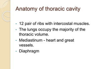
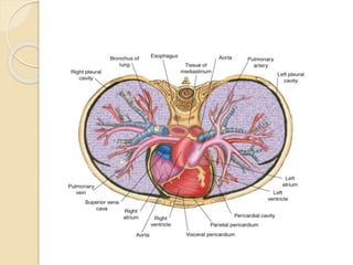
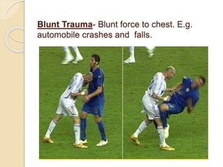
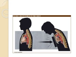
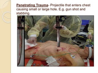
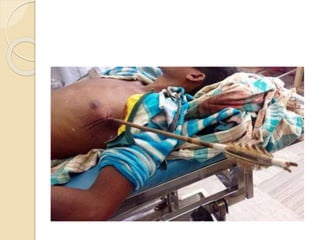
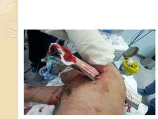
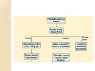
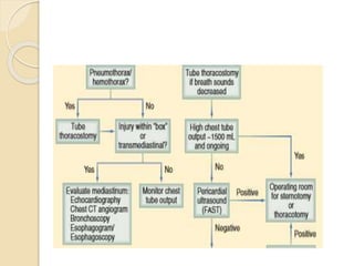
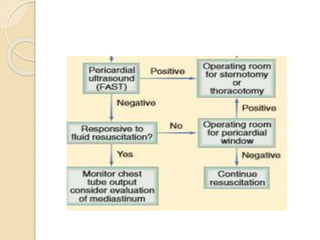
CHEST INJURY- BLUNT/ Trauma Surgery
Dear viewers,
Greetings from “Surgical Educator”
Today I have uploaded a video on CHEST INJURY- BLUNT- an important topic in trauma. Even the blunt chest trauma can turn into penetrating one because of jagged edges of the broken ribs. I haven’t talked elaborately but have included the essential minimum an undergraduate medical student should know. I have talked about pathophysiology, clinical approach, symptoms, signs, investigations, different individual types of Chest injuries and management of all the varieties of Chest injuries. My aim is, after watching this video all of you should be able to arrive at a correct working diagnosis of the type of chest injury and should also be able to institute immediate lifesaving treatment to the patients if there is a need. You can watch the video in the following links:
surgicaleducator.blogspot.com
youtube.com/c/surgicaleducator
Thank you for watching the video.
chest trauma is one of the leading cause of death in poly trauma patients. ER doctor should be aware of how to suspect and how to deal with life threatening conditions resulting from chest trauma
Mediastinal injury includes cardiac, thoracic aorta, pulmonary vessels and SVC injury. Pelvic includes pelvic fracture. GI injury includes upper and lower GI injuries. Abdominal vascular includes abdominal aorta, IVC and other named vascular injury.
These simplified slides by Dr. Sidra Arshad present an overview of the non-respiratory functions of the respiratory tract.
Learning objectives:
1. Enlist the non-respiratory functions of the respiratory tract
2. Briefly explain how these functions are carried out
3. Discuss the significance of dead space
4. Differentiate between minute ventilation and alveolar ventilation
5. Describe the cough and sneeze reflexes
Study Resources:
1. Chapter 39, Guyton and Hall Textbook of Medical Physiology, 14th edition
2. Chapter 34, Ganong’s Review of Medical Physiology, 26th edition
3. Chapter 17, Human Physiology by Lauralee Sherwood, 9th edition
4. Non-respiratory functions of the lungs https://academic.oup.com/bjaed/article/13/3/98/278874
TEST BANK for Operations Management, 14th Edition by William J. Stevenson, Ve...kevinkariuki227
TEST BANK for Operations Management, 14th Edition by William J. Stevenson, Verified Chapters 1 - 19, Complete Newest Version.pdf
Prix Galien International 2024 Forum ProgramLevi Shapiro
June 20, 2024, Prix Galien International and Jerusalem Ethics Forum in ROME. Detailed agenda including panels:
- ADVANCES IN CARDIOLOGY: A NEW PARADIGM IS COMING
- WOMEN’S HEALTH: FERTILITY PRESERVATION
- WHAT’S NEW IN THE TREATMENT OF INFECTIOUS,
ONCOLOGICAL AND INFLAMMATORY SKIN DISEASES?
- ARTIFICIAL INTELLIGENCE AND ETHICS
- GENE THERAPY
- BEYOND BORDERS: GLOBAL INITIATIVES FOR DEMOCRATIZING LIFE SCIENCE TECHNOLOGIES AND PROMOTING ACCESS TO HEALTHCARE
- ETHICAL CHALLENGES IN LIFE SCIENCES
- Prix Galien International Awards Ceremony
Tom Selleck Health: A Comprehensive Look at the Iconic Actor’s Wellness Journeygreendigital
Tom Selleck, an enduring figure in Hollywood. has captivated audiences for decades with his rugged charm, iconic moustache. and memorable roles in television and film. From his breakout role as Thomas Magnum in Magnum P.I. to his current portrayal of Frank Reagan in Blue Bloods. Selleck's career has spanned over 50 years. But beyond his professional achievements. fans have often been curious about Tom Selleck Health. especially as he has aged in the public eye.
Follow us on: Pinterest
Introduction
Many have been interested in Tom Selleck health. not only because of his enduring presence on screen but also because of the challenges. and lifestyle choices he has faced and made over the years. This article delves into the various aspects of Tom Selleck health. exploring his fitness regimen, diet, mental health. and the challenges he has encountered as he ages. We'll look at how he maintains his well-being. the health issues he has faced, and his approach to ageing .
Early Life and Career
Childhood and Athletic Beginnings
Tom Selleck was born on January 29, 1945, in Detroit, Michigan, and grew up in Sherman Oaks, California. From an early age, he was involved in sports, particularly basketball. which played a significant role in his physical development. His athletic pursuits continued into college. where he attended the University of Southern California (USC) on a basketball scholarship. This early involvement in sports laid a strong foundation for his physical health and disciplined lifestyle.
Transition to Acting
Selleck's transition from an athlete to an actor came with its physical demands. His first significant role in "Magnum P.I." required him to perform various stunts and maintain a fit appearance. This role, which he played from 1980 to 1988. necessitated a rigorous fitness routine to meet the show's demands. setting the stage for his long-term commitment to health and wellness.
Fitness Regimen
Workout Routine
Tom Selleck health and fitness regimen has evolved. adapting to his changing roles and age. During his "Magnum, P.I." days. Selleck's workouts were intense and focused on building and maintaining muscle mass. His routine included weightlifting, cardiovascular exercises. and specific training for the stunts he performed on the show.
Selleck adjusted his fitness routine as he aged to suit his body's needs. Today, his workouts focus on maintaining flexibility, strength, and cardiovascular health. He incorporates low-impact exercises such as swimming, walking, and light weightlifting. This balanced approach helps him stay fit without putting undue strain on his joints and muscles.
Importance of Flexibility and Mobility
In recent years, Selleck has emphasized the importance of flexibility and mobility in his fitness regimen. Understanding the natural decline in muscle mass and joint flexibility with age. he includes stretching and yoga in his routine. These practices help prevent injuries, improve posture, and maintain mobilit
Explore natural remedies for syphilis treatment in Singapore. Discover alternative therapies, herbal remedies, and lifestyle changes that may complement conventional treatments. Learn about holistic approaches to managing syphilis symptoms and supporting overall health.
Flu Vaccine Alert in Bangalore Karnatakaaddon Scans
As flu season approaches, health officials in Bangalore, Karnataka, are urging residents to get their flu vaccinations. The seasonal flu, while common, can lead to severe health complications, particularly for vulnerable populations such as young children, the elderly, and those with underlying health conditions.
Dr. Vidisha Kumari, a leading epidemiologist in Bangalore, emphasizes the importance of getting vaccinated. "The flu vaccine is our best defense against the influenza virus. It not only protects individuals but also helps prevent the spread of the virus in our communities," he says.
This year, the flu season is expected to coincide with a potential increase in other respiratory illnesses. The Karnataka Health Department has launched an awareness campaign highlighting the significance of flu vaccinations. They have set up multiple vaccination centers across Bangalore, making it convenient for residents to receive their shots.
To encourage widespread vaccination, the government is also collaborating with local schools, workplaces, and community centers to facilitate vaccination drives. Special attention is being given to ensuring that the vaccine is accessible to all, including marginalized communities who may have limited access to healthcare.
Residents are reminded that the flu vaccine is safe and effective. Common side effects are mild and may include soreness at the injection site, mild fever, or muscle aches. These side effects are generally short-lived and far less severe than the flu itself.
Healthcare providers are also stressing the importance of continuing COVID-19 precautions. Wearing masks, practicing good hand hygiene, and maintaining social distancing are still crucial, especially in crowded places.
Protect yourself and your loved ones by getting vaccinated. Together, we can help keep Bangalore healthy and safe this flu season. For more information on vaccination centers and schedules, residents can visit the Karnataka Health Department’s official website or follow their social media pages.
Stay informed, stay safe, and get your flu shot today!
New Directions in Targeted Therapeutic Approaches for Older Adults With Mantl...i3 Health
i3 Health is pleased to make the speaker slides from this activity available for use as a non-accredited self-study or teaching resource.
This slide deck presented by Dr. Kami Maddocks, Professor-Clinical in the Division of Hematology and
Associate Division Director for Ambulatory Operations
The Ohio State University Comprehensive Cancer Center, will provide insight into new directions in targeted therapeutic approaches for older adults with mantle cell lymphoma.
STATEMENT OF NEED
Mantle cell lymphoma (MCL) is a rare, aggressive B-cell non-Hodgkin lymphoma (NHL) accounting for 5% to 7% of all lymphomas. Its prognosis ranges from indolent disease that does not require treatment for years to very aggressive disease, which is associated with poor survival (Silkenstedt et al, 2021). Typically, MCL is diagnosed at advanced stage and in older patients who cannot tolerate intensive therapy (NCCN, 2022). Although recent advances have slightly increased remission rates, recurrence and relapse remain very common, leading to a median overall survival between 3 and 6 years (LLS, 2021). Though there are several effective options, progress is still needed towards establishing an accepted frontline approach for MCL (Castellino et al, 2022). Treatment selection and management of MCL are complicated by the heterogeneity of prognosis, advanced age and comorbidities of patients, and lack of an established standard approach for treatment, making it vital that clinicians be familiar with the latest research and advances in this area. In this activity chaired by Michael Wang, MD, Professor in the Department of Lymphoma & Myeloma at MD Anderson Cancer Center, expert faculty will discuss prognostic factors informing treatment, the promising results of recent trials in new therapeutic approaches, and the implications of treatment resistance in therapeutic selection for MCL.
Target Audience
Hematology/oncology fellows, attending faculty, and other health care professionals involved in the treatment of patients with mantle cell lymphoma (MCL).
Learning Objectives
1.) Identify clinical and biological prognostic factors that can guide treatment decision making for older adults with MCL
2.) Evaluate emerging data on targeted therapeutic approaches for treatment-naive and relapsed/refractory MCL and their applicability to older adults
3.) Assess mechanisms of resistance to targeted therapies for MCL and their implications for treatment selection
HOT NEW PRODUCT! BIG SALES FAST SHIPPING NOW FROM CHINA!! EU KU DB BK substit...GL Anaacs
Contact us if you are interested:
Email / Skype : kefaya1771@gmail.com
Threema: PXHY5PDH
New BATCH Ku !!! MUCH IN DEMAND FAST SALE EVERY BATCH HAPPY GOOD EFFECT BIG BATCH !
Contact me on Threema or skype to start big business!!
Hot-sale products:
NEW HOT EUTYLONE WHITE CRYSTAL!!
5cl-adba precursor (semi finished )
5cl-adba raw materials
ADBB precursor (semi finished )
ADBB raw materials
APVP powder
5fadb/4f-adb
Jwh018 / Jwh210
Eutylone crystal
Protonitazene (hydrochloride) CAS: 119276-01-6
Flubrotizolam CAS: 57801-95-3
Metonitazene CAS: 14680-51-4
Payment terms: Western Union,MoneyGram,Bitcoin or USDT.
Deliver Time: Usually 7-15days
Shipping method: FedEx, TNT, DHL,UPS etc.Our deliveries are 100% safe, fast, reliable and discreet.
Samples will be sent for your evaluation!If you are interested in, please contact me, let's talk details.
We specializes in exporting high quality Research chemical, medical intermediate, Pharmaceutical chemicals and so on. Products are exported to USA, Canada, France, Korea, Japan,Russia, Southeast Asia and other countries.
- Video recording of this lecture in English language: https://youtu.be/lK81BzxMqdo
- Video recording of this lecture in Arabic language: https://youtu.be/Ve4P0COk9OI
- Link to download the book free: https://nephrotube.blogspot.com/p/nephrotube-nephrology-books.html
- Link to NephroTube website: www.NephroTube.com
- Link to NephroTube social media accounts: https://nephrotube.blogspot.com/p/join-nephrotube-on-social-media.html
2. Introduction
Thoracic injuries are directly
responsible for 25% of all trauma
deaths and are a major contributory
factor to mortality in a further 25%.
Although many of these deaths occur
almost immediately, there is a
significant group of patients that may
be salvaged with early effective
management.
3. The majority (approximately 90%) of
all patients who sustain thoracic
trauma can be managed
conservatively, with no more than a
chest drain, monitoring and
analgesia.
Few patients require surgery, and an
emergency department thoracotomy
is indicated in only a very small
minority.
4. A reproducible and safe approach to
the diagnosis and management of
chest injury is taught by the ATLS
course of the American College of
Surgeons.
5. Anatomy of thoracic cavity
• 12 pair of ribs with intercostal muscles.
• The lungs occupy the majority of the
thoracic volume.
• Mediastinum - heart and great
vessels.
• Diaphragm
15. The main consequences of chest
trauma occur as a result of its
combined effects on respiratory and
haemodynamic function.
The commonest manifestation of
thoracic trauma is hypoxia
16. Mechanism of injury is important in so
far as blunt and penetrating injuries
have different pathophysiologies and
clinical courses.
Most blunt injuries are managed non-
operatively or with simple
interventions such as intubation and
ventilation and chest tube insertion.
17. Diagnosis of blunt injuries may be
more difficult and require additional
investigations such as CT scanning
(when the patient is STABLE).
In contrast, penetrating injuries are
more likely to require operation, and
complex investigations are required
infrequently.
18. • Impairments in ventilatory efficiency
Chest movement compromise due to
pain
air in pleural space
asymmetrical movement
Bleeding in pleural space
Ineffective diaphragm contraction
19. ◦ Impairments in gas exchange
Atelectasis
Pulmonary contusion
Respiratory tract disruption
20. Causes of hypoxia in chest
trauma
Haemorrhage;
Lung collapse and compression;
Ventilatory or cardiac failure;
Pulmonary contusion;
Changes in intrathoracic pressure;
and
Mediastinal displacement.
21. Profound hypovolaemia in chest
trauma due to
Great vessel damage,
Pulmonary hilar injury
Cardiac or pericardial
laceration without tamponade.
Hypovolaemia produces a low cardiac
output state, which further contributes
to the pathophysiological
consequences of chest injury.
22. Hypovolaemia produces a low cardiac
output state, which further contributes
to the pathophysiological
consequences of chest injury.
Pulmonary contusion is one of the
main factors responsible for the
increased morbidity and mortality
associated with chest trauma.
23. It is a progressive condition,
Alveolar haemorrhage and oedema
Interstitial fluid accumulation
Decreased alveolar membrane
diffusion.
25. Importantly, there is a Ventilation-
perfusion mismatch' (alveoli are
perfused, but are unavailable for gas
exchange because they are full of
blood).
This contributes significantly to the
hypoxaemia, especially in the early
stages following trauma.
26. Later, hypoxia-induced pulmonary
vasoconstriction will divert the blood
away from the non-ventilated alveoli
A loss of mechanical function of the
chest wall will also result in hypoxia
27. If the chest wall is sufficiently
disrupted, the patient may be unable
spontaneously to generate sufficient
movement of air to allow adequate
gas transfer.
28. Cardiac output may be directly
reduced by
Decreased myocardial contractility (e.g.
myocardial contusion),
Cardiac disruption (e.g. a tear in a cardiac valve),
Reduced venous filling (e.g. in cardiac
tamponade),
With changes in intrathoracic pressure (tension
pneumothorax).
29.
30. ASSESSMENT
On arrival in the emergency
department, decisions and action
need to be taken without delay.
Important information may be obtained
from the ambulance service relating to
the patient's history and mechanism of
injury.
31. The sequence of questions as follow
Mechanism of injury
Injuries found and suspected
Signs (respiratory rate, SpO2, pulse,
blood pressure)
Treatment given pre-hospital.
32. In every case, the system of a primary
survey with simultaneous resuscitation
is followed.
In the stable patient, once this has
been completed, a secondary survey
can be performed.
33. Certain wounds or bruising patterns
highlight the likelihood of underlying
injury
for example a seat-belt mark on the
chest wall may arouse suspicion of
fractured ribs, lung contusion, or
solid organ injury in the abdomen,
34. Penetrating wound medial to the
nipple or the scapula suggests
possible damage to the heart (with
potential cardiac tamponade), the
great vessels, or the hilar
structures.
However, major intrathoracic injuries
may occur without obvious external
damage
35. Additionally, some injuries point to
possible associated more serious
pathology
for example, fractures of the first
and second ribs are associated
with major vessel injury.
36. The Advanced Trauma Life Support
(ATLS) course of the American
College of Surgeons Committee on
Trauma was developed in the late
1970s,
based on the premise that appropriate
and timely care can significantly
improve the outcome for the injured
patient.
37. ATLS provides a structured approach
to the trauma patient with standard
algorithms of care
It emphasizes the “golden hour”
concept that timely, prioritized
interventions are necessary to prevent
death and disability.
38. The initial management of seriously
injured patients consists of phases
that include
Primary survey/concurrent
Resuscitation,
Secondary survey/diagnostic
evaluation,
Tertiary survey.
39. The first step in patient management is
performing the primary survey, the goal
of which is to identify and treat
conditions that constitute an
immediate threat to life.
The ATLS course refers to the primary
survey as assessment of the “ABCs”
(Airway with cervical spine protection,
Breathing, and Circulation).
40. Although the concepts within the
primary survey are presented in a
sequential fashion, in reality they are
pursued simultaneously in coordinated
team resuscitation.
Life-threatening injuries must be
identified and treated before being
distracted by the secondary survey.
41. AIRWAY
It is necessary to recognize and
address major injuries affecting the
airway during the primary survey.
Airway patency and air exchange
should be assessed by listening for air
movement at the patient’s nose,
mouth, and lung fields; inspecting the
oropharynx for foreign-body
obstruction.
42. Laryngeal injury can accompany major
thoracic trauma.
Patients who have an abnormal
voice, abnormal breathing sounds,
tachypnea, or altered mental status
require further airway evaluation
43. Endotracheal intubation is indicated in
Patients with apnea
Inability to protect the airway due to
altered mental status
Impending airway compromise due to
inhalation injury, hematoma, facial
bleeding, soft tissue swelling, or
aspiration;
Inability to maintain oxygenation.
44. Altered mental status is the most
common indication for intubation.
Options for endotracheal intubation
include nasotracheal, orotracheal, or
operative routes.
45. Patients in whom attempts at intubation
have failed or who are precluded from
intubation due to extensive facial injuries
require operative establishment
(cricothyroidotmy/tracheostomy) of an
airway.
In patients under the age of 11,
cricothyroidotomy is relatively
contraindicated due to the risk of
subglottic stenosis, and tracheostomy
should be performed.
46. Emergent tracheostomy is indicated in
patients with laryngotracheal
separation or laryngeal fractures, in
whom cricothyroidotomy may cause
further damage or result in complete
loss
47. BREATHING
Before examining the chest, the neck
should be carefully examined for
wounds,
bleeding,
tracheal deviation,
laryngeal crepitus,
jugular vein engorgement
48. If a cervical collar is already in place, it
should ideally be removed temporarily
to allow examination, Bt
Do not forget to examine the neck
49. The chest must be completely
exposed so that respiratory movement
and quality of ventilation can be
assessed.
The mechanics of breathing can be
disrupted by major airway obstruction,
haemothorax or pneumothorax, pain
or pulmonary contusion.
50. Impending hypoxia is sometimes
indicated by subtle changes in the
breathing pattern, which may become
shallow and rapid.
Visual inspection and palpation of the
chest wall may reveal
deformity,
contusion,
abrasion,
penetrating injury,
52. CIRCULATION
The pulse should be assessed for
quality, rate and regularity.
The peripheral circulation is assessed
by skin colour, temperature and
capillary return.
Venous distension in the neck may not
always be present in a patient with
cardiac tamponade who has
hypovolaemia.
53. Circulation maintained by
IV fluids (crystalloids,PCV )
External control of any visible
hemorrhage should be achieved
promptly while circulating volume is
restored.
Manual compression of open wounds
with ongoing bleeding should be done
with a single 4 × 4 gauze and a gloved
hand.
54. During the circulation section of the
primary survey, two life-threatening
injuries must be identified promptly:
(a) massive hemothorax, (b) cardiac
tamponade.
Two critical tools used to differentiate
these in trauma patient are chest
radiograph and focused abdominal
sonography for trauma (FAST)
55. A massive hemothorax is defined as
>1500 mL of blood or, in the pediatric
population, >25% of the patient’s
blood volume in the pleural space
Although it may be estimated on
chest radiograph, tube thoracostomy
is the only reliable means to quantify
the amount of hemothorax.
56. After blunt trauma, a major
hemothorax usually is due to
Multiple rib fractures with severed
intercostal arteries
Lacerated lung parenchyma
57. Cardiac tamponade occurs most
commonly after penetrating thoracic
wounds, although occasionally blunt
rupture of the heart, particularly the
atrial appendage, is seen.
Acutely, <100 mL of pericardial blood
may cause pericardial tamponade.
58. The classic Beck’s triad—
dilated neck veins,
muffled heart tones, and a
decline in arterial pressure
is usually not appreciated in the
trauma bay because of the noisy
environment and associated
hypovolemia.
59. DISABILITY
The hypoxic patient will initially be
confused. Primary head injury will also
cause an altered mental state, which
will be compounded by hypoxia or
hypercarbia.
60. Immediately life-threatening injuries to be
identified during the primary survey
Airway
Airway obstruction
Airway injury
Breathing
Tension pneumothorax
Open pneumothorax
Flail chest
62. Secondary Survey
Once the immediate threats to life
have been addressed, a thorough
history is obtained and the patient is
examined in a systematic fashion
Adjuncts to the physical examination
include
vital sign and CVP monitoring,
ECG monitoring
nasogastric tube placement,
70. Evaluation
Most thoracic injuries can be identified
with a physical examination and plain
chest radiography.
Physical examination will reveal
superficial injuries, including chest wall
defects and penetrating wounds.
Chest radiography is performed on all
significantly injured patients at risk for
thoracic injuries.
71. The chest radiograph easily identifies
the presence of a pneumothorax or
hemothorax, as well as rib and sternal
fractures.
The appearance of the mediastinum
may suggest a thoracic aortic injury.
An ultrasound of the pericardium is a
component of the FAST examination,
which may reveal pericardial blood.
72. In recent years, thoracic CT
angiography has emerged as a
valuable tool in the evaluation of blunt
thoracic trauma.
CT provides visualization of the chest
wall and hemithoraces, allowing
determination of rib fractures,
pneumothoraces and hemothoraces,
and pulmonary contusion.
73. Chest CT angiography is able to
identify transection of the aortic wall,
as well as lower grade injuries that
involve only the aortic intima.