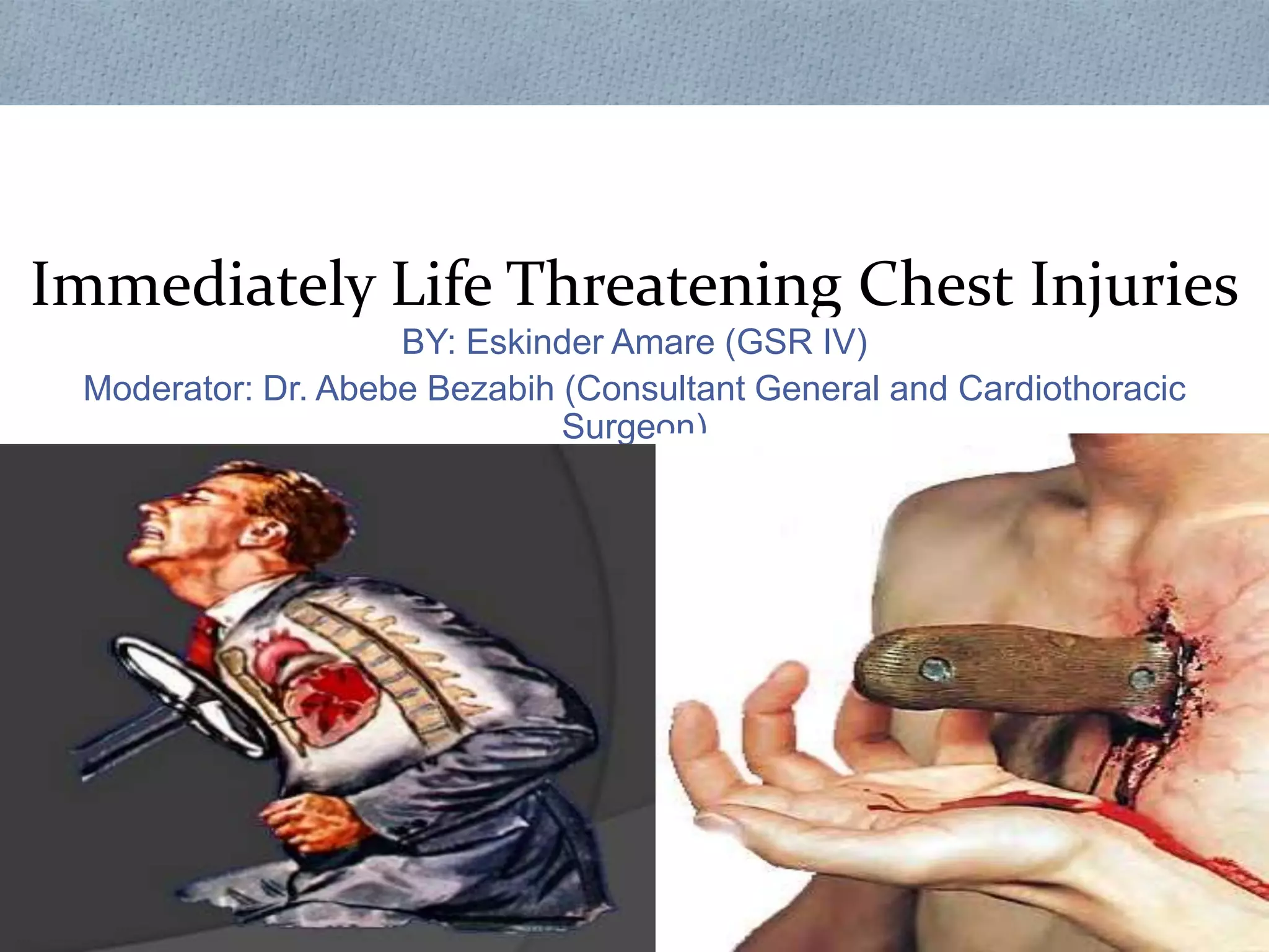
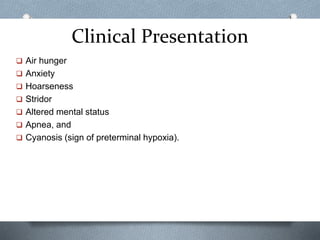
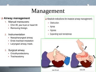
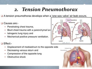
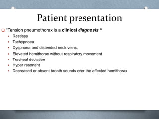
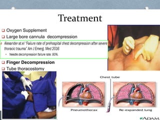
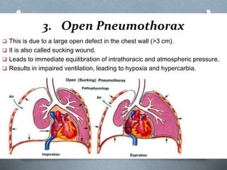
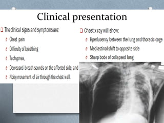
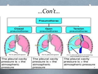
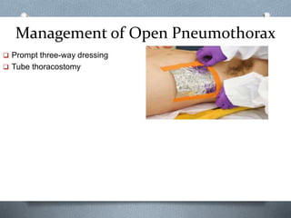
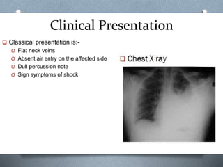
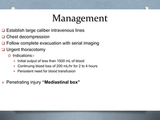
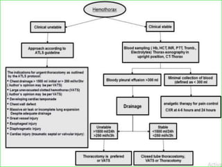
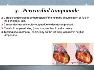
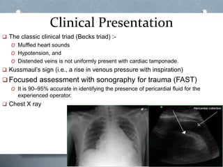
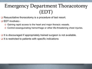
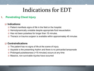
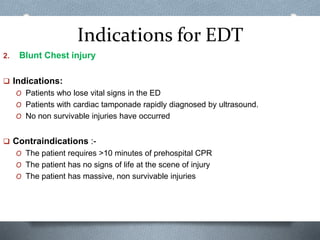
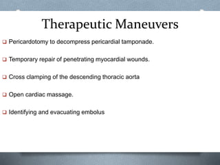
Immediately Life Threatening Chest Injuries
BY: Eskinder Amare (GSR IV)
Moderator: Dr. Abebe Bezabih (Consultant General and Cardiothoracic
Surgeon)
Outline
 Introduction
 Classification of chest injury
 Immediately life threatening conditions:-
O Airway obstruction
O Tension pneumothorax
O Open pneumothorax
O Massive haemothorax
O Pericardial tamponade
O Tracheobronchial injuries
 Emergency Department Thoracotomy (EDT)
 Summary
 References
Introduction
 Trauma is known to be the leading cause of death in the first four decades of
life.
 Chest trauma is second most common causes of traumatic deaths.
 The key to a good outcome is early physiological resuscitation followed by a
correct diagnosis.
 Operative intervention is required:-
O Less than 10% of blunt chest injuries and
O Only 15% to 30% of penetrating chest injuries
Trends of Chest injury Management in
Our Setup
…Con’t…
 The three commonest cause of death in chest injury are:
O Hypoxemia
O Hypovolemia
O Tamponade
 Management of thoracic trauma patient consists of:-
1. Primary survey with resuscitation of vital function
O A: Airway -with cervical spine control
O B: Breathing
O C: Circulation
O D: Disability
O E: Exposure
2. Detailed secondary survey, and
3. Definitive care
Classification of Chest Trauma
The ‘deadly dozen’ threats to life from chest injury.
Immediately life threatening 1. Airway obstruction
2. Tension pneumothorax
3. Open pneumothorax
4. Massive haemothorax
5. Pericardial tamponade
6. Tracheobronchial injuries
Potentially life threatening 1. Flail chest
2. Aortic injuries
3. Myocardial contusion
4. Rupture of diaphragm
5. Oesophageal injuries
6. Pulmonary contusion
1. Airway Obstruction
 Airway is the first priority in treating trauma patients.
 It can be a primary problem or the result of other injury.
 Common causes of airway obstruction are:-
O Tongue
O Avulsed teeth
O Dentures
O Secretions, and blood
O Expanding hematoma
O Airway edema
Clinical Presentation
 Air hunger
 Anxiety
 Hoarseness
 Stridor
 Altered mental status
 Apnea, and
 Cyanosis (sign of preterminal hypoxia).
Management
 Airway management
1. Manual maneuvers:
 Chin lift, jaw trust or head tilt
 Removing foreign.
2. Instrumentation
 Nasopharyngeal airway
 Endo tracheal intubation
 Laryngeal airway mask.
3. Surgical airway
 Cricothyroidectomy
 Tracheostomy
2. Tension Pneumothorax
 A tension pneumothorax develops when a ‘one way valve’ air leak occurs.
 Causes are:-
 Penetrating chest trauma,
 Blunt chest trauma with a parenchymal lung injury
 Iatrogenic lung injury and
 Mechanical positive pressure ventilation.
 Effect:-
 Displacement of mediastinum to the opposite side
 Decreasing venous return and
 Compression of the opposite lung
 Obstructive shock
Patient presentation
 “Tension pneumothorax is a clinical diagnosis “
 Restless
 Tachypnoea
 Dyspnoea and distended neck veins.
 Elevated hemithorax without respiratory movement
 Tracheal deviation
 Hyper resonant
 Decreased or absent breath sounds over the affected hemithorax.
Treatment
 Oxygen Supplement
 Large bore cannula decompression
 Finger Decompression
 Tube thoracostomy
3. Open Pneumothorax
 This is due to a large open defect in the chest wall (>3 cm).
 It is also called sucking wound.
 Leads to immediate equilibration of intrathoracic and atmospheric pressure.
 Results in impaired ventilation, leading to hypoxia and hypercarbia.
Clinical presentation
…Con’t…
Management of Open Pneumothorax
 Prompt three-way dressing
 Tube thoracostomy
Place of Conservative Management
for Pneumothorax
4. Massive Haemothorax
 Massive hemothorax results from the rapid accumulation of more than 1500
mL or one third of blood or more in the pleural cavity.
 It is most commonly caused by:-
O Penetrating chest injury
O Blunt trauma
 Intercostal and internal mammary vessels are most commonly injured
 Bleeding from parenchymal lacerations often stops on its own
 A left-sided massive hemothorax is more common than the right-sided one.
Clinical Presentation
 Classical presentation is:-
O Flat neck veins
O Absent air entry on the affected side
O Dull percussion note
O Sign symptoms of shock
Management
 Establish large caliber intravenous lines
 Chest decompression
 Follow complete evacuation with serial imaging
 Urgent thoracotomy
O Indications:-
 Initial output of less than 1500 mL of blood
 Continuing blood loss of 200 mL/hr for 2 to 4 hours
 Persistent need for blood transfusion
 Penetrating injury “Mediastinal box”
Place of Size of Chest Tube for Hemothorax
5. Pericardial tamponade
 Cardiac tamponade is compression of the heart by accumulation of fluid in
the pericardial sac.
 Causes decreased cardiac output due to decreased preload.
 Results from penetrating (commonly) or blunt cardiac injury.
 Tension pneumothorax, particularly on the left side, can mimic cardiac
tamponade.
Clinical Presentation
 The classic clinical triad (Becks triad) :-
O Muffled heart sounds
O Hypotension, and
O Distended veins is not uniformly present with cardiac tamponade.
 Kussmaul’s sign (i.e., a rise in venous pressure with inspiration)
 Focused assessment with sonography for trauma (FAST)
O It is 90–95% accurate in identifying the presence of pericardial fluid for the
experienced operator.
 Chest X ray
Management
 Administration of intravenous fluid
 Needle pericardiocentesis
 Subxiphoid window
 Open surgery
O Sternotomy or
O Left thoracotomy
6. Tracheobronchial Injuries
 Injury to the trachea or a major bronchus is an unusual but potentially fatal
condition.
 Less than 1% of all injured patients sustain intrathoracic tracheobronchial
injury.
 Majority of tracheobronchial tree injuries occur within 1 inch (2.54 cm) of the
carina.
 These injuries can be severe, and the majority of patients die at the scene.
 Can result from blunt or penetrating injury.
 Intubation can potentially cause or worsen an injury to the trachea or
proximal bronchi.
Clinical presentation
 Hemoptysis
 Cervical subcutaneous emphysema
 Tension pneumothorax, and/or cyanosis.
 Incomplete expansion of the lung after tube thoracostomy.
 Bronchoscopy:- is gold standard and confirms the diagnosis
Management
 Expectant management
 Intubation;-
O Fiber optically assisted intubation or
O Selective intubation of the unaffected bronchus.
 Endoscopy, bronchoscopically directed fibrin glue sealing
 Surgical repair:-
O Is the mainstay of management
O Indication for surgery:-
 Tracheobronchial injuries greater than 1/3 the circumference.
 Persistent air leak
Management
 Surgical repair principles:-
 Approach:-
 Collar incision:-
• Proximal half of trachea
 Right posterolateral thoracotomy:-
• Distal half of the trachea
• Right mainstem bronchus, and
• Proximal left mainstem bronchus
 left posterolateral thoracotomy.
• For distal left mainstem bronchus
Management
 Devitalized tissue is debrided
 Dissection should be limited to the area of injury
 Primary end-to-end anastomosis with 3-0 PDS suture is performed.
 Suture lines should be encircled with vascularized tissue
Emergency Department Thoracotomy
(EDT)
 Resuscitative thoracotomy is a procedure of last resort.
 EDT involves:-
O Gaining rapid access to the heart and major thoracic vessels.
O Control exsanguinating hemorrhage or other life-threatening chest injuries.
 It is discouraged if appropriately trained surgeon is not available.
 It is restricted to patients with specific indications
Indications for EDT
1. Penetrating Chest Injury
 Indications:
O Patient manifests signs of life in the field or the hospital.
O Hemodynamically unstable despite appropriate fluid resuscitation.
O Has not been pulseless for longer than 15 minutes.
O Thoracic or trauma surgeon is available within approximately 45 minutes
 Contraindications
O The patient has no signs of life at the scene of injury
O Asystole is the presenting rhythm and there is no pericardial tamponade
O Prolonged pulselessness (>15 minutes) occurs at any time
O Massive, non survivable injuries have occurred
Indications for EDT
2. Blunt Chest injury
 Indications:
O Patients who lose vital signs in the ED
O Patients with cardiac tamponade rapidly diagnosed by ultrasound.
O No non survivable injuries have occurred
 Contraindications :-
O The patient requires >10 minutes of prehospital CPR
O The patient has no signs of life at the scene of injury
O The patient has massive, non survivable injuries
Therapeutic Maneuvers
 Pericardotomy to decompress pericardial tamponade.
 Temporary repair of penetrating myocardial wounds.
 Cross clamping of the descending thoracic aorta
 Open cardiac massage.
 Identifying and evacuating embolus
Summary
 Chest injury contributes for 75% of trauma related deaths.
 The three common cause of death in such patients are hypoxia, hypovolemia
and cardiac tamponade.
 Patient with thoracic trauma should be approached according to ATLS
principles.
 primary survey should address the six lethal threats of chest injury.
 Only 10-15% of patients with thoracic injury require surgical intervention.
References
1. Advanced Trauma Life Support® Student Course Manual, 10th edition
2. Bailey and love’s short practice of surgery, 27th edition.
3. Mattox Trauma , 9th edition
4. Schwartz’s Principles of Surgery 11th edition.
5. Yamamoto L. at.el “Thoracic trauma: the deadly dozen” Crit Care Nurs Q. 2005 Jan-
Mar;28(1):22-40.
6. John F. Resuscitative thoracotomy: Up To Date : Jan 2021.
7. Mahoozi HR, Volmerig J, Hecker E (2016) Modern Management of Traumatic
Hemothorax. J Trauma Treat 5: 326.
8. A. Adem at.el “Chest injuries in Tikur Anbessa Hospital, Addis Ababa: a
three year experience” East and CentralAfrican Journal of Surgery Vol. 6, No. 1
9. Walker SP, Barratt SL, Thompson J, Maskell NA. Conservative Management in
Traumatic Pneumothoraces: An Observational Study. Chest. 2018
Apr;153(4):946-953.
References
10. Kaserer A, Stein P, Simmen HP, Spahn DR, Neuhaus V. Failure rate of
prehospital chest decompression after severe thoracic trauma. Am J Emerg
Med. 2017 Mar;35(3):469-474.
11. Gilbert RW, Fontebasso AM, Park L, Tran A, Lampron J. The management of
occult hemothorax in adults with thoracic trauma: A systematic review and
meta-analysis. J Trauma Acute Care Surg. 2020 Dec;89(6):1225-1232.
Thank You!

Chest 12. Chest Trauma.pptx

  • 1.
    Immediately Life ThreateningChest Injuries BY: Eskinder Amare (GSR IV) Moderator: Dr. Abebe Bezabih (Consultant General and Cardiothoracic Surgeon)
  • 2.
    Outline  Introduction  Classificationof chest injury  Immediately life threatening conditions:- O Airway obstruction O Tension pneumothorax O Open pneumothorax O Massive haemothorax O Pericardial tamponade O Tracheobronchial injuries  Emergency Department Thoracotomy (EDT)  Summary  References
  • 3.
    Introduction  Trauma isknown to be the leading cause of death in the first four decades of life.  Chest trauma is second most common causes of traumatic deaths.  The key to a good outcome is early physiological resuscitation followed by a correct diagnosis.  Operative intervention is required:- O Less than 10% of blunt chest injuries and O Only 15% to 30% of penetrating chest injuries
  • 4.
    Trends of Chestinjury Management in Our Setup
  • 5.
    …Con’t…  The threecommonest cause of death in chest injury are: O Hypoxemia O Hypovolemia O Tamponade  Management of thoracic trauma patient consists of:- 1. Primary survey with resuscitation of vital function O A: Airway -with cervical spine control O B: Breathing O C: Circulation O D: Disability O E: Exposure 2. Detailed secondary survey, and 3. Definitive care
  • 6.
  • 7.
    Classification of ChestTrauma The ‘deadly dozen’ threats to life from chest injury. Immediately life threatening 1. Airway obstruction 2. Tension pneumothorax 3. Open pneumothorax 4. Massive haemothorax 5. Pericardial tamponade 6. Tracheobronchial injuries Potentially life threatening 1. Flail chest 2. Aortic injuries 3. Myocardial contusion 4. Rupture of diaphragm 5. Oesophageal injuries 6. Pulmonary contusion
  • 8.
    1. Airway Obstruction Airway is the first priority in treating trauma patients.  It can be a primary problem or the result of other injury.  Common causes of airway obstruction are:- O Tongue O Avulsed teeth O Dentures O Secretions, and blood O Expanding hematoma O Airway edema
  • 9.
    Clinical Presentation  Airhunger  Anxiety  Hoarseness  Stridor  Altered mental status  Apnea, and  Cyanosis (sign of preterminal hypoxia).
  • 10.
    Management  Airway management 1.Manual maneuvers:  Chin lift, jaw trust or head tilt  Removing foreign. 2. Instrumentation  Nasopharyngeal airway  Endo tracheal intubation  Laryngeal airway mask. 3. Surgical airway  Cricothyroidectomy  Tracheostomy
  • 11.
    2. Tension Pneumothorax A tension pneumothorax develops when a ‘one way valve’ air leak occurs.  Causes are:-  Penetrating chest trauma,  Blunt chest trauma with a parenchymal lung injury  Iatrogenic lung injury and  Mechanical positive pressure ventilation.  Effect:-  Displacement of mediastinum to the opposite side  Decreasing venous return and  Compression of the opposite lung  Obstructive shock
  • 12.
    Patient presentation  “Tensionpneumothorax is a clinical diagnosis “  Restless  Tachypnoea  Dyspnoea and distended neck veins.  Elevated hemithorax without respiratory movement  Tracheal deviation  Hyper resonant  Decreased or absent breath sounds over the affected hemithorax.
  • 13.
    Treatment  Oxygen Supplement Large bore cannula decompression  Finger Decompression  Tube thoracostomy
  • 14.
    3. Open Pneumothorax This is due to a large open defect in the chest wall (>3 cm).  It is also called sucking wound.  Leads to immediate equilibration of intrathoracic and atmospheric pressure.  Results in impaired ventilation, leading to hypoxia and hypercarbia.
  • 15.
  • 16.
  • 17.
    Management of OpenPneumothorax  Prompt three-way dressing  Tube thoracostomy
  • 18.
    Place of ConservativeManagement for Pneumothorax
  • 19.
    4. Massive Haemothorax Massive hemothorax results from the rapid accumulation of more than 1500 mL or one third of blood or more in the pleural cavity.  It is most commonly caused by:- O Penetrating chest injury O Blunt trauma  Intercostal and internal mammary vessels are most commonly injured  Bleeding from parenchymal lacerations often stops on its own  A left-sided massive hemothorax is more common than the right-sided one.
  • 20.
    Clinical Presentation  Classicalpresentation is:- O Flat neck veins O Absent air entry on the affected side O Dull percussion note O Sign symptoms of shock
  • 21.
    Management  Establish largecaliber intravenous lines  Chest decompression  Follow complete evacuation with serial imaging  Urgent thoracotomy O Indications:-  Initial output of less than 1500 mL of blood  Continuing blood loss of 200 mL/hr for 2 to 4 hours  Persistent need for blood transfusion  Penetrating injury “Mediastinal box”
  • 24.
    Place of Sizeof Chest Tube for Hemothorax
  • 25.
    5. Pericardial tamponade Cardiac tamponade is compression of the heart by accumulation of fluid in the pericardial sac.  Causes decreased cardiac output due to decreased preload.  Results from penetrating (commonly) or blunt cardiac injury.  Tension pneumothorax, particularly on the left side, can mimic cardiac tamponade.
  • 26.
    Clinical Presentation  Theclassic clinical triad (Becks triad) :- O Muffled heart sounds O Hypotension, and O Distended veins is not uniformly present with cardiac tamponade.  Kussmaul’s sign (i.e., a rise in venous pressure with inspiration)  Focused assessment with sonography for trauma (FAST) O It is 90–95% accurate in identifying the presence of pericardial fluid for the experienced operator.  Chest X ray
  • 27.
    Management  Administration ofintravenous fluid  Needle pericardiocentesis  Subxiphoid window  Open surgery O Sternotomy or O Left thoracotomy
  • 28.
    6. Tracheobronchial Injuries Injury to the trachea or a major bronchus is an unusual but potentially fatal condition.  Less than 1% of all injured patients sustain intrathoracic tracheobronchial injury.  Majority of tracheobronchial tree injuries occur within 1 inch (2.54 cm) of the carina.  These injuries can be severe, and the majority of patients die at the scene.  Can result from blunt or penetrating injury.  Intubation can potentially cause or worsen an injury to the trachea or proximal bronchi.
  • 29.
    Clinical presentation  Hemoptysis Cervical subcutaneous emphysema  Tension pneumothorax, and/or cyanosis.  Incomplete expansion of the lung after tube thoracostomy.  Bronchoscopy:- is gold standard and confirms the diagnosis
  • 30.
    Management  Expectant management Intubation;- O Fiber optically assisted intubation or O Selective intubation of the unaffected bronchus.  Endoscopy, bronchoscopically directed fibrin glue sealing  Surgical repair:- O Is the mainstay of management O Indication for surgery:-  Tracheobronchial injuries greater than 1/3 the circumference.  Persistent air leak
  • 31.
    Management  Surgical repairprinciples:-  Approach:-  Collar incision:- • Proximal half of trachea  Right posterolateral thoracotomy:- • Distal half of the trachea • Right mainstem bronchus, and • Proximal left mainstem bronchus  left posterolateral thoracotomy. • For distal left mainstem bronchus
  • 32.
    Management  Devitalized tissueis debrided  Dissection should be limited to the area of injury  Primary end-to-end anastomosis with 3-0 PDS suture is performed.  Suture lines should be encircled with vascularized tissue
  • 33.
    Emergency Department Thoracotomy (EDT) Resuscitative thoracotomy is a procedure of last resort.  EDT involves:- O Gaining rapid access to the heart and major thoracic vessels. O Control exsanguinating hemorrhage or other life-threatening chest injuries.  It is discouraged if appropriately trained surgeon is not available.  It is restricted to patients with specific indications
  • 34.
    Indications for EDT 1.Penetrating Chest Injury  Indications: O Patient manifests signs of life in the field or the hospital. O Hemodynamically unstable despite appropriate fluid resuscitation. O Has not been pulseless for longer than 15 minutes. O Thoracic or trauma surgeon is available within approximately 45 minutes  Contraindications O The patient has no signs of life at the scene of injury O Asystole is the presenting rhythm and there is no pericardial tamponade O Prolonged pulselessness (>15 minutes) occurs at any time O Massive, non survivable injuries have occurred
  • 35.
    Indications for EDT 2.Blunt Chest injury  Indications: O Patients who lose vital signs in the ED O Patients with cardiac tamponade rapidly diagnosed by ultrasound. O No non survivable injuries have occurred  Contraindications :- O The patient requires >10 minutes of prehospital CPR O The patient has no signs of life at the scene of injury O The patient has massive, non survivable injuries
  • 36.
    Therapeutic Maneuvers  Pericardotomyto decompress pericardial tamponade.  Temporary repair of penetrating myocardial wounds.  Cross clamping of the descending thoracic aorta  Open cardiac massage.  Identifying and evacuating embolus
  • 37.
    Summary  Chest injurycontributes for 75% of trauma related deaths.  The three common cause of death in such patients are hypoxia, hypovolemia and cardiac tamponade.  Patient with thoracic trauma should be approached according to ATLS principles.  primary survey should address the six lethal threats of chest injury.  Only 10-15% of patients with thoracic injury require surgical intervention.
  • 38.
    References 1. Advanced TraumaLife Support® Student Course Manual, 10th edition 2. Bailey and love’s short practice of surgery, 27th edition. 3. Mattox Trauma , 9th edition 4. Schwartz’s Principles of Surgery 11th edition. 5. Yamamoto L. at.el “Thoracic trauma: the deadly dozen” Crit Care Nurs Q. 2005 Jan- Mar;28(1):22-40. 6. John F. Resuscitative thoracotomy: Up To Date : Jan 2021. 7. Mahoozi HR, Volmerig J, Hecker E (2016) Modern Management of Traumatic Hemothorax. J Trauma Treat 5: 326. 8. A. Adem at.el “Chest injuries in Tikur Anbessa Hospital, Addis Ababa: a three year experience” East and CentralAfrican Journal of Surgery Vol. 6, No. 1 9. Walker SP, Barratt SL, Thompson J, Maskell NA. Conservative Management in Traumatic Pneumothoraces: An Observational Study. Chest. 2018 Apr;153(4):946-953.
  • 39.
    References 10. Kaserer A,Stein P, Simmen HP, Spahn DR, Neuhaus V. Failure rate of prehospital chest decompression after severe thoracic trauma. Am J Emerg Med. 2017 Mar;35(3):469-474. 11. Gilbert RW, Fontebasso AM, Park L, Tran A, Lampron J. The management of occult hemothorax in adults with thoracic trauma: A systematic review and meta-analysis. J Trauma Acute Care Surg. 2020 Dec;89(6):1225-1232.
  • 40.

Editor's Notes

  • #11 Cricothyrotomy is most commonly performed when a "can't intubate, can't oxygenate" (CICO) situation.
  • #13 Patients who are spontaneously breathing often manifest extreme tachypnea and air hunger, whereas patients who are mechanically ventilated manifest hemodynamic collapse.
  • #15 Because air tends to follow the path of least resistance, when the opening in the chest wall is approximately two-thirds the diameter of the trachea
  • #22 Do not perform thoracotomy unless a surgeon, qualified by training and experience,is present.
  • #26 Accumulation of a relatively small amount of blood into the non­distensible pericardial sac can produce compression of the heart and obstruction of the venous return, leading to decreased filling of the cardiac chambers during diastole
  • #27 Concomitant hemothorax may account for both false positive and false negative exams. Remember that tamponade can develop at any time during the resuscitation phase, and repeat FAST exams may be necessary.
  • #28 Because complications are common with blind insertion techniques, pericardiocentesis should represent a lifesaving measure of last resort in a setting where no qualified surgeon is available to perform a thoracotomy or sternotomy. Ultrasound guidance can facilitate accurate insertion of the large, over-the-needle catheter into the pericardial space.
  • #29 Those who reach the hospital alive have a high mortality rate from associated injuries, inadequate airway, or development of a tension pneumothorax or tension pneumopericardium.
  • #30 Cervical tracheal injuries are often obvious on physical examination.
  • #33 Tracheal repair is accomplished with interrupted absorbable sutures.