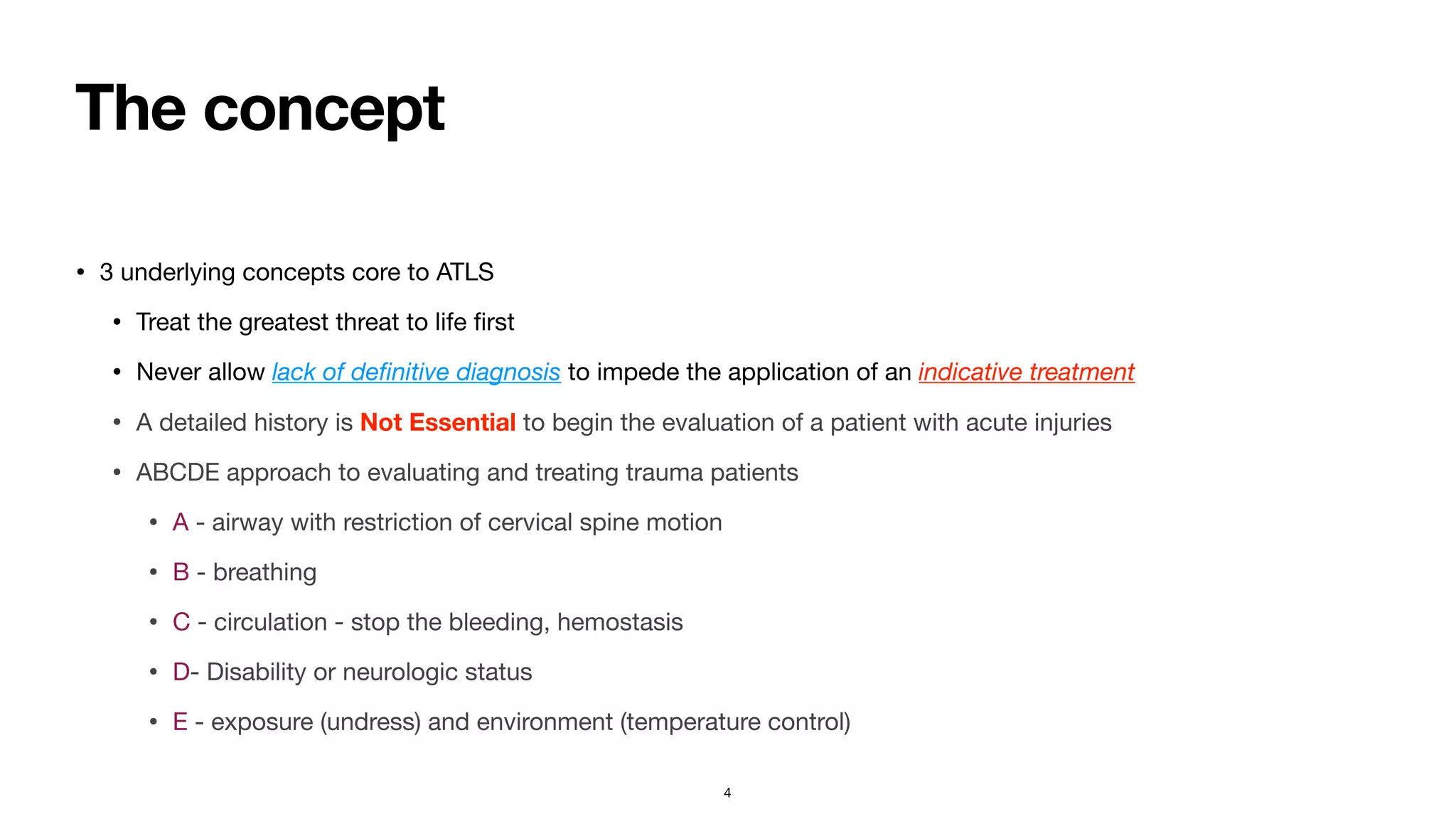
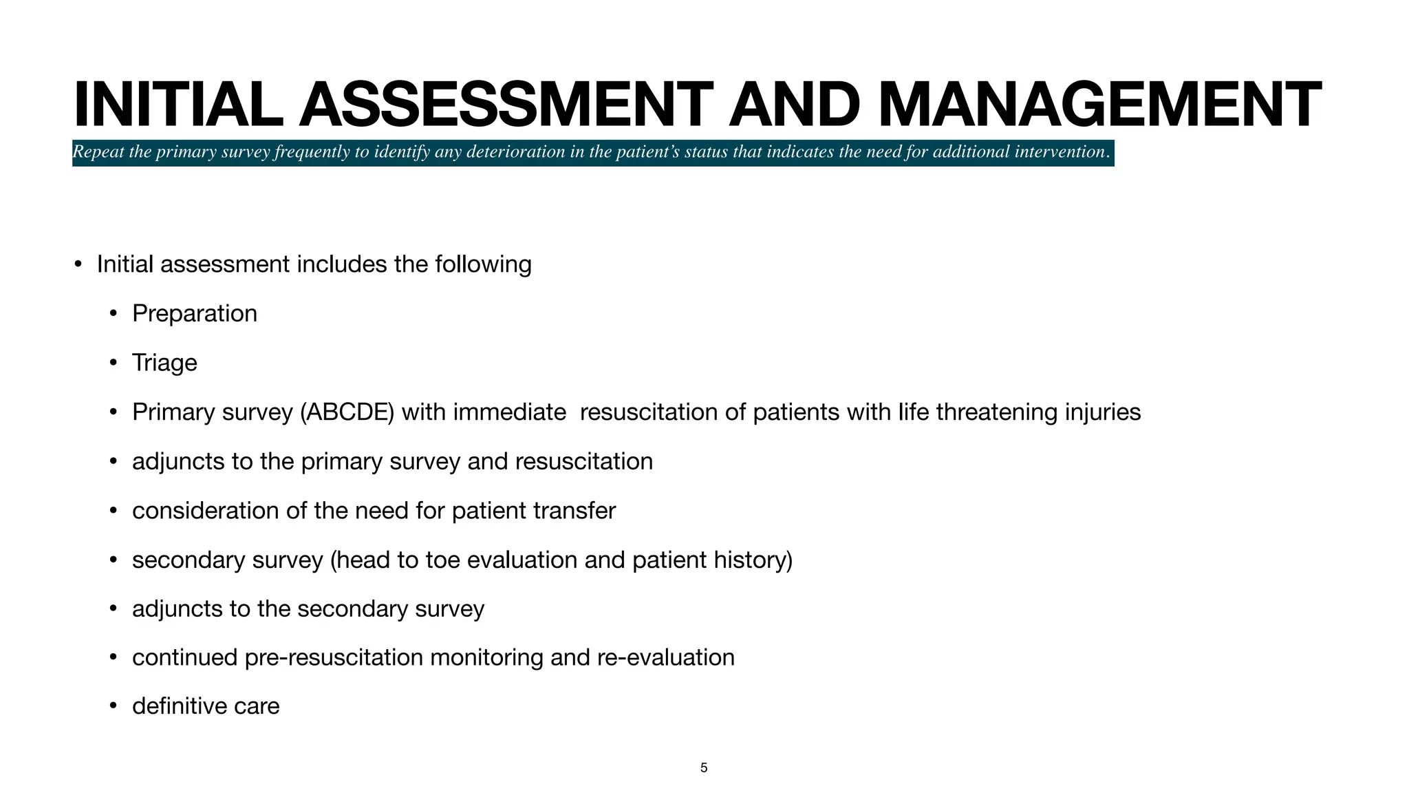
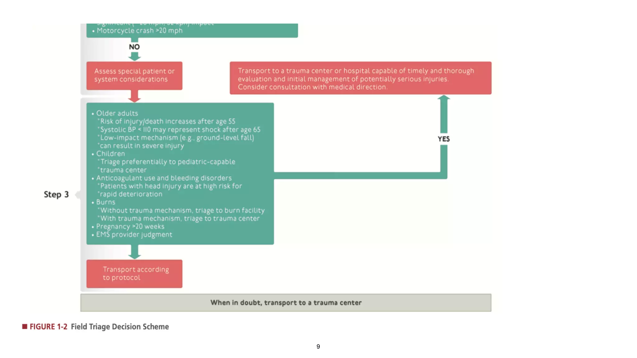
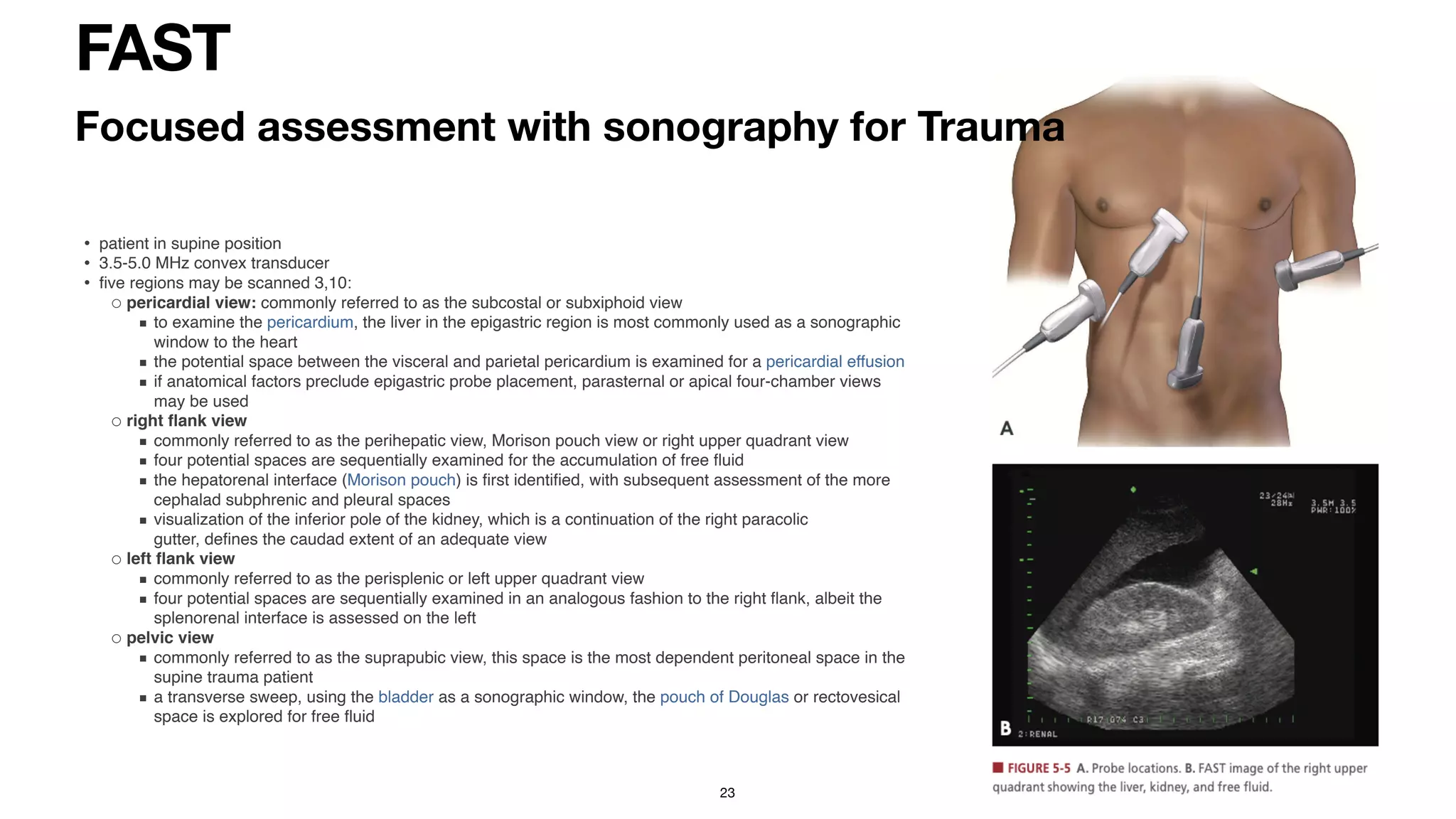
The document provides information on the Advanced Trauma Life Support (ATLS) approach to evaluating and managing trauma patients. It discusses the history and concepts of ATLS, including focusing first on treating life-threatening injuries in the order of airway, breathing, circulation, disability, and exposure (ABCDE). The summary describes the primary and secondary surveys in ATLS for initial assessment and management of trauma patients. It also highlights key components such as hemorrhage control, use of the FAST exam, and damage control resuscitation principles.
INITIAL ASSESSMENT OF TRAUMA PATIENTS....(INSPIRED FROM CTLS AND ATLS GUIDELI...Prerna Biswal
THIS PRESENTATION WAS MADE AT IMA HOUSE IN BHUBANESWAR,ODISHA, BY DR.NIBEDITA PANI,HOD ,DEPT. OF ANAESTHESIOLOGY AND DR.PRERNA BISWAL,PG,ANAESTHESIOLOGY,SCBMCH,CUTTACK,
INITIAL ASSESSMENT OF TRAUMA PATIENTS....(INSPIRED FROM CTLS AND ATLS GUIDELI...Prerna Biswal
THIS PRESENTATION WAS MADE AT IMA HOUSE IN BHUBANESWAR,ODISHA, BY DR.NIBEDITA PANI,HOD ,DEPT. OF ANAESTHESIOLOGY AND DR.PRERNA BISWAL,PG,ANAESTHESIOLOGY,SCBMCH,CUTTACK,
This is a presentation which contains basics of polytrauma management,ATLS, triage, critical decision making skills, application of Glasgow coma scale and complications of different management strategies, if not applied properly.
INITIAL ASSESSMENT AND INTENSIVE CARE IN TRAUMA.pptxMahima Shanker
CONTENTS-
INTRODUCTION
TRIMODAL DISTRIBUTION
GOLDEN HOUR
PREPARATION
TRIAGE
BASIC LIFE SUPPORT
A B C D E
SECONDARY SURVEY
DIAGNOSIS OF MAXILLOFACIAL INJURIES
performing a successful triage at the hospital level. triaging for infants, children, and adults.
nevertheless, the triage area must be well secured. the area must be signed. babies less than one-month-old must be seen immediately by a physician without delay in a queue. triaging must be carried out by an adequately trained caregiver.
2018 ATLS PROTOCOL
FOCUSING ON THE PRIMORDIAL MANAGEMENT OF PT IN THE APPROACH OF IMPROVING TRAUMA PT MANAGEMENT AND REDUCING MORTALITY OF TRAUMA PT AT OUR RESPECTIVE HEALTH FACILITIES AS DOCTORS AND CLINICIANS WORKING IN THE EMERGENCY DEPARTMENT.
GIVEN THE NECESSARY EQUIPMENT AND FAVOURABLE AMBIENT WORKING ENVIRONMENT WE SHOULD BE ABLE TO OFFER OUR HUMANITY RACE QUALITY SERVICES BEARING IN MIND THAT LIFE COME FIRST AND ALL THE OTHER ATTRIBUTES IN LIFE FOLLOWS
Struggling with intense fears that disrupt your life? At Renew Life Hypnosis, we offer specialized hypnosis to overcome fear. Phobias are exaggerated fears, often stemming from past traumas or learned behaviors. Hypnotherapy addresses these deep-seated fears by accessing the subconscious mind, helping you change your reactions to phobic triggers. Our expert therapists guide you into a state of deep relaxation, allowing you to transform your responses and reduce anxiety. Experience increased confidence and freedom from phobias with our personalized approach. Ready to live a fear-free life? Visit us at Renew Life Hypnosis..
We understand the unique challenges pickleball players face and are committed to helping you stay healthy and active. In this presentation, we’ll explore the three most common pickleball injuries and provide strategies for prevention and treatment.
India Clinical Trials Market: Industry Size and Growth Trends [2030] Analyzed...Kumar Satyam
According to TechSci Research report, "India Clinical Trials Market- By Region, Competition, Forecast & Opportunities, 2030F," the India Clinical Trials Market was valued at USD 2.05 billion in 2024 and is projected to grow at a compound annual growth rate (CAGR) of 8.64% through 2030. The market is driven by a variety of factors, making India an attractive destination for pharmaceutical companies and researchers. India's vast and diverse patient population, cost-effective operational environment, and a large pool of skilled medical professionals contribute significantly to the market's growth. Additionally, increasing government support in streamlining regulations and the growing prevalence of lifestyle diseases further propel the clinical trials market.
Growing Prevalence of Lifestyle Diseases
The rising incidence of lifestyle diseases such as diabetes, cardiovascular diseases, and cancer is a major trend driving the clinical trials market in India. These conditions necessitate the development and testing of new treatment methods, creating a robust demand for clinical trials. The increasing burden of these diseases highlights the need for innovative therapies and underscores the importance of India as a key player in global clinical research.
One of the most developed cities of India, the city of Chennai is the capital of Tamilnadu and many people from different parts of India come here to earn their bread and butter. Being a metropolitan, the city is filled with towering building and beaches but the sad part as with almost every Indian city
Navigating Challenges: Mental Health, Legislation, and the Prison System in B...Guillermo Rivera
This conference will delve into the intricate intersections between mental health, legal frameworks, and the prison system in Bolivia. It aims to provide a comprehensive overview of the current challenges faced by mental health professionals working within the legislative and correctional landscapes. Topics of discussion will include the prevalence and impact of mental health issues among the incarcerated population, the effectiveness of existing mental health policies and legislation, and potential reforms to enhance the mental health support system within prisons.
Navigating Challenges: Mental Health, Legislation, and the Prison System in B...
Approach to a trauma patient - Advanced Trauma Life Support
1. DR. PARTHA SARATHI GHOSH
MBBS, MD Anaesthesiology
Department of Critical Care Medicine
Manipal Hospital White
fi
eld
APPROACH TO A TRAUMA
PATIENT
1
2. ATLS
• Advanced Trauma Life Support (ATLS)
• Developed in 1976, following a plane crash in which Dr. James K Styner crashed in Nebraska - wife killed instantly, 3 children
sustained critical injuries. Initially triaged in the
fi
eld,
fl
agged down a car to transport to nearest hospital, which was closed.
Upon opening, facilities inadequate and inappropriate .
• Creators of ATLS had seen how well coordinated e
ff
orts of trained providers to revive trauma patients in the battle
fi
elds,
improved survival of injured
• 1973 Emergency Medical Services (EMS) act - established guidelines and funding for regional EMS development over the next
25 years
• Wartime experience showed advantage of rapid evacuation and early de
fi
nitive treatment of casualties
• became apparent - how crucial it was to coordinate
fi
eld treatment and transportation of the injured to a trauma care facility
• Implementation of such systems, lead to decrease in “preventable death”
• The ATLS course was conducted nationally for the
fi
rst time under the auspices of the American College of Surgeons in
January 1980. International promulgation of the course began in 1980
• The text for the course is revised every 4th year.
• India (Association for Trauma Care of India)
2
4. The concept
• 3 underlying concepts core to ATLS
• Treat the greatest threat to life
fi
rst
• Never allow lack of de
fi
nitive diagnosis to impede the application of an indicative treatment
• A detailed history is Not Essential to begin the evaluation of a patient with acute injuries
• ABCDE approach to evaluating and treating trauma patients
• A - airway with restriction of cervical spine motion
• B - breathing
• C - circulation - stop the bleeding, hemostasis
• D- Disability or neurologic status
• E - exposure (undress) and environment (temperature control)
4
5. INITIAL ASSESSMENT AND MANAGEMENT
Repeat the primary survey frequently to identify any deterioration in the patient’s status that indicates the need for additional intervention.
• Initial assessment includes the following
• Preparation
• Triage
• Primary survey (ABCDE) with immediate resuscitation of patients with life threatening injuries
• adjuncts to the primary survey and resuscitation
• consideration of the need for patient transfer
• secondary survey (head to toe evaluation and patient history)
• adjuncts to the secondary survey
• continued pre-resuscitation monitoring and re-evaluation
• de
fi
nitive care
5
6. Preparation
• Preparation for trauma patient occurs in two di
ff
erent clinical settings
• In the
fi
eld
• in the hospital
• In the prehospital phase, events are coordinated from the
fi
eld to the clinicians at the
receiving hospital.
• coordination with prehospital agencies and personnel can greatly expedite
treatment in the
fi
eld.
• During the hospital phase, preparations made to facilitate rapid trauma patient
resuscitation
6
7. Field Triage Decision Scheme
• Helps prehospital providers to minimise scene time.
• Emphasis placed on obtaining and reporting information needed for triage at the hospital,
including information like
• time of injury
• events related to the injury - mechanism suggests the degree of injury as well as speci
fi
c
injuries the patient needs evaluated and treated
• patient history
• Periodic multidisciplinary review of patient care through a quality improvement process is
an essential component of each hospital’s trauma program.
•
7
10. Hospital Phase
• Advance planning for arrival of trauma patient is essential.
• Team leader to ensure smooth handover of the patient from the Pre-hospital
team to those at the receiving centre for trauma.
• Critical aspects of hospital preparation include the following:
• A resuscitation area is available for trauma patients.
• Properly functioning airway equipment (e.g., laryngoscopes and endotracheal tubes) is organized, tested, and
strategically placed to be easily accessible.
• Warmed intravenous crystalloid solutions are immediately available for infusion, as are appropriate monitoring devices.
• A protocol to summon additional medical assistance is in place, as well as a means to ensure prompt responses by
laboratory and radiology personnel.
• Transfer agreements with veri
fi
ed trauma centers are established and operational.
• Due to concerns about communicable diseases, CDC recommends use of standard protection equipment
when coming into contact with bodily
fl
uids.
10
11. Triage
• Involves the sorting of patients based on the resources required for treatment and resources
readily available.
• Order of treatment based on ABC (airway with cervical spine protection, breathing and
circulation with haemorrhage control)
• Other factors include - severity of injury, ability to survive, available resources
• Sort out the patient in the
fi
eld to determine appropriate receiving medical facility.
• Trauma team activation for severely injured
• pre-hospital personnel and their directors should ensure that appropriate patients arrive at
the correct centres
• Prehospital trauma scoring is thus a useful tool to assist in the triaging of patients.
11
12. Prehospital Trauma Scoring
• Correct triaging essential for the e
ff
ective functioning of regional trauma systems
• to prevent over or under triage
• Commonly used scoring systems
• Glasgow Coma Score - assesses level of consciousness, motor response contributes the greatest to the discriminatory power of the score
• ABC score
• simplest
• penetrating trauma mechanism, SBP<90, HR>120, positive FAST
• Trauma Score (TS) - gcs, respiratory rate, resp e
ff
ort, SBP, capillary re
fi
ll
• Revised Trauma Score - 3 variables - GCS, SBP, RR
• Disadvantage - under triaging
• Pediatric Trauma Score (PTS)
• RTS may not apply to pediatric populations
• 6 measures - child’s weight, SBP, level of consciousness, presence of fracture, presence of open wound, state of the airway
• Geriatric Trauma Outcome Score (GTOS)
• Age, ISS (injury severity score), 24 hour transfusion requirement
12
13. Primary Survey
with simultaneous resuscitation
• Clinicians can quickly assess A, B, C, and D in a trauma patient (10-second assessment) by
identifying themselves, asking the patient for his or her name, and asking what happened.
• An appropriate response suggests that there is no major airway compromise (i.e., ability to speak clearly),
breathing is not severely compromised (i.e., ability to generate air movement to permit speech), and the level
of consciousness is not markedly decreased (i.e., alert enough to describe what happened).
• Failure to respond to these questions suggests abnormalities in A, B, C, or D that warrant urgent assessment
and management.
•
13
14. with restriction of cervical spine motion
• Assess airway patency
• inspect for foreign bodies,
• facial or mandibular or tracheal/laryngeal
fractures
• suctioning to clear accumulated blood,
secretions
• begin measures to establish airway latency
while restricting cervical spine motion
• Quickest way to assess - if patient is able
to communicate verbally, most likely airway
is secure - but assess repeatedly
• if GCS<8 or lower better to secure airway
Airway maintenance
14
Cervical spine motion restriction technique. When the cervical collar is
removed, a member of the trauma team manually stabilizes the patient’s head
and neck.
15. • Jaw thrust or chin lift manoeuvres
initially may often be su
ffi
cient as an
intervention
• if unconscious and no gag re
fl
ex,
oropharyngeal airway maybe helpful
but temporarily
• If in doubt, ESTABLISH a de
fi
nitive
airway
• Should be knowledgeable about the
di
ff
erences in adult and pediatric
airways
• Nonpurposeful motor movements,
suggest need for de
fi
nitive airway
• While assessing and managing a
patient’s airway, take great care to
prevent excessive movement of the
cervical spine.
• Based on the mechanism of trauma,
assume that a spinal injury exists
• The spine must be protected from
excessive mobility to prevent
development of or progression of a
de
fi
cit
•
15
17. MILS (manual inline stabilisation) of cervical spine
• ILS is performed by an assistant during airway management to maintain a neutral position and
prevent inadvertent movement of the head and neck, by either:
•crouching beside the intubator with hands placed on the patient’s mastoid processes or cradling
the occiput
•standing beside the patient in front of the intubator with hands placed on the sides of the patient’s
head and forearms resting on the patient’s chest
•traction must not be applied
•note that there is no universal definition of neutral position
MILS is replaced by a cervical collar, lateral blocks/ sand bags, and head and chin straps once the
airway is secure
•hard collars should not be used during airway management
•nearly 2/3 of patients on a hardboard with collar, straps and sandbags have grade 3 or 4 airways
•hard collars also limit mouth opening
•while MILS worsens laryngoscopic view 45% of the time, a lower proportion (22%) have grade 3
airways
•MILS decreases cervical spine movements more effectively that collars during airway
management, though it is unclear if that translates into injury at the sire of movement
•56% of patients improve their Cormack-Lehane grade when their hard collar is switched to MILS
17
18. Breathing and Ventilation
• Adequate gas exchange is key to maximise oxygenation and carbon dioxide elimination
• Expose patient neck and chest
• assess Jugular venous distension
• position of the trachea
• chest wall excursion
• Auscultate to ensure air entry in the lungs
• Visual inspection and palpation can detect injuries to the chest wall, which compromise ventilation
• Percussion although helpful, may not be possible in the noisy environment around resuscitation
area
18
19. Injuries impairing Breathing & Ventilation
• During primary survey
• Tension pneumothorax
• Massive hemothorax
• Open pneumothorax
• Tracheal or bronchial injuries
• During secondary survey
• Simple pneumothorax,
• simple hemothorax,
• fractured ribs,
•
fl
ail chest, and
• pulmonary contusion
• can compromise ventilation to a lesser degree and are usually identi
fi
ed during the secondary survey
• A simple pneumothorax can be converted to a tension pneumothorax when a patient is intubated and
positive pressure ventilation is provided before decompressing the pneumothorax with a chest tube.
•
19
20. C: Circulation with Hemorrhage control
Blood volume, Cardiac Output, Hemorrhage
• Hemorrhage is the predominant cause of preventable deaths after injury
• Crucial steps
• Identifying
• quickly controlling hemorrhage
• initiating resuscitation
• Once tension pneumothorax has been excluded as a cause of shock, consider that hypotension following injury is due to
blood loss until proven otherwise
• Elements to observe
• Level of consciousness
• Skin perfusion
• Pulse
20
21. Hemorrhagic Shock
• abnormality of the circulatory system resulting in inadequate organ perfusion and tissue
oxygenation
• Early circulatory responses to shock
• progressive vasoconstriction of peripheries to preserve blood
fl
ow to vital organs
• Tachycardia to maintain cardiac output in view of volume depletion
• The most e
ff
ective method of restoring adequate cardiac output, end-organ perfusion,
and tissue oxygenation is to restore venous return to normal by locating and stopping
the source of bleeding.
• Volume repletion will allow recovery from the shock state only when the bleeding has
stopped.
21
22. Bleeding (Hemorrhage)
External or Internal
• Identify source - external or internal
• External identi
fi
ed during primary survey
• Managed by pressure application. Torniquets are useful but carry risk of ischemic injury
• Internal haemorrhage
• Major areas - chest, abdomen, retroperitoneum, pelvis and long bones
• Identi
fi
ed by physical examination + imaging
• Chest Xray
• Pelvic Xray
• Focused Assessment with sonography for trauma (FAST)
• Diagnostic peritoneal lavage (DPL)
• Immediate management includes chest decompression, pelvic stabilisation device, extremity splints
• De
fi
nitive management requires
• surgical / radiological interventions
• replacement of intravascular volume (1:1:1 PRBC:Platelet:FFP)
• Damage control resuscitation
22
23. FAST
• patient in supine positio
n
• 3.5-5.0 MHz convex transduce
r
•
fi
ve regions may be scanned 3,10
:
◦ pericardial view: commonly referred to as the subcostal or subxiphoid vie
w
▪ to examine the pericardium, the liver in the epigastric region is most commonly used as a sonographic
window to the hear
t
▪ the potential space between the visceral and parietal pericardium is examined for a pericardial effusion
▪ if anatomical factors preclude epigastric probe placement, parasternal or apical four-chamber views
may be use
d
◦ right
fl
ank vie
w
▪ commonly referred to as the perihepatic view, Morison pouch view or right upper quadrant vie
w
▪ four potential spaces are sequentially examined for the accumulation of free
fl
ui
d
▪ the hepatorenal interface (Morison pouch) is
fi
rst identi
fi
ed, with subsequent assessment of the more
cephalad subphrenic and pleural space
s
▪ visualization of the inferior pole of the kidney, which is a continuation of the right paracolic
gutter, de
fi
nes the caudad extent of an adequate vie
w
◦ left
fl
ank vie
w
▪ commonly referred to as the perisplenic or left upper quadrant vie
w
▪ four potential spaces are sequentially examined in an analogous fashion to the right
fl
ank, albeit the
splenorenal interface is assessed on the lef
t
◦ pelvic vie
w
▪ commonly referred to as the suprapubic view, this space is the most dependent peritoneal space in the
supine trauma patien
t
▪ a transverse sweep, using the bladder as a sonographic window, the pouch of Douglas or rectovesical
space is explored for free
fl
uid
23
Focused assessment with sonography for Trauma
24. eFAST
extended Focused assessment with Sonography for TRAUMA
• in addition to views with FAST
,
• anterior pleural view
s
◦the anterior pleura is assessed for the presence or absence of lung sliding as a sensitive, but
non-speci
fi
c, indicator of a traumatic pneumothora
x
◦the probe is placed in a sagittal orientation in the midclavicular line between the clavicle and
diaphrag
m
◦anterior and lateral interrogation of interspaces 5-8 bilaterally is recommended
24
25. Damage control resuscitation
• 3 arms -
• Damage control surgery
• Permissive hypotension
• Hemostatic resuscitation
• Prevent “Lethal Triad”
• Acidosis
• coagulopathy
• hypothermia
• Survival is given preference over morbidity
25
26. Classes of Hemorrhagic shock
26
Class I
Class II
MILD
Class III
MODERATE
Class IV
SEVERE
Approx. blood loss <15% blood loss 15-30% blood loss 31-40% >40%
Glasgow Coma Scale
score
GCS drop GCS drop
Urine output - - Urine output drop Urine output drop
Respiratory rate Tachypnea Tachypnea
Heart rate Tachycardia Tachycardia Tachycardia
Blood pressure Hypotension Hypotension
Need for blood
products
Monitor Possible
Blood products
needed
Massive Transfusion
Protocol
Base De
fi
cit 0 to -2 mEq/L -2 to -6 mEq/L -6 to -10 mEq/L -10 mEq/L or less
27. D: Disability (neurologic evaluation)
• Establish
• Patient’s level of consciousness
• Pupillary size and reaction
• Presence of lateralising signs
• Determine spinal cord injury level
• Glasgow Coma Scale - quick and objective method to determine level of consciousness
• Motor score of GCS correlates with outcome
• Decreased level of consciousness may indicate
• decreased cerebral oxygenation
• Perfusion
• direct cerebral injury
• Hypoglycemia, alcohol, narcotics, also alter
27
28. E-Exposure and Environmental control
• Completely undress patient during Primary survey to facilitate thorough
examination and assessment
• after that, cover patient with warm blankets or external warming device to
prevent hypothermia
• Warm IV
fl
uids before administration
• The patient’s body temperature is a higher priority than the comfort of the
healthcare providers, and the temperature of the resuscitation area should be
increased to minimize the loss of body heat
28
29. Adjuncts to primary survey with resuscitation
• Continuous ECG monitoring
• to monitor dysrythmias, pulseless electrical activity - cardiac tamponade, tension pneumothorax, profound hypovolemia
• Assesment of respiratory rate
• Pulse-oximetry
• EtCO2 to monitor et tubes in position
• Urinary catheterisation to monitor urine output, volume status and renal perfusion
• Avoid transurethral bladder catheterisation if urethral injury - blood at meatus or perineal ecchymosis
• Gastric catheterisation - decompress stomach, decrease risk of aspiration, check for upper GI haemorrhage
• Blood in GI aspirate - oropharyngeal blood, traumatic insertion, actual injury of upper digestive tract
• Avoid tube nasally if fracture fo cribriform plate suspected - insert orally
• X-ray examination and diagnostic studies
• AP chest and pelvic
fi
lms
• FAST, eFAST, DPL - intraabdominal blood, pneumothorax, hemothorax
• involve surgeons
• Transfer to de
fi
nitive care
29
30. Special populations
• Children
• Pregnant women - hCG, fetal assessment,
• Geriatric - multiple comorbidities
• obese
• athletes - do not manifest early signs of shock due to excellent conditioning
30
31. Secondary Survey
• begins after completion of primary survey, resuscitative e
ff
orts underway and
improvement of patient’s vital functions.
• Head to toe evaluation of the patient - complete history and physical examination
• History
• AMPLE
• Allergies
• Medications currently used
• Past illness / pregnancy
• Last meal
• Events / Environment related to the injury
31
36. Cervical spine and neck
• patients with maxillofacial and head trauma should be presumed to have a cervical spine injury
• restrict cervical spine motion
• evaluate with CT / radiographs
• NEXUS criteria / Canadian C-Spine rule
• Examination - inspection, palpation, auscultation
• c-spine tenderness
• subcutaneous emphysema
• tracheal deviation
• laryngeal fracture
• auscultate carotid arteries for bruit
• look for seatbelt mark
36
38. Chest
• Visual examination of the chest - anterior and posterior
• open pneumothorax
• large
fl
ail segments
• A chest x-ray or eFAST can con
fi
rm the presence of a hemothorax or
simple pneumothorax. Rib fractures may be present, but they may not be
visible on an x-ray. A widened mediastinum and other radiographic signs
can suggest an aortic rupture
38
39. Abdomen and Pelvis
• Abdomen and pelvic injuries must be identi
fi
ed and treated aggressively
• identifying speci
fi
c injury less important than determining whether operative
intervention is required
• early involvement of surgeon essential
• Damage control surgery
• Pelvic fractures can be suspected by the identi
fi
cation of ecchymosis over the
iliac wings, pubis, labia, or scrotum
• Pain on palpation of the pelvic ring is an important
fi
nding in alert patients.
In addition, assessment of peripheral pulses can identify vascular injuries.
•
39
40. Perineum, Rectum and Vagina
• Examine perineum for contusions, hematomas, lacerations, urethral bleeding
• Rectal examination to assess for presence of blood within bowel lumen,
integrity of rectal wall and quality of sphincter tone
• Vaginal examination in patients at risk of vaginal injury
• Pregnancy tests to be performed in females of child bearing age
40
41. Musculoskeletal System
• Inspect extremities for contusions and deformities
• palpation of bones and examining for tenderness, occult fractures
• Ligament ruptures produce joint instability
• Muscle tendon injuries cause Restriction of movement
• Impaired sensation, loss of voluntary movement - nerve injury, ischemia,
compartment syndrome
• Examine the patient’s back
•
41
42. Neurological system
• motor and sensory evaluation of the extremities
• revaluation of patient’s consciousness, pupillary size and response
• GCS scores facilitates detection of early changes and trends in the
neurological status
• Early consultation with neurosurgeon for patients with head injury
•
42
43. Adjuncts to Secondary Survey
• Specialized diagnostic tests may be performed during the secondary survey to identify speci
fi
c
injuries. These include additional x-ray examinations of the spine and extremities; CT scans of
the head, chest, abdomen, and spine; contrast urography and angiography; transesophageal
ultrasound; bronchoscopy; esophagoscopy; and other diagnostic procedures (n FIGURE 1-7).
• During the secondary survey, complete cervical and thoracolumbar spine imaging may be
obtained if the patient’s care is not compromised and the mechanism of injury suggests the
possibility of spinal injury. Many trauma centers forego plain
fi
lms and use CT instead for
detecting spine injury. Restriction of spinal motion should be maintained until spine injury has
been excluded. An AP chest
fi
lm and additional
fi
lms pertinent to the site(s) of suspected injury
should be obtained.
• Often these procedures require transportation of the patient to other areas of the hospital, where
equipment and personnel to manage life-threatening contingencies may not be immediately
available. Therefore, these specialized tests should not be performed until the patient has been
carefully examined and his or her hemodynamic status has been normalized. Missed injuries can
be minimized by maintaining a high index of suspicion and providing continuous monitoring of
the patient’s status during performance of additional testing.
43