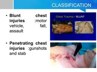
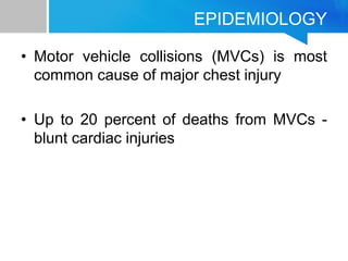
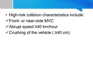
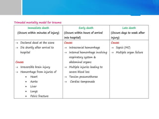
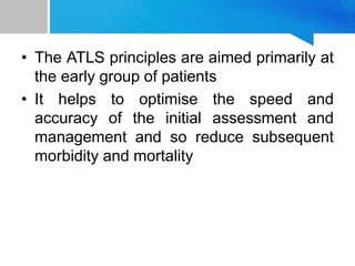
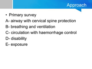
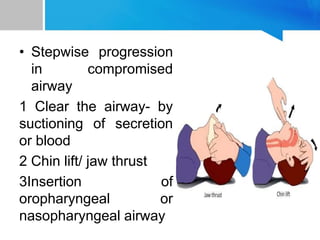
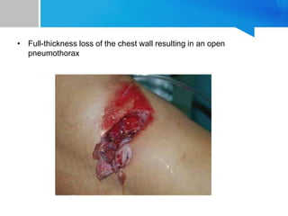
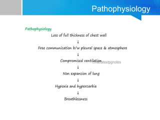
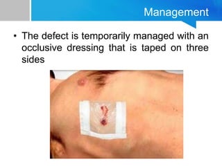
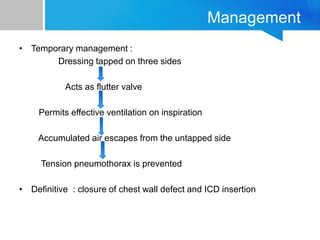
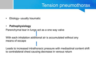
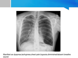
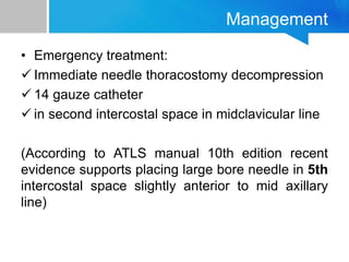
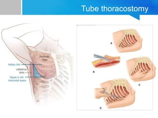
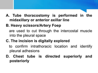
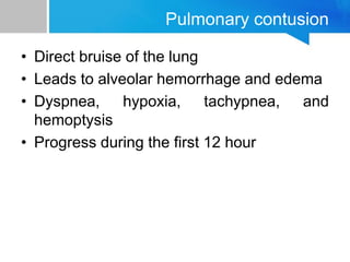
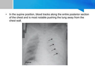
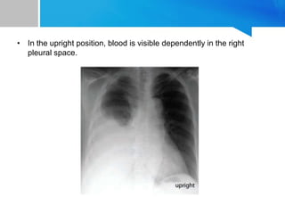
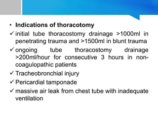
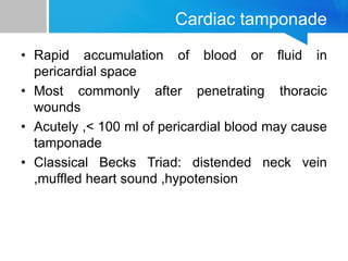
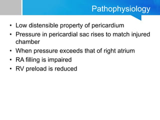
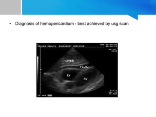
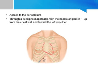
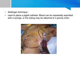
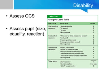
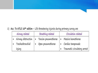
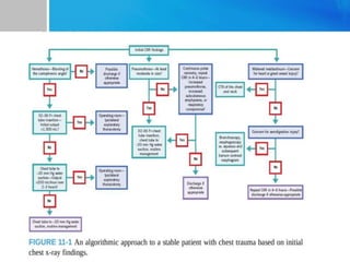
Dr Lalit K Shah
Resident 1st year
General Surgery
Chest Trauma
Introduction
• Trauma is the study of medical problems
associated with physical injury
• Injury is the adverse effect of a physical
force upon a person
• The most common force involved in most
injuries are mechanical
• Trauma is most common cause of death
and disability in the first four decades of
life
• Third most common cause of death
regardless of age
• Overall chest injuries are responsible for
20-25 % of deaths
CLASSIFICATION
• Blunt chest
injuries :motor
vehicle, fall,
assault
• Penetrating chest
injuries :gunshots
and stab
EPIDEMIOLOGY
• Motor vehicle collisions (MVCs) is most
common cause of major chest injury
• Up to 20 percent of deaths from MVCs -
blunt cardiac injuries
• High-risk occupant characteristics include:
Age ≥60
Front-seat occupancy
Not wearing a seatbelt
• High-risk collision characteristics include:
Front- or near-side MVC
Abrupt speed ≥40 km/hour
Crushing of the vehicle ( ≥40 cm)
• The ATLS principles are aimed primarily at
the early group of patients
• It helps to optimise the speed and
accuracy of the initial assessment and
management and so reduce subsequent
morbidity and mortality
Approach
• Primary survey
A- airway with cervical spine protection
B- breathing and ventilation
C- circulation with haemorrhage control
D- disability
E- exposure
Airway
• In all trauma patients cervical spine injury
should be suspected and cervical spine
immobilization should be done
• Assess the patency of airway
• Indication of airway management
- obstruction of airway
- mental status depression
- noisy breathing
- facial trauma
- GCS less than or equal to 8
• Stepwise progression
in compromised
airway
1 Clear the airway- by
suctioning of secretion
or blood
2 Chin lift/ jaw thrust
3Insertion of
oropharyngeal or
nasopharyngeal airway
Breathing
• Assess breathing by visualizing chest
movement
• Assess rate and depth by percussion and
auscultation
• 02 saturation
• Adequate oxygenation and ventilation
• Conditions constitute an immediate threat
to life d/t inadequate ventilation during
primary survey :
Tension pneumothorax
Open pneumothorax
Flail chest with pulmonary contusion
Massive hemothorax
Major air leak d/t Tracheobrochial injury
Pneumothorax
• Spontaneous
-Primary and secondary
• Traumatic
-Penetrating ,Blunt
• Iatrogenic
-Mechanical ventilation,Needle puncture
Open pneumothorax
• “sucking chest wound”
• Occurs with full thickness loss of chest
wall
• Free communication between pleural
space and atmosphere
• Full-thickness loss of the chest wall resulting in an open
pneumothorax
Pathophysiology
Management
• The defect is temporarily managed with an
occlusive dressing that is taped on three
sides
Management
• Temporary management :
Dressing tapped on three sides
Acts as flutter valve
Permits effective ventilation on inspiration
Accumulated air escapes from the untapped side
Tension pneumothorax is prevented
• Definitive : closure of chest wall defect and ICD insertion
Tension pneumothorax
• Etiology- usually traumatic
• Pathophysiology
Parenchymal tear in lungs act as a one way valve
With each inhalation additional air is accumulated without any
means of escape
Leads to increased intrathoracic pressure with mediastinal content shift
to contralateral chest causing decrease in venous return
Manifest as dyspnea,tachypnea,chest pain,hypoxia,diminished/absent breathe
sound
Management
• Emergency treatment:
 Immediate needle thoracostomy decompression
 14 gauze catheter
 in second intercostal space in midclavicular line
(According to ATLS manual 10th edition recent
evidence supports placing large bore needle in 5th
intercostal space slightly anterior to mid axillary
line)
Conclusions: In a cadaveric model, needle thoracostomy was successfully placed in
100% of attempts at the fifth intercostal space but in only 58% at the traditional
second intercostal position. On average, the chest wall was 1 cm thinner at this
position and may improve successful needle placement. Live patient validation of
Tube thoracostomy
A. Tube thoracostomy is performed in the
midaxillary or anterior axillar line
B. Heavy scissors/Artery Foep
are used to cut through the intercostal muscle
into the pleural space
C. The incision is digitally explored
to confirm intrathoracic location and identify
pleural adhesions
D. Chest tube is directed superiorly and
posteriorly
Pulmonary contusion
• Direct bruise of the lung
• Leads to alveolar hemorrhage and edema
• Dyspnea, hypoxia, tachypnea, and
hemoptysis
• Progress during the first 12 hour
Management
• Close monitor and frequent clinical re
evaluation
• Pain control
• Pulmonary hygiene
• Hypoxia or difficulty ventilating require
airway management
Flail chest
• Two or more consecutive ribs are each fractured
in two or more places (ATLS 10th edition)
• occurs when segment of chest wall doesnt have
bony continuity with rest of thoracic cage
• Dignosed clinically- ask the patient to cough
chest cavity moves in while the flail segment
moves out (paradoxical motion)
Pressure within the chest is negative during inspiration, causing the flail segment to
retract.
With expiration, intrathoracic pressure becomes positive and the flail segment bulges.
Management
• O2 administration + adequate analgesia for pain
control + physiotherapy
• For flail chest + respiratory failure- Noninvasive
positive airway pressure by mask
• Opearative management :
• Open reduction and internal fixation can
decrease :
1. Mortality in flail chest
2. Duration of mechanical ventilation
3. Reduce hospital and intensive care length of
stay
Circulation
• Four life threatning injuries
• Should be identified prompltly during circulation
section of primary survey
1.Massive hemothorax
2.Cardiac tamponade
3.Massive hemoperitoneum
4.Mechanically unstable pelvic fracture with
bleeding
Hemothorax
• Collection of blood in pleural space
• Most common cause- Trauma
• Massive Hemothorax , defined as >1500 ml of
blood
• In peaditric population, >25% of blood volume in
pleural space
Pathophysiology
• Multiple rib fractures with severed intercostal
vessels
• Bleeding from lacerated lung parenchyma
• A great vessel or pulmonary hilar vessel injury
after penetrating trauma
• In the supine position, blood tracks along the entire posterior section
of the chest and is most notable pushing the lung away from the
chest wall.
• In the upright position, blood is visible dependently in the right
pleural space.
Management
• Tube thoracostomy for rexpansion of lung
• Massive hemothorax is an indication for
operative approach
• Indications of thoracotomy
 initial tube thoracostomy drainage >1000ml in
penetrating trauma and >1500ml in blunt trauma
 ongoing tube thoracostomy drainage
>200ml/hour for consecutive 3 hours in non-
coagulopathic patients
 Tracheobronchial injury
 Pericardial tamponade
 massive air leak from chest tube with inadequate
ventilation
Cardiac tamponade
• Rapid accumulation of blood or fluid in
pericardial space
• Most commonly after penetrating thoracic
wounds
• Acutely ,< 100 ml of pericardial blood may cause
tamponade
• Classical Becks Triad: distended neck vein
,muffled heart sound ,hypotension
Pathophysiology
• Low distensible property of pericardium
• Pressure in pericardial sac rises to match injured
chamber
• When pressure exceeds that of right atrium
• RA filling is impaired
• RV preload is reduced
• Diagnosis of hemopericardium - best achieved by usg scan
Management
• Pericardiocentesis :succesful in decompression
of tamponade (80 % cases)
• Failure : Due to presence of clotted blood in
pericardium
• Persistent SBP <60 mm of hg – Resuscitative
thoractomy
• Access to the pericardium
• Through a subxiphoid approach, with the needle angled 45° up
from the chest wall and toward the left shoulder.
• Seldinger technique :
• used to place a pigtail catheter. Blood can be repeatedly aspirated
with a syringe, or the tubing may be attached to a gravity drain.
Disability
• Assess GCS
• Assess pupil (size,
equality, reaction)
Exposure
• All clothing of patient should be removed
for adequate examination
• core body temperature should be obtained
• Keep the patient warm
References
• Bailey & Love 27th edition
• Sabiston Textbook Of Surgery 21st edition
• ATLS guidlines 10th edition
• Washington manual of surgery 8th edition
• Pubmed
THANK YOU

chest trauma

  • 1.
    Dr Lalit KShah Resident 1st year General Surgery Chest Trauma
  • 2.
    Introduction • Trauma isthe study of medical problems associated with physical injury • Injury is the adverse effect of a physical force upon a person • The most common force involved in most injuries are mechanical
  • 3.
    • Trauma ismost common cause of death and disability in the first four decades of life • Third most common cause of death regardless of age • Overall chest injuries are responsible for 20-25 % of deaths
  • 6.
    CLASSIFICATION • Blunt chest injuries:motor vehicle, fall, assault • Penetrating chest injuries :gunshots and stab
  • 7.
    EPIDEMIOLOGY • Motor vehiclecollisions (MVCs) is most common cause of major chest injury • Up to 20 percent of deaths from MVCs - blunt cardiac injuries
  • 8.
    • High-risk occupantcharacteristics include: Age ≥60 Front-seat occupancy Not wearing a seatbelt
  • 9.
    • High-risk collisioncharacteristics include: Front- or near-side MVC Abrupt speed ≥40 km/hour Crushing of the vehicle ( ≥40 cm)
  • 11.
    • The ATLSprinciples are aimed primarily at the early group of patients • It helps to optimise the speed and accuracy of the initial assessment and management and so reduce subsequent morbidity and mortality
  • 12.
    Approach • Primary survey A-airway with cervical spine protection B- breathing and ventilation C- circulation with haemorrhage control D- disability E- exposure
  • 13.
    Airway • In alltrauma patients cervical spine injury should be suspected and cervical spine immobilization should be done • Assess the patency of airway
  • 14.
    • Indication ofairway management - obstruction of airway - mental status depression - noisy breathing - facial trauma - GCS less than or equal to 8
  • 15.
    • Stepwise progression incompromised airway 1 Clear the airway- by suctioning of secretion or blood 2 Chin lift/ jaw thrust 3Insertion of oropharyngeal or nasopharyngeal airway
  • 16.
    Breathing • Assess breathingby visualizing chest movement • Assess rate and depth by percussion and auscultation • 02 saturation • Adequate oxygenation and ventilation
  • 17.
    • Conditions constitutean immediate threat to life d/t inadequate ventilation during primary survey : Tension pneumothorax Open pneumothorax Flail chest with pulmonary contusion Massive hemothorax Major air leak d/t Tracheobrochial injury
  • 18.
    Pneumothorax • Spontaneous -Primary andsecondary • Traumatic -Penetrating ,Blunt • Iatrogenic -Mechanical ventilation,Needle puncture
  • 19.
    Open pneumothorax • “suckingchest wound” • Occurs with full thickness loss of chest wall • Free communication between pleural space and atmosphere
  • 20.
    • Full-thickness lossof the chest wall resulting in an open pneumothorax
  • 21.
  • 22.
    Management • The defectis temporarily managed with an occlusive dressing that is taped on three sides
  • 23.
    Management • Temporary management: Dressing tapped on three sides Acts as flutter valve Permits effective ventilation on inspiration Accumulated air escapes from the untapped side Tension pneumothorax is prevented • Definitive : closure of chest wall defect and ICD insertion
  • 24.
    Tension pneumothorax • Etiology-usually traumatic • Pathophysiology Parenchymal tear in lungs act as a one way valve With each inhalation additional air is accumulated without any means of escape Leads to increased intrathoracic pressure with mediastinal content shift to contralateral chest causing decrease in venous return
  • 25.
    Manifest as dyspnea,tachypnea,chestpain,hypoxia,diminished/absent breathe sound
  • 26.
    Management • Emergency treatment: Immediate needle thoracostomy decompression  14 gauze catheter  in second intercostal space in midclavicular line (According to ATLS manual 10th edition recent evidence supports placing large bore needle in 5th intercostal space slightly anterior to mid axillary line)
  • 27.
    Conclusions: In acadaveric model, needle thoracostomy was successfully placed in 100% of attempts at the fifth intercostal space but in only 58% at the traditional second intercostal position. On average, the chest wall was 1 cm thinner at this position and may improve successful needle placement. Live patient validation of
  • 29.
  • 30.
    A. Tube thoracostomyis performed in the midaxillary or anterior axillar line B. Heavy scissors/Artery Foep are used to cut through the intercostal muscle into the pleural space C. The incision is digitally explored to confirm intrathoracic location and identify pleural adhesions D. Chest tube is directed superiorly and posteriorly
  • 31.
    Pulmonary contusion • Directbruise of the lung • Leads to alveolar hemorrhage and edema • Dyspnea, hypoxia, tachypnea, and hemoptysis • Progress during the first 12 hour
  • 33.
    Management • Close monitorand frequent clinical re evaluation • Pain control • Pulmonary hygiene • Hypoxia or difficulty ventilating require airway management
  • 34.
    Flail chest • Twoor more consecutive ribs are each fractured in two or more places (ATLS 10th edition) • occurs when segment of chest wall doesnt have bony continuity with rest of thoracic cage • Dignosed clinically- ask the patient to cough chest cavity moves in while the flail segment moves out (paradoxical motion)
  • 35.
    Pressure within thechest is negative during inspiration, causing the flail segment to retract. With expiration, intrathoracic pressure becomes positive and the flail segment bulges.
  • 36.
    Management • O2 administration+ adequate analgesia for pain control + physiotherapy • For flail chest + respiratory failure- Noninvasive positive airway pressure by mask
  • 37.
    • Opearative management: • Open reduction and internal fixation can decrease : 1. Mortality in flail chest 2. Duration of mechanical ventilation 3. Reduce hospital and intensive care length of stay
  • 38.
    Circulation • Four lifethreatning injuries • Should be identified prompltly during circulation section of primary survey 1.Massive hemothorax 2.Cardiac tamponade 3.Massive hemoperitoneum 4.Mechanically unstable pelvic fracture with bleeding
  • 40.
    Hemothorax • Collection ofblood in pleural space • Most common cause- Trauma • Massive Hemothorax , defined as >1500 ml of blood • In peaditric population, >25% of blood volume in pleural space
  • 41.
    Pathophysiology • Multiple ribfractures with severed intercostal vessels • Bleeding from lacerated lung parenchyma • A great vessel or pulmonary hilar vessel injury after penetrating trauma
  • 42.
    • In thesupine position, blood tracks along the entire posterior section of the chest and is most notable pushing the lung away from the chest wall.
  • 43.
    • In theupright position, blood is visible dependently in the right pleural space.
  • 44.
    Management • Tube thoracostomyfor rexpansion of lung • Massive hemothorax is an indication for operative approach
  • 45.
    • Indications ofthoracotomy  initial tube thoracostomy drainage >1000ml in penetrating trauma and >1500ml in blunt trauma  ongoing tube thoracostomy drainage >200ml/hour for consecutive 3 hours in non- coagulopathic patients  Tracheobronchial injury  Pericardial tamponade  massive air leak from chest tube with inadequate ventilation
  • 46.
    Cardiac tamponade • Rapidaccumulation of blood or fluid in pericardial space • Most commonly after penetrating thoracic wounds • Acutely ,< 100 ml of pericardial blood may cause tamponade • Classical Becks Triad: distended neck vein ,muffled heart sound ,hypotension
  • 47.
    Pathophysiology • Low distensibleproperty of pericardium • Pressure in pericardial sac rises to match injured chamber • When pressure exceeds that of right atrium • RA filling is impaired • RV preload is reduced
  • 48.
    • Diagnosis ofhemopericardium - best achieved by usg scan
  • 49.
    Management • Pericardiocentesis :succesfulin decompression of tamponade (80 % cases) • Failure : Due to presence of clotted blood in pericardium • Persistent SBP <60 mm of hg – Resuscitative thoractomy
  • 50.
    • Access tothe pericardium • Through a subxiphoid approach, with the needle angled 45° up from the chest wall and toward the left shoulder.
  • 51.
    • Seldinger technique: • used to place a pigtail catheter. Blood can be repeatedly aspirated with a syringe, or the tubing may be attached to a gravity drain.
  • 52.
    Disability • Assess GCS •Assess pupil (size, equality, reaction)
  • 53.
    Exposure • All clothingof patient should be removed for adequate examination • core body temperature should be obtained • Keep the patient warm
  • 56.
    References • Bailey &Love 27th edition • Sabiston Textbook Of Surgery 21st edition • ATLS guidlines 10th edition • Washington manual of surgery 8th edition • Pubmed
  • 57.