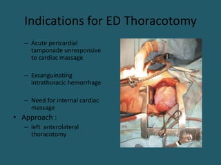
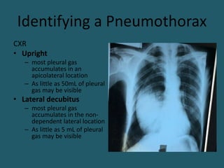
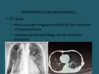
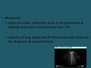
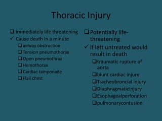
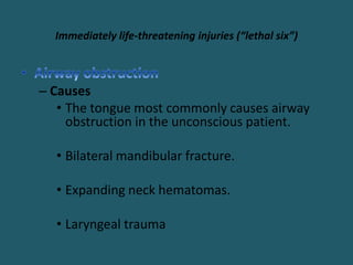
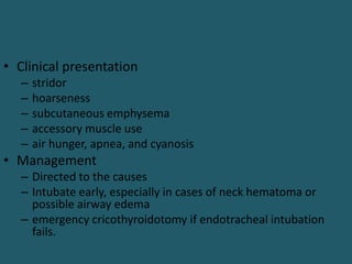
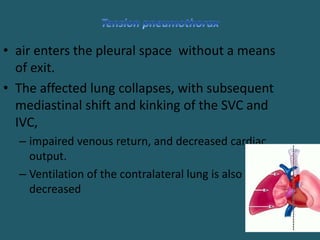
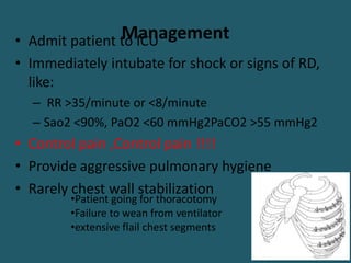
Introduction
• Thoracic injuries account for ~ 25% of all trauma
deaths
– Immediate (within seconds to minutes),
• disruption of the heart or great vessel injury
– Early (minutes to hours)
• Airway obstruction, tension pneumothorax,
pulmonary contusion, or cardiac tamponade
– Late (days to weeks) after injury
• Pulmonary complications, sepsis, and missed injuries
• Most patients with thoracic injury are managed
nonoperatively( 85% )
– analgesia,
– pulmonary hygiene,
– endotracheal intubation, and
– tube thoracostomy.
• Only 10% to 15% of patients with chest trauma will
require thoracotomy or sternotomy
Indications for Urgent Thoracotomy
• Chest drainage >1,500 mL initial or >200 mL/hr for 4
consecutive hrs
• Large unevacuated clotted hemothorax
• Developing cardiac tamponade
• Chest wall defect
• Massive air leak or incomplete lung expansion despite
adequate drainage
• Great vessel injury
• Esophageal injury
• Diaphragmatic laceration
• Traumatic septal or valvular injury
Indications for ED Thoracotomy
– Acute pericardial
tamponade unresponsive
to cardiac massage
– Exsanguinating
intrathoracic hemorrhage
– Need for internal cardiac
massage
• Approach :
– left anterolateral
thoracotomy
Identifying a Pneumothorax
CXR
• Upright
– most pleural gas
accumulates in an
apicolateral location
– As little as 50mL of pleural
gas may be visible
• Lateral decubitus
– most pleural gas
accumulates in the non-
dependent lateral location
– As little as 5 mL of pleural
gas may be visible
IDENTIFYING A PNEUMOTHORAX…
• CT Scan
– most accurate imaging modality for the detection
of pneumothorax
– complex pleural pathology can be optimally
displayed
• ultrasound
– highly accurate , extremely quick to be performed at
bedside and much more sensitive than CXR
– absence of lung sliding and B lines (comet tail) allows for
the diagnosis of pneumothorax
Thoracic Injury
 immediately life threatening
 Cause death in a minute
airway obstruction
Tension pneumothorax
Open pneumothrax
Hemothorax
Cardiac tamponade
Flail chest
Potentially life-
threatening
 If left untreated would
result in death
traumatic rupture of
aorta
blunt cardiac injury
Tracheobroncial injury
Diaphragmaticinjury
Esophagealperforation
pulmonarycontusion
Immediately life-threatening injuries (“lethal six”)
– Causes
• The tongue most commonly causes airway
obstruction in the unconscious patient.
• Bilateral mandibular fracture.
• Expanding neck hematomas.
• Laryngeal trauma
• Clinical presentation
– stridor
– hoarseness
– subcutaneous emphysema
– accessory muscle use
– air hunger, apnea, and cyanosis
• Management
– Directed to the causes
– Intubate early, especially in cases of neck hematoma or
possible airway edema
– emergency cricothyroidotomy if endotracheal intubation
fails.
• air enters the pleural space without a means
of exit.
• The affected lung collapses, with subsequent
mediastinal shift and kinking of the SVC and
IVC,
– impaired venous return, and decreased cardiac
output.
– Ventilation of the contralateral lung is also
decreased
• Most common causes
– Penetrating injury to the chest
– Blunt trauma with parenchymal lung injury
– Mechanical ventilation with high airway pressure
– Spontaneous pneumothorax with blebs that failed to
seal
• Diagnosis
• Hx and P/E
– Severe respiratory distress
– Hypotension
– absence of breath sounds
– Hyperresonance to percussion
– Neck vein distention (absent in hypovolemic pt)
– Tracheal deviation (late finding)
– Cyanosis
treatment
 Immediately decompress by
inserting a 12- or 14-gauge
i.v. canula into the 2nd ICS in
the MCL
 this converts the tension
pneumothorax into a simple
pneumothorax
 Follow immediately with
tube thoracostomy
• caused by stab injury or destructive penetrating
wound (shotgun)
• Large open defect >3 cm cause equilibration b/n
intrathoracic & atmospheric pressure
• If the opening >2/3rd of the Ø of the trachea, then
air follows the path of least resistance through the
chest wall with each inspiration
• Signs and symptoms are usually proportional to the
size of the defect.
• Mediastinal flutter
– Swinging of the mediastinum
• Pendulum air
– Expiratory air from the normal lung filling the
collapsed lung
• Management
 Close the chest wall defect with occlusive dressing
taped on three sides to act as a flutter-type valve.
Followed by tube thoracostomy
Large defects may require flap closure.
• is defined as >1500 mL of blood or, in the pediatric population, 1/3rd
of the pt's blood volume in the pleural space
• Common in penetrating trauma with hilar or systemic vessel
disruption
• Intercostal and internal mammary vessels are most
commonly injured
• Each hemithorax can hold up to 3 L of blood
• Hilar or great vessel disruption will present with severe shock.
Diagnosis
• Hemorrhagic shock
• Unilateral absence or diminution of breath sounds
• Unilateral dullness to percussion
• CXR will show unilateral “white out” (opacification)
• Treatment
– Establish large-bore i.v. access and have blood available
for infusion before decompression
– tube thoracostomy with a large tube (36F or 40F) in 5th
ICS
– A second chest tube is occasionally necessary to
adequately drain the hemothorax.
• Thoracotomy is indicated for:
– 1,500 mL blood evacuated initially or drainage of ≥20
mL/kg
– Ongoing bleeding of >200 mL/hr for 4 hrs(generally >3
mL/kg/hr)
– Failure to completely drain hemothorax, despite at least
two functioning and appropriately positioned chest
tubes
– Pt presents after a delay may be observed if output
ceases & the lung is re-expanded
– when ≥2 consecutive ribs are #d in two or more
locations
– leads to paradoxical motion of that chest wall segment
– flail segment classically involves anterior (costochondral
cartilage) or lateral rib
– Morbidity and mortality are generally related to the lung
parenchymal injury rather than the chest wall injury
Diagnosis
• is clinical, not radiographic
• Chest wall must be observed for several respiratory
cycles and during coughing
• flail segment, underlying pulmonary contusion, and
chest splinting due to pain
– all exacerbate hypoxemia….severe RD
Management
• Admit patient to ICU
• Immediately intubate for shock or signs of RD,
like:
– RR >35/minute or <8/minute
– Sao2 <90%, PaO2 <60 mmHg2PaCO2 >55 mmHg2
• Control pain ,Control pain !!!!
• Provide aggressive pulmonary hygiene
• Rarely chest wall stabilization
•Patient going for thoracotomy
•Failure to wean from ventilator
•extensive flail chest segments
• is commonly the result of penetrating trauma, but it
can also be seen in blunt chest trauma.
• Mostly ventricular, right > left
• pericardial sac does not acutely distend; 75 to 100
mL of blood can produce tamponade physiology in
the adult.
Diagnosis
• Classic signs:
– Increase in JVP,
– hypotension, and Beck's triad are present in only 33% of pts
– muffled heart sound
• Pulsus paradoxus
• Kussmaul's sign
– is a hard and true sign
• shock or ongoing hypotension without blood loss suggest this injury
• FAST , ECHO cardiography, chest CT,
• Pericardial window
Treatment
• Assess the need for intubation, oxygenate, and
start volume resuscitation.
• Pericardiocentesis can be used as a temporizing
maneuver .
– prevent subendocardial ischemia and lethal arrhythmias.
• emergent left anterolateral thoracotomy
• urgent sternotomy
Potentially life-threatening injuries (“hidden six”)
Traumatic rupture of the aorta
• defined as a tear in the wall of the aorta that is contained
by the adventitia of artery and the parietal pleura
• The mechanism of injury :
– rapid deceleration, such as falls from significant height, high-
speed motor vehicle crashes
• Laceration is usually located near the ligamentum
arteriosum (85%), distal to the left subclavian artery
• Diagnosis
• Clinical signs
• Asymmetry in upper extremity BP and upper extremity HTN
• Widened pulse pressure
• Chest wall contusion
• 50% pts with great vessel injury from blunt trauma have no
external signs of blunt chest injury.
 CXR
Widened mediastinum (supine CXR >8 cm; upright CXR >6 cm)
this is the most consistent finding
Aortography
Gold standard
2% to 3% are falsely negative
Chest CT
Mediastinal hematomas
Negative scans rule out aortic injury with a 92% sensitivity
Transesophageal echocardiogram
excellent alternative for unstable patients
• Management
• Control and prevent hypertension
– goal for SBP should be ~100mm/Hg and HR<100
beats/minute
– Beta blockade
• repair
• Most pts with major AW injuries die at the scene
as a result of asphyxia.
• Those who survive to reach the hospital are
usually in extremis.
• Of major bronchial injuries,
– 80% occur within 2.5 cm of carina
– Main stem 86%
– More common in right side
Diagnosis
– Intrapleural laceration (more distal airway injuries)
• massive pneumothorax that does not reexpand with chest tube
• Severe RD
– Extra pleural rupture into the mediastinum (thoracic
trachea and proximal bronchi)
• pneumomediastinum and subcutaneous emphysema
• RD may be minimal
– Imaging
• CXR
• Bronchoscopy
Management
• Surgical
– Extra thoracic - collar incision
– Intrathoracic- thoracotomy
– Injuries <1/3rd of the luminal circumference may be
considered for nonoperative management if
• pneumothorax and associated air leak that were present
resolve after insertion of a chest tube and
• the lung expands completely
• pts don’t require ppv
• only 3.8% of cases of blunt
chest trauma
• asymptomatic myocardial
muscle contusion to
clinically significant
dysrhythmia, septal defects
or valvular injury
Cardiac injuries…
diagnosis
• FAST
• CXR
• ECG
• ECHO
CARDIOGRAP
HY
treatment
• Treat dysrhythmias and put the pt on
continues monitoring
• Blunt trauma.
– classically large (~10 cm), radial, and located
posterolaterally
– herniation of the abdominal viscera typically occurs
acutely~ 66%
– The left hemidiaphragm is involved in 65% to 80% of
cases.
– are markers for severe intraabdominal injuries
• Penetrating trauma.
– Wounds are smaller (~3 cm) but tend to enlarge
over time
–herniation of the abdominal viscera typically
occurs after acute setting
–Left-sided injuries still predominate
• Diagnosis
• Hx and P/E
• CXR is diagnostic in only 25% to 50% of cases of blunt
trauma.
– elevated, obscured, or irregular diaphragmatic dome, and a
blunted CP angle
• CT scan
• Barium meal or SB follow through
• FAST
• Thoracoscopy, laparotomy, VATS
Treatment
• operative repair
– do not heal spontaneously & produce herniation
• Via laparotomy,
– Acute repair (<7-10days)
• Via thoracotomy
– in chronic herniation.
– posterolateral injury to the right hemidiaphragm
• Prosthetic material or flaps are rarely needed to
close the defect.
• Most injuries result from penetrating trauma.
• Blunt injury is rare (<0.1% incidence)
– caused by a rapid elevation in intraluminal pressure
during compression of the chest or abdomen
• more commonly injured in the neck
• Clinical presentation
– Odenophagia, subcutaneous emphysema, mediastinitis
Diagnosis
• Any possibility of injury need aggressive
investigation
• Esophagoscopy
– disruption of the mucosa both studies will detect
almost all injuries
• esophagogram
– extravasation of contrast
• CT scan may have a role in determining trajectory
in stable patients.
• Thoracentesis
– exudate with a high amylase.
Treatment
• Injuries identified within 24hrs
– Debridement and Primary repair + mediastinal drainage
• More than 24hrs
– Primary repair buttressed with vascularized autologous
tissue(parietal pleura & intercostal muscle)
– cervical esophagostomy , distal part stapled, gastrostomy and
feeding jejunostomy , mediastinal drainage for
• Critical pts and advanced mediastinitis or
• severe associated injuries
– T-tube drainage
• in critical pt and friable esophagus
– Esophagectomy for destructive injuries & gastric pull-up
• It is the most common potentially lethal chest injury
with mortality rate of 22% -30%
• Caused by h’ge into alveolar & interstitial spaces
and tissue damage
• Commonly, this accompanies multiple ribs # due to
blunt chest trauma
• It can also accompany a penetrating injury esp.
blast injuries from high-velocity missiles.
• Children may have it in the absence of rib # b/s of the resilience of
the chest wall.
Diagnosis
– Hemoptysis or blood in
the endotracheal tube
– CXR findings are
typically delayed in
appearance and
nonsegmental
– CT
• Treatment
• Largely supportive
– supplemental O2, pulmonary toilet, and analgesia
– Judicious fluid administration
– intubate and mechanically ventilate
• pulmonary resection
– is required in anatomic or nonanatomic patterns to
manage larger segments of injured lung tissue
Other thoracic injuries
• Rib Fractures
– most common major thoracic injuries…40%
– Factors that have the most impact on outcome of a pt is
• number of ribs fractured,
• age of the patient, and
• underlying pulmonary status
– One should always keep in mind that mortality from isolated rib fractures can
be as high as 5% in children and 20% in the elderly
– Diagnosis
• pain exacerbated by deep breathing
• CXR
• Bone scans
Rib Fractures….
• presence of ≥3 ribs # suggests the need for
hospitalization
– Management is directed at pain control
– repeat CXR at 6hrs after presentation to look for delayed
pneumothorax development (7%)
• There has been renewed interest in the operative fixation of rib
#
– optimal indications remains incompletely defined
– associated benefit
Pulmonary Hematoma
• difficult to differentiate from pulmonary contusion
• 24 to 48 hrs after the injury, it typically develops into a
discrete mass with distinct margins
• does not interfere with gas exchange and is reabsorbed
in time
• Treatment
– observation
– rarely it may become secondarily infected and present as an
abscess requiring drainage
Sternal Fracture
• Associated with severe blunt anterior trauma
• Mortality: 25-45%
– Myocardial contusion
– Pericardial tamponade
– Cardiac rupture
– Pulmonary contusion
• Treatment
• Pain control
• Plating
– severely displaced fractures
Mediastinal and Subcutaneous Emphysema
• Type of air entry
– Extrapleural( upper airway, tracheobronchial tree, esophagus)
• allows air to leak into mediastinum and then up to the soft tissues of
anterior neck
– Intrapleural (lung parenchyma)
• leakage typically creates a pneumothorax and then air leaks through the
parietal pleura and into the thoracic wall
– Source undetermined in 15%
• Typically, the more distal the injury, the more likely it is to
cause decompensation
• bronchoscopy , esophagoscopy and CXR
Mediastinal and Subcutaneous Emphysema…
• Management
– Mostly they are benign and self limited and can be treated with high flow
oxygen
• Facilitates re-absorption of nitrogen from tissues
– Key is identifying underlying injury
– Massive accumulations may be uncomfortable to a patient
• Decompression incisions in the skin are rarely if ever indicated
THANK YOU!!

4._management_of_chest_injury_2015/23.pptx

  • 2.
    Introduction • Thoracic injuriesaccount for ~ 25% of all trauma deaths – Immediate (within seconds to minutes), • disruption of the heart or great vessel injury – Early (minutes to hours) • Airway obstruction, tension pneumothorax, pulmonary contusion, or cardiac tamponade – Late (days to weeks) after injury • Pulmonary complications, sepsis, and missed injuries
  • 3.
    • Most patientswith thoracic injury are managed nonoperatively( 85% ) – analgesia, – pulmonary hygiene, – endotracheal intubation, and – tube thoracostomy. • Only 10% to 15% of patients with chest trauma will require thoracotomy or sternotomy
  • 4.
    Indications for UrgentThoracotomy • Chest drainage >1,500 mL initial or >200 mL/hr for 4 consecutive hrs • Large unevacuated clotted hemothorax • Developing cardiac tamponade • Chest wall defect • Massive air leak or incomplete lung expansion despite adequate drainage • Great vessel injury • Esophageal injury • Diaphragmatic laceration • Traumatic septal or valvular injury
  • 5.
    Indications for EDThoracotomy – Acute pericardial tamponade unresponsive to cardiac massage – Exsanguinating intrathoracic hemorrhage – Need for internal cardiac massage • Approach : – left anterolateral thoracotomy
  • 6.
    Identifying a Pneumothorax CXR •Upright – most pleural gas accumulates in an apicolateral location – As little as 50mL of pleural gas may be visible • Lateral decubitus – most pleural gas accumulates in the non- dependent lateral location – As little as 5 mL of pleural gas may be visible
  • 7.
    IDENTIFYING A PNEUMOTHORAX… •CT Scan – most accurate imaging modality for the detection of pneumothorax – complex pleural pathology can be optimally displayed
  • 8.
    • ultrasound – highlyaccurate , extremely quick to be performed at bedside and much more sensitive than CXR – absence of lung sliding and B lines (comet tail) allows for the diagnosis of pneumothorax
  • 9.
    Thoracic Injury  immediatelylife threatening  Cause death in a minute airway obstruction Tension pneumothorax Open pneumothrax Hemothorax Cardiac tamponade Flail chest Potentially life- threatening  If left untreated would result in death traumatic rupture of aorta blunt cardiac injury Tracheobroncial injury Diaphragmaticinjury Esophagealperforation pulmonarycontusion
  • 10.
    Immediately life-threatening injuries(“lethal six”) – Causes • The tongue most commonly causes airway obstruction in the unconscious patient. • Bilateral mandibular fracture. • Expanding neck hematomas. • Laryngeal trauma
  • 11.
    • Clinical presentation –stridor – hoarseness – subcutaneous emphysema – accessory muscle use – air hunger, apnea, and cyanosis • Management – Directed to the causes – Intubate early, especially in cases of neck hematoma or possible airway edema – emergency cricothyroidotomy if endotracheal intubation fails.
  • 12.
    • air entersthe pleural space without a means of exit. • The affected lung collapses, with subsequent mediastinal shift and kinking of the SVC and IVC, – impaired venous return, and decreased cardiac output. – Ventilation of the contralateral lung is also decreased
  • 13.
    • Most commoncauses – Penetrating injury to the chest – Blunt trauma with parenchymal lung injury – Mechanical ventilation with high airway pressure – Spontaneous pneumothorax with blebs that failed to seal
  • 14.
    • Diagnosis • Hxand P/E – Severe respiratory distress – Hypotension – absence of breath sounds – Hyperresonance to percussion – Neck vein distention (absent in hypovolemic pt) – Tracheal deviation (late finding) – Cyanosis
  • 15.
    treatment  Immediately decompressby inserting a 12- or 14-gauge i.v. canula into the 2nd ICS in the MCL  this converts the tension pneumothorax into a simple pneumothorax  Follow immediately with tube thoracostomy
  • 16.
    • caused bystab injury or destructive penetrating wound (shotgun) • Large open defect >3 cm cause equilibration b/n intrathoracic & atmospheric pressure • If the opening >2/3rd of the Ø of the trachea, then air follows the path of least resistance through the chest wall with each inspiration • Signs and symptoms are usually proportional to the size of the defect.
  • 17.
    • Mediastinal flutter –Swinging of the mediastinum • Pendulum air – Expiratory air from the normal lung filling the collapsed lung
  • 18.
    • Management  Closethe chest wall defect with occlusive dressing taped on three sides to act as a flutter-type valve. Followed by tube thoracostomy Large defects may require flap closure.
  • 19.
    • is definedas >1500 mL of blood or, in the pediatric population, 1/3rd of the pt's blood volume in the pleural space • Common in penetrating trauma with hilar or systemic vessel disruption • Intercostal and internal mammary vessels are most commonly injured • Each hemithorax can hold up to 3 L of blood • Hilar or great vessel disruption will present with severe shock.
  • 20.
    Diagnosis • Hemorrhagic shock •Unilateral absence or diminution of breath sounds • Unilateral dullness to percussion • CXR will show unilateral “white out” (opacification)
  • 21.
    • Treatment – Establishlarge-bore i.v. access and have blood available for infusion before decompression – tube thoracostomy with a large tube (36F or 40F) in 5th ICS – A second chest tube is occasionally necessary to adequately drain the hemothorax.
  • 22.
    • Thoracotomy isindicated for: – 1,500 mL blood evacuated initially or drainage of ≥20 mL/kg – Ongoing bleeding of >200 mL/hr for 4 hrs(generally >3 mL/kg/hr) – Failure to completely drain hemothorax, despite at least two functioning and appropriately positioned chest tubes – Pt presents after a delay may be observed if output ceases & the lung is re-expanded
  • 23.
    – when ≥2consecutive ribs are #d in two or more locations – leads to paradoxical motion of that chest wall segment – flail segment classically involves anterior (costochondral cartilage) or lateral rib – Morbidity and mortality are generally related to the lung parenchymal injury rather than the chest wall injury
  • 24.
    Diagnosis • is clinical,not radiographic • Chest wall must be observed for several respiratory cycles and during coughing • flail segment, underlying pulmonary contusion, and chest splinting due to pain – all exacerbate hypoxemia….severe RD
  • 25.
    Management • Admit patientto ICU • Immediately intubate for shock or signs of RD, like: – RR >35/minute or <8/minute – Sao2 <90%, PaO2 <60 mmHg2PaCO2 >55 mmHg2 • Control pain ,Control pain !!!! • Provide aggressive pulmonary hygiene • Rarely chest wall stabilization •Patient going for thoracotomy •Failure to wean from ventilator •extensive flail chest segments
  • 26.
    • is commonlythe result of penetrating trauma, but it can also be seen in blunt chest trauma. • Mostly ventricular, right > left • pericardial sac does not acutely distend; 75 to 100 mL of blood can produce tamponade physiology in the adult.
  • 27.
    Diagnosis • Classic signs: –Increase in JVP, – hypotension, and Beck's triad are present in only 33% of pts – muffled heart sound • Pulsus paradoxus • Kussmaul's sign – is a hard and true sign • shock or ongoing hypotension without blood loss suggest this injury • FAST , ECHO cardiography, chest CT, • Pericardial window
  • 28.
    Treatment • Assess theneed for intubation, oxygenate, and start volume resuscitation. • Pericardiocentesis can be used as a temporizing maneuver . – prevent subendocardial ischemia and lethal arrhythmias. • emergent left anterolateral thoracotomy • urgent sternotomy
  • 29.
    Potentially life-threatening injuries(“hidden six”) Traumatic rupture of the aorta • defined as a tear in the wall of the aorta that is contained by the adventitia of artery and the parietal pleura • The mechanism of injury : – rapid deceleration, such as falls from significant height, high- speed motor vehicle crashes • Laceration is usually located near the ligamentum arteriosum (85%), distal to the left subclavian artery
  • 30.
    • Diagnosis • Clinicalsigns • Asymmetry in upper extremity BP and upper extremity HTN • Widened pulse pressure • Chest wall contusion • 50% pts with great vessel injury from blunt trauma have no external signs of blunt chest injury.
  • 31.
     CXR Widened mediastinum(supine CXR >8 cm; upright CXR >6 cm) this is the most consistent finding Aortography Gold standard 2% to 3% are falsely negative Chest CT Mediastinal hematomas Negative scans rule out aortic injury with a 92% sensitivity Transesophageal echocardiogram excellent alternative for unstable patients
  • 32.
    • Management • Controland prevent hypertension – goal for SBP should be ~100mm/Hg and HR<100 beats/minute – Beta blockade • repair
  • 33.
    • Most ptswith major AW injuries die at the scene as a result of asphyxia. • Those who survive to reach the hospital are usually in extremis. • Of major bronchial injuries, – 80% occur within 2.5 cm of carina – Main stem 86% – More common in right side
  • 34.
    Diagnosis – Intrapleural laceration(more distal airway injuries) • massive pneumothorax that does not reexpand with chest tube • Severe RD – Extra pleural rupture into the mediastinum (thoracic trachea and proximal bronchi) • pneumomediastinum and subcutaneous emphysema • RD may be minimal – Imaging • CXR • Bronchoscopy
  • 35.
    Management • Surgical – Extrathoracic - collar incision – Intrathoracic- thoracotomy – Injuries <1/3rd of the luminal circumference may be considered for nonoperative management if • pneumothorax and associated air leak that were present resolve after insertion of a chest tube and • the lung expands completely • pts don’t require ppv
  • 36.
    • only 3.8%of cases of blunt chest trauma • asymptomatic myocardial muscle contusion to clinically significant dysrhythmia, septal defects or valvular injury
  • 37.
    Cardiac injuries… diagnosis • FAST •CXR • ECG • ECHO CARDIOGRAP HY treatment • Treat dysrhythmias and put the pt on continues monitoring
  • 38.
    • Blunt trauma. –classically large (~10 cm), radial, and located posterolaterally – herniation of the abdominal viscera typically occurs acutely~ 66% – The left hemidiaphragm is involved in 65% to 80% of cases. – are markers for severe intraabdominal injuries
  • 39.
    • Penetrating trauma. –Wounds are smaller (~3 cm) but tend to enlarge over time –herniation of the abdominal viscera typically occurs after acute setting –Left-sided injuries still predominate
  • 40.
    • Diagnosis • Hxand P/E • CXR is diagnostic in only 25% to 50% of cases of blunt trauma. – elevated, obscured, or irregular diaphragmatic dome, and a blunted CP angle • CT scan • Barium meal or SB follow through • FAST • Thoracoscopy, laparotomy, VATS
  • 41.
    Treatment • operative repair –do not heal spontaneously & produce herniation • Via laparotomy, – Acute repair (<7-10days) • Via thoracotomy – in chronic herniation. – posterolateral injury to the right hemidiaphragm • Prosthetic material or flaps are rarely needed to close the defect.
  • 42.
    • Most injuriesresult from penetrating trauma. • Blunt injury is rare (<0.1% incidence) – caused by a rapid elevation in intraluminal pressure during compression of the chest or abdomen • more commonly injured in the neck • Clinical presentation – Odenophagia, subcutaneous emphysema, mediastinitis
  • 43.
    Diagnosis • Any possibilityof injury need aggressive investigation • Esophagoscopy – disruption of the mucosa both studies will detect almost all injuries • esophagogram – extravasation of contrast • CT scan may have a role in determining trajectory in stable patients. • Thoracentesis – exudate with a high amylase.
  • 44.
    Treatment • Injuries identifiedwithin 24hrs – Debridement and Primary repair + mediastinal drainage • More than 24hrs – Primary repair buttressed with vascularized autologous tissue(parietal pleura & intercostal muscle) – cervical esophagostomy , distal part stapled, gastrostomy and feeding jejunostomy , mediastinal drainage for • Critical pts and advanced mediastinitis or • severe associated injuries – T-tube drainage • in critical pt and friable esophagus – Esophagectomy for destructive injuries & gastric pull-up
  • 45.
    • It isthe most common potentially lethal chest injury with mortality rate of 22% -30% • Caused by h’ge into alveolar & interstitial spaces and tissue damage • Commonly, this accompanies multiple ribs # due to blunt chest trauma • It can also accompany a penetrating injury esp. blast injuries from high-velocity missiles. • Children may have it in the absence of rib # b/s of the resilience of the chest wall.
  • 46.
    Diagnosis – Hemoptysis orblood in the endotracheal tube – CXR findings are typically delayed in appearance and nonsegmental – CT
  • 47.
    • Treatment • Largelysupportive – supplemental O2, pulmonary toilet, and analgesia – Judicious fluid administration – intubate and mechanically ventilate • pulmonary resection – is required in anatomic or nonanatomic patterns to manage larger segments of injured lung tissue
  • 48.
    Other thoracic injuries •Rib Fractures – most common major thoracic injuries…40% – Factors that have the most impact on outcome of a pt is • number of ribs fractured, • age of the patient, and • underlying pulmonary status – One should always keep in mind that mortality from isolated rib fractures can be as high as 5% in children and 20% in the elderly – Diagnosis • pain exacerbated by deep breathing • CXR • Bone scans
  • 49.
    Rib Fractures…. • presenceof ≥3 ribs # suggests the need for hospitalization – Management is directed at pain control – repeat CXR at 6hrs after presentation to look for delayed pneumothorax development (7%) • There has been renewed interest in the operative fixation of rib # – optimal indications remains incompletely defined – associated benefit
  • 50.
    Pulmonary Hematoma • difficultto differentiate from pulmonary contusion • 24 to 48 hrs after the injury, it typically develops into a discrete mass with distinct margins • does not interfere with gas exchange and is reabsorbed in time • Treatment – observation – rarely it may become secondarily infected and present as an abscess requiring drainage
  • 51.
    Sternal Fracture • Associatedwith severe blunt anterior trauma • Mortality: 25-45% – Myocardial contusion – Pericardial tamponade – Cardiac rupture – Pulmonary contusion • Treatment • Pain control • Plating – severely displaced fractures
  • 52.
    Mediastinal and SubcutaneousEmphysema • Type of air entry – Extrapleural( upper airway, tracheobronchial tree, esophagus) • allows air to leak into mediastinum and then up to the soft tissues of anterior neck – Intrapleural (lung parenchyma) • leakage typically creates a pneumothorax and then air leaks through the parietal pleura and into the thoracic wall – Source undetermined in 15% • Typically, the more distal the injury, the more likely it is to cause decompensation • bronchoscopy , esophagoscopy and CXR
  • 53.
    Mediastinal and SubcutaneousEmphysema… • Management – Mostly they are benign and self limited and can be treated with high flow oxygen • Facilitates re-absorption of nitrogen from tissues – Key is identifying underlying injury – Massive accumulations may be uncomfortable to a patient • Decompression incisions in the skin are rarely if ever indicated
  • 54.