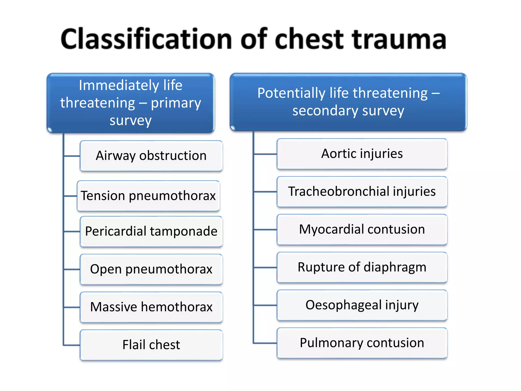
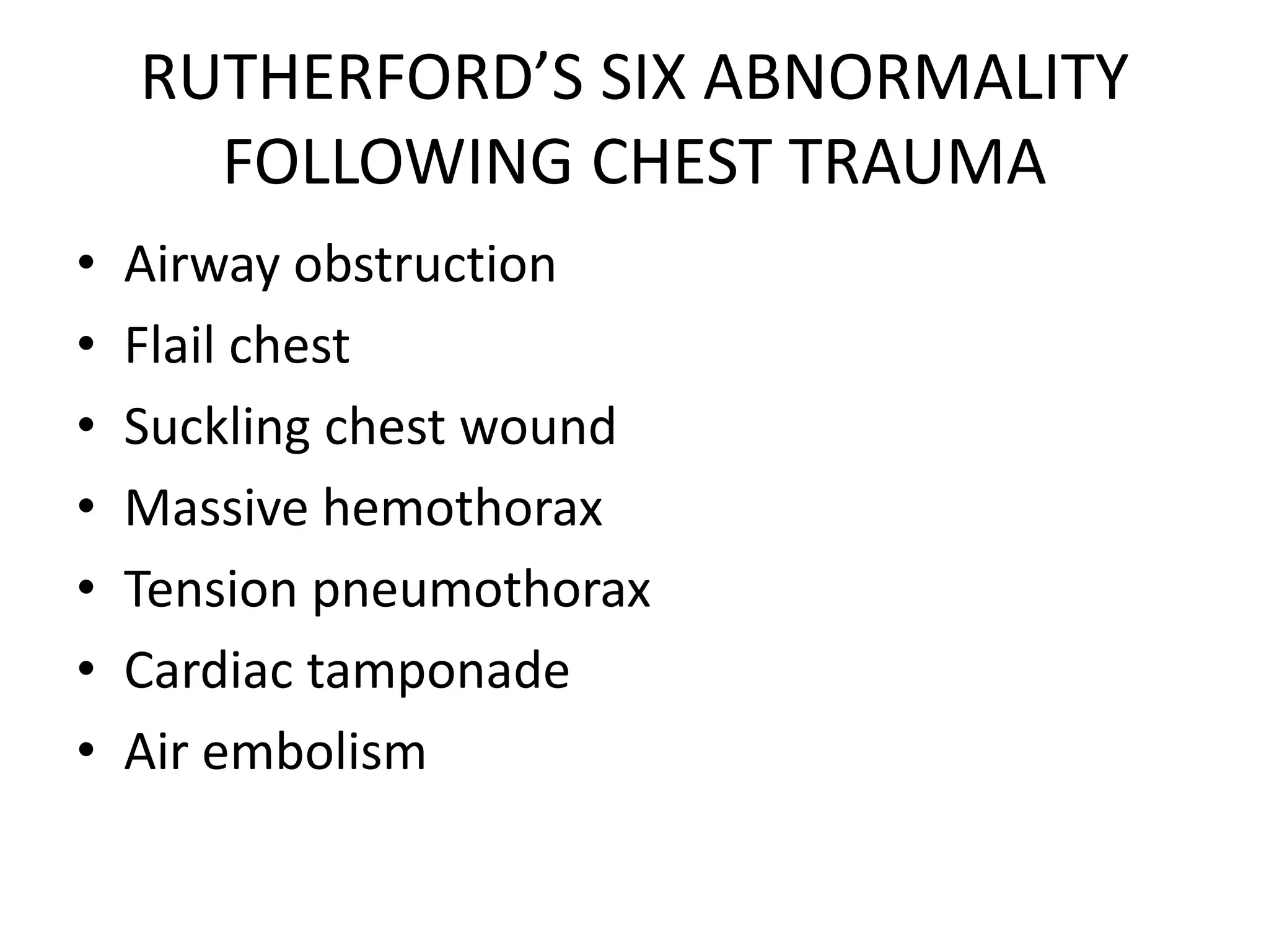
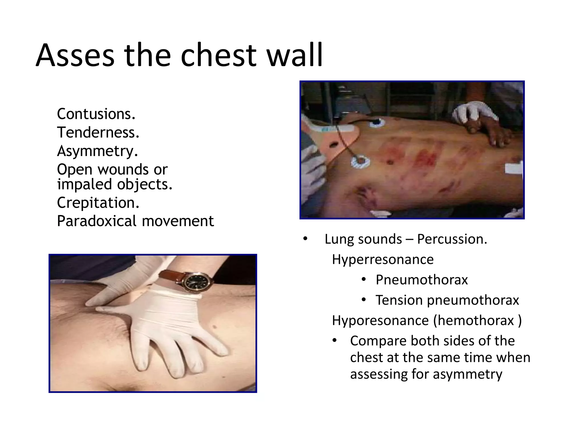
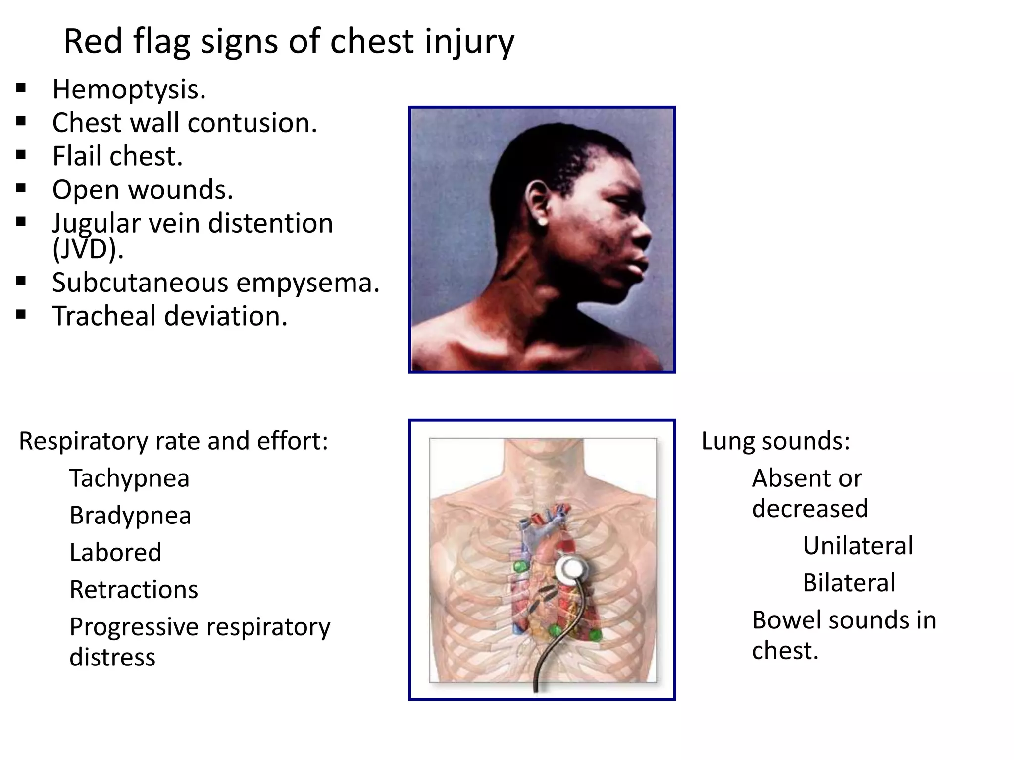
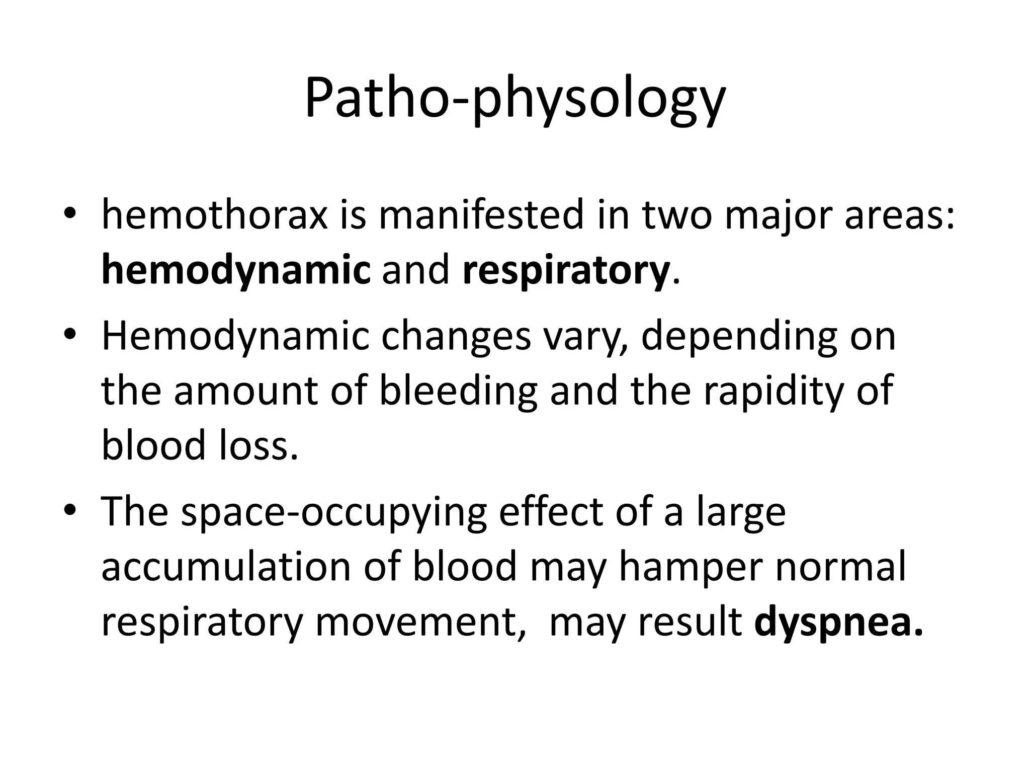
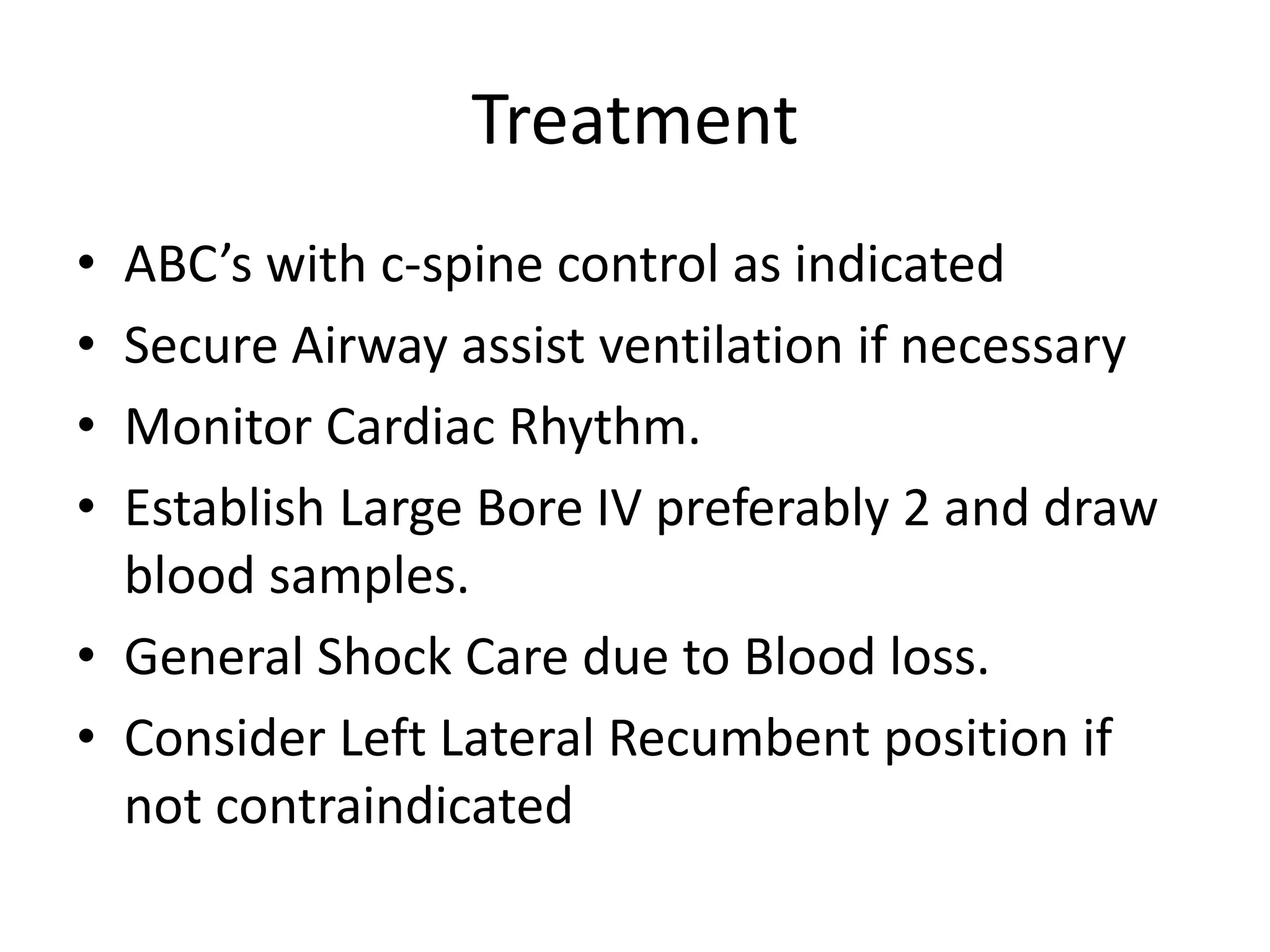
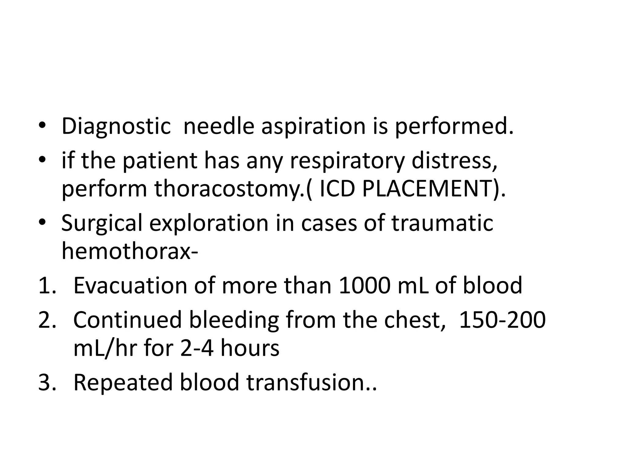
CHEST TRAUMA
Emergency Management
Objectives
• Anatomy of Thorax
• Main Causes of Chest Injuries
• S/S of Chest Injuries
• Different Types of Chest Injuries
• Emergency department care of Chest Injuries
Anatomy of the chest
• It provides protection to vital organs ( heart
and major vessels, lungs, liver) and provides
stability for movement of the shoulder girdles
and upper arms.
• thoracic skeleton consists of 12 thoracic
vertebrae bones, Twelve ribs , neurovascular
bundles arising from the spine (Brachial
plexus), Muscles…
GROSS ANATOMY
Physiology
BRAIN STEM
• Respiratory centres pons
and medulla
-autonomic
-central chemoreceptors(h+)
-periphral
chemoreceptors(paco2)
Respiratory mechanic
-muscular forces
-mechanic’s of ventilation
• Compliance
• Gas liquid interference
• Airway resistance
EPIDEMIOLGY
• Chest trauma ranks third behind head and
extremity trauma in major accidents in the
United States.
• The motor vehicle accident is the most common
etiology (70 per cent).
• Many of these injuries are of moderate severity
and rarely require surgical intervention.
• *chest -[33.33%] in RTA cases.
• The majority of such injuries may be diagnosed
with relatively simple tests such as chest
radiographs.
• *JRFMT-2016Month :December Volume :2
Issue:2 Page :8-13
Main Causes of Chest Trauma
• *Blunt Trauma- Blunt force to chest.
• Penetrating Trauma- Projectile that enters
chest causing small or large hole.
• Compression Injury- Chest is caught between
two objects and chest is compressed.
“DEADLY DOZEN”
Immediately life
threatening – primary
survey
Airway obstruction
Tension pneumothorax
Pericardial tamponade
Open pneumothorax
Massive hemothorax
Flail chest
Potentially life threatening –
secondary survey
Aortic injuries
Tracheobronchial injuries
Myocardial contusion
Rupture of diaphragm
Oesophageal injury
Pulmonary contusion
RUTHERFORD’S SIX ABNORMALITY
FOLLOWING CHEST TRAUMA
• Airway obstruction
• Flail chest
• Suckling chest wound
• Massive hemothorax
• Tension pneumothorax
• Cardiac tamponade
• Air embolism
Asses the chest wall
 Contusions.
 Tenderness.
 Asymmetry.
 Open wounds or
impaled objects.
 Crepitation.
 Paradoxical movement
• Lung sounds – Percussion.
Hyperresonance
• Pneumothorax
• Tension pneumothorax
Hyporesonance (hemothorax )
• Compare both sides of the
chest at the same time when
assessing for asymmetry
Red flag signs of chest injury
 Hemoptysis.
 Chest wall contusion.
 Flail chest.
 Open wounds.
 Jugular vein distention
(JVD).
 Subcutaneous empysema.
 Tracheal deviation.
Respiratory rate and effort:
Tachypnea
Bradypnea
Labored
Retractions
Progressive respiratory
distress
Lung sounds:
Absent or
decreased
Unilateral
Bilateral
Bowel sounds in
chest.
Airway obstruction
• Injury of chest can disrupt the airway
continuity in it course through the upper
mediastinum.
• Devastating injury.
Patho-physology
• Airway obstruction injuries the patient via
hypoventilation and elevation of pco2.
• Impairs minute volume..
Diagnosis
• Clinical diagnosis …
• Chest x-ray
• Abg
• Rigid bronchoscopy
Treatment
• Upper airway obstruction- treat the cause for
emergency (tracheostomy).
• Lower airway obstruction- bronchoscopy.
Basic techniques
• Chin lift
• Jaw thrust
Adjuncts
• Oropharyngeal airway (Guedel)
• Nasopharyngeal airway
• Supraglottic airway devices
• Laryngeal mask airway (LMA)
• I-gel
• Laryngeal tube (LT)
• Orotracheal intubation
• Surgical airway
• Needle or surgical
cricothyroidotomy
• Mechanical ventilation
Flail chest
 The breaking of 2
or more ribs in 2
or more places
Patho-physology
• Tidal volume is wasted in moving the chest
wall in and out.
• Increased work of breathing
• hypoventilation
Diagnosis
• Shortness of Breath
• Paradoxical Movement
• Bruising/Swelling
• Crepitus( Grinding of bone ends on palpation)
Flail chest x-ray
Treatment
• ABC’s with c-spine control as indicated
• High Flow oxygen that may include BVM
• Monitor Cardiac Rhythm
• Establish IV access
• Bulky Dressing for splint of Flail Chest.
• Intubation and positive pressure ventilation.
Bulky Dressing for splint of Flail Chest
Use Trauma bandage
and Triangular
Bandages to splint ribs.
Can also place a bag of
D5W on area and tape
down.
• Observe for signs of Pneumothorax or Tension
Pneumothorax.
• Late complication pulmonary contusion.
Suckling chest wound
• Suckling wound results from a loss of integrity
of the chest wall so that air is free to move in
and out of pleural space to atmosphere.
• Example- chest tube
Patho-physology
• Tidal volume wasted
• Bigger the defect- more ventilator burden
• Hypoventilation and high pco2
Diagnosis
• Clinical diagnosis
• Supportive …ABG ,x-ray
Treatment
• Chest tube place and Occlusive dressing.
Massive hemothorax
• Occurs when pleural space fills with blood
• Usually occurs due to lacerated blood vessel in
thorax
• As blood increases, it puts pressure on heart
and other vessels in chest cavity
• Each Lung can hold 1.5 liters of blood
Patho-physology
• hemothorax is manifested in two major areas:
hemodynamic and respiratory.
• Hemodynamic changes vary, depending on
the amount of bleeding and the rapidity of
blood loss.
• The space-occupying effect of a large
accumulation of blood may hamper normal
respiratory movement, may result dyspnea.
Hemothorax
Where does the blood come from.
Hemothorax
May put pressure on the heart
S/S of Hemothorax
• Anxiety/Restlessness
• Tachypnea
• Signs of Shock
• Frothy, Bloody Sputum
• Diminished Breath Sounds on Affected Side
• Tachycardia
• Flat Neck Veins
• Plain chest radiograph.
• USG AND CT THORAX may sometimes be
required for identification and quantification
of a hemothorax .
• Nontraumatic hemothorax- diagnostic needle
aspiration is performed.
• pleural effusion with a hematocrit value more
than 50% of that of the circulating hematocrit
is considered a hemothorax.
Right hemothorax.
Treatment
• ABC’s with c-spine control as indicated
• Secure Airway assist ventilation if necessary
• Monitor Cardiac Rhythm.
• Establish Large Bore IV preferably 2 and draw
blood samples.
• General Shock Care due to Blood loss.
• Consider Left Lateral Recumbent position if
not contraindicated
• Diagnostic needle aspiration is performed.
• if the patient has any respiratory distress,
perform thoracostomy.( ICD PLACEMENT).
• Surgical exploration in cases of traumatic
hemothorax-
1. Evacuation of more than 1000 mL of blood
2. Continued bleeding from the chest, 150-200
mL/hr for 2-4 hours
3. Repeated blood transfusion..
Insertion of chest tube.
Tension pneumothorax
• presence of air or gas in the pleural cavity.
• life-threatening condition that develops when
air is trapped in the pleural cavity under
positive pressure, displacing mediastinal
structures and compromising
cardiopulmonary function.
Each time we inhale,
the lung collapses further. There
is no place for the air to
escape..
left-sided tension pneumothorax,
Patho-physology
• disruption involves the visceral pleura,
parietal pleura, or the tracheobronchial tree.
• injured tissue forms a one-way valve...
pressure rises within the affected hemithorax.
Leads to hypoxia.
• mediastinum to shift toward the contralateral
side impair the venous return to the right
atrium.
S/S of Tension Pneumothorax
• Anxiety/Restlessness
• Severe Dyspnea
• Absent Breath sounds
on affected side
• Tachypnea
• Tachycardia
• Accessory Muscle Use
• JVD
• Narrowing Pulse
Pressures
• Hypotension
• Tracheal Deviation
(late if seen at all)
• Tension pneumothorax primarily is a clinical
diagnosis .
• chest radiography CT THORAX.
• ABG analysis may be useful in evaluating
hypoxia and hypercarbia and respiratory
acidosis.
Treatment
• ABC’s with c-spine as indicated
• High Flow oxygen including BVM
• Monitor Cardiac Rhythm
• Establish IV access and Draw Blood Samples
• Treat for S/S of Shock
• Needle Decompression of Affected Side
Needle Decompression
• Locate 2-3 Intercostal space midclavicular line
• Cleanse area using aseptic technique
• Insert catheter ( 14g or larger) at least 3” in length
over the top of the 3rd rib( nerve, artery, vein lie
along bottom of rib)
• Remove Stylette and listen for rush of air
• Place Flutter valve over catheter
• Reassess for Improvement
Needle Decompression
Classic examination findings
Cardiac tamponade
• accumulation of fluid in the pericardial space.
• reduced ventricular filling and subsequent
hemodynamic compromise.
• complications of which include pulmonary
edema, shock, and death.
Patho-physology
• The pericardium, which is the membrane
surrounding the heart, is composed of 2
layers.
• thicker parietal pericardium & thinner
visceral pericardium
• The pericardial space normally contains 20-
50mL of fluid.
Reddy -3 phases of hemodynamic
changes
• Phase I - pericardial fluid impairs relaxation and filling
of the ventricles, requiring a higher filling pressure; the
left and right ventricular filling pressures are higher
than the intrapericardial pressure.
• Phase II - With further fluid accumulation, the
pericardial pressure increases above the ventricular
filling pressure, resulting in reduced cardiac output
• Phase III - A further decrease in cardiac output occurs,
which is due to the equilibration of pericardial and left
ventricular (LV) filling pressures
• The amount of pericardial fluid needed to
impair diastolic filling of the heart depends on
the rate of fluid accumulation and the
compliance of the pericardium.
• Rapid accumulation of as little as 150mL of
fluid -impede cardiac output
• 1000 mL of fluid may accumulate over a
longer period without any significant effect.
S/S of Pericardial Tamponade
• Distended Neck Veins
• Increased Heart Rate
• Respiratory Rate increases
• Narrowing Pulse Pressures
• Hypotension
• Chest radiography findings may show
cardiomegaly .
• Echocardiography
• 12-lead electrocardiogram-
• Sinus tachycardia
• Low-voltage QRS complexes
• Electrical alternans -
• PR segment depression
Pericardial Tamponade
sinus tachycardia with electrical
alternans
Treatment
• ABC’s with c-spine control as indicated
• High Flow oxygen which may include BVM
• Cardiac Monitor
• Large Bore IV access
• Treat S/S of shock
• What patient needs is pericardiocentesis
Pericardiocentesis
• Using aseptic technique, Insert at least 3” needle at
the angle of the Xiphoid Cartilage at the 7th rib
• Advance needle at 45 degree towards the clavicle
while aspirating syringe till blood return is seen
• Continue to Aspirate till syringe is full then discard
blood and attempt again till signs of no more blood
• Closely monitor patient due to small about of blood
aspirated can cause a rapid change in blood pressure
Pericardiocentesis
systemic Air embolism
• Systemic air or gas embolism has been
increasingly recognized as a complication of
serious chest trauma and often presents with
catastrophic circulatory and cerebral events.
• The classic findings are hemoptysis, sudden
cardiac or cerebral dysfunction after initiation
of PPV, air in retinal vessels, and air in arterial
aspirations.
• Several diagnostic tools (TEE, Doppler, CT) can
detect intracardiac and cerebral air, but they
may not be necessary to confirm the diagnosis
of SAE.
• Spontaneous ventilation is preferred .
• When PPV is necessary ,isolate the injured
lung.
ADDITIONAL POINTS
• Stab wound any where
in “thoracic mantle”
should be suspected of
producing cardiac
injury.
• Gun wounds anywhere in vicinity of the chest
should be suspected of producing cardiac
injury because of the unpredictable of bullets
in the body.
• Finger control can stop bleeding from most
wounds of the atria ,ventricles and aorta.
application of clamps and instruments
urgently is usually not necessary and can
cause severe injury.
• Because of the position of the diaphragm in
expiration, injuries anywhere below the
nipples may produce intra-abdominal as well
as intrathoaracic injury.
• Emergency room thoracotomy is indicated
only for penetrating cardiac injuries in
patients in extremis.
• The approach to transmediastinal gun shot
wounds does not always mandate surgery.
• “focussed assessment with sonography in
trauma” can detect blood not only in
pericardium but also in abdomen and chest.
•Thank you

Chest trauma (Emergency Medicine)

  • 1.
  • 2.
    Objectives • Anatomy ofThorax • Main Causes of Chest Injuries • S/S of Chest Injuries • Different Types of Chest Injuries • Emergency department care of Chest Injuries
  • 3.
    Anatomy of thechest • It provides protection to vital organs ( heart and major vessels, lungs, liver) and provides stability for movement of the shoulder girdles and upper arms. • thoracic skeleton consists of 12 thoracic vertebrae bones, Twelve ribs , neurovascular bundles arising from the spine (Brachial plexus), Muscles…
  • 4.
  • 5.
    Physiology BRAIN STEM • Respiratorycentres pons and medulla -autonomic -central chemoreceptors(h+) -periphral chemoreceptors(paco2) Respiratory mechanic -muscular forces -mechanic’s of ventilation • Compliance • Gas liquid interference • Airway resistance
  • 7.
    EPIDEMIOLGY • Chest traumaranks third behind head and extremity trauma in major accidents in the United States. • The motor vehicle accident is the most common etiology (70 per cent). • Many of these injuries are of moderate severity and rarely require surgical intervention.
  • 8.
    • *chest -[33.33%]in RTA cases. • The majority of such injuries may be diagnosed with relatively simple tests such as chest radiographs. • *JRFMT-2016Month :December Volume :2 Issue:2 Page :8-13
  • 9.
    Main Causes ofChest Trauma • *Blunt Trauma- Blunt force to chest. • Penetrating Trauma- Projectile that enters chest causing small or large hole. • Compression Injury- Chest is caught between two objects and chest is compressed.
  • 10.
    “DEADLY DOZEN” Immediately life threatening– primary survey Airway obstruction Tension pneumothorax Pericardial tamponade Open pneumothorax Massive hemothorax Flail chest Potentially life threatening – secondary survey Aortic injuries Tracheobronchial injuries Myocardial contusion Rupture of diaphragm Oesophageal injury Pulmonary contusion
  • 11.
    RUTHERFORD’S SIX ABNORMALITY FOLLOWINGCHEST TRAUMA • Airway obstruction • Flail chest • Suckling chest wound • Massive hemothorax • Tension pneumothorax • Cardiac tamponade • Air embolism
  • 12.
    Asses the chestwall  Contusions.  Tenderness.  Asymmetry.  Open wounds or impaled objects.  Crepitation.  Paradoxical movement • Lung sounds – Percussion. Hyperresonance • Pneumothorax • Tension pneumothorax Hyporesonance (hemothorax ) • Compare both sides of the chest at the same time when assessing for asymmetry
  • 13.
    Red flag signsof chest injury  Hemoptysis.  Chest wall contusion.  Flail chest.  Open wounds.  Jugular vein distention (JVD).  Subcutaneous empysema.  Tracheal deviation. Respiratory rate and effort: Tachypnea Bradypnea Labored Retractions Progressive respiratory distress Lung sounds: Absent or decreased Unilateral Bilateral Bowel sounds in chest.
  • 14.
    Airway obstruction • Injuryof chest can disrupt the airway continuity in it course through the upper mediastinum. • Devastating injury.
  • 15.
    Patho-physology • Airway obstructioninjuries the patient via hypoventilation and elevation of pco2. • Impairs minute volume..
  • 16.
    Diagnosis • Clinical diagnosis… • Chest x-ray • Abg • Rigid bronchoscopy
  • 17.
    Treatment • Upper airwayobstruction- treat the cause for emergency (tracheostomy). • Lower airway obstruction- bronchoscopy.
  • 18.
    Basic techniques • Chinlift • Jaw thrust
  • 19.
    Adjuncts • Oropharyngeal airway(Guedel) • Nasopharyngeal airway • Supraglottic airway devices • Laryngeal mask airway (LMA) • I-gel • Laryngeal tube (LT) • Orotracheal intubation • Surgical airway • Needle or surgical cricothyroidotomy • Mechanical ventilation
  • 20.
    Flail chest  Thebreaking of 2 or more ribs in 2 or more places
  • 21.
    Patho-physology • Tidal volumeis wasted in moving the chest wall in and out. • Increased work of breathing • hypoventilation
  • 22.
    Diagnosis • Shortness ofBreath • Paradoxical Movement • Bruising/Swelling • Crepitus( Grinding of bone ends on palpation)
  • 23.
  • 24.
    Treatment • ABC’s withc-spine control as indicated • High Flow oxygen that may include BVM • Monitor Cardiac Rhythm • Establish IV access • Bulky Dressing for splint of Flail Chest. • Intubation and positive pressure ventilation.
  • 25.
    Bulky Dressing forsplint of Flail Chest Use Trauma bandage and Triangular Bandages to splint ribs. Can also place a bag of D5W on area and tape down.
  • 26.
    • Observe forsigns of Pneumothorax or Tension Pneumothorax. • Late complication pulmonary contusion.
  • 27.
    Suckling chest wound •Suckling wound results from a loss of integrity of the chest wall so that air is free to move in and out of pleural space to atmosphere. • Example- chest tube
  • 28.
    Patho-physology • Tidal volumewasted • Bigger the defect- more ventilator burden • Hypoventilation and high pco2
  • 29.
    Diagnosis • Clinical diagnosis •Supportive …ABG ,x-ray
  • 30.
    Treatment • Chest tubeplace and Occlusive dressing.
  • 31.
    Massive hemothorax • Occurswhen pleural space fills with blood • Usually occurs due to lacerated blood vessel in thorax • As blood increases, it puts pressure on heart and other vessels in chest cavity • Each Lung can hold 1.5 liters of blood
  • 32.
    Patho-physology • hemothorax ismanifested in two major areas: hemodynamic and respiratory. • Hemodynamic changes vary, depending on the amount of bleeding and the rapidity of blood loss. • The space-occupying effect of a large accumulation of blood may hamper normal respiratory movement, may result dyspnea.
  • 33.
    Hemothorax Where does theblood come from.
  • 34.
  • 35.
    S/S of Hemothorax •Anxiety/Restlessness • Tachypnea • Signs of Shock • Frothy, Bloody Sputum • Diminished Breath Sounds on Affected Side • Tachycardia • Flat Neck Veins
  • 36.
    • Plain chestradiograph. • USG AND CT THORAX may sometimes be required for identification and quantification of a hemothorax . • Nontraumatic hemothorax- diagnostic needle aspiration is performed. • pleural effusion with a hematocrit value more than 50% of that of the circulating hematocrit is considered a hemothorax.
  • 37.
  • 38.
    Treatment • ABC’s withc-spine control as indicated • Secure Airway assist ventilation if necessary • Monitor Cardiac Rhythm. • Establish Large Bore IV preferably 2 and draw blood samples. • General Shock Care due to Blood loss. • Consider Left Lateral Recumbent position if not contraindicated
  • 39.
    • Diagnostic needleaspiration is performed. • if the patient has any respiratory distress, perform thoracostomy.( ICD PLACEMENT). • Surgical exploration in cases of traumatic hemothorax- 1. Evacuation of more than 1000 mL of blood 2. Continued bleeding from the chest, 150-200 mL/hr for 2-4 hours 3. Repeated blood transfusion..
  • 40.
  • 41.
    Tension pneumothorax • presenceof air or gas in the pleural cavity. • life-threatening condition that develops when air is trapped in the pleural cavity under positive pressure, displacing mediastinal structures and compromising cardiopulmonary function.
  • 42.
    Each time weinhale, the lung collapses further. There is no place for the air to escape..
  • 43.
  • 44.
    Patho-physology • disruption involvesthe visceral pleura, parietal pleura, or the tracheobronchial tree. • injured tissue forms a one-way valve... pressure rises within the affected hemithorax. Leads to hypoxia. • mediastinum to shift toward the contralateral side impair the venous return to the right atrium.
  • 45.
    S/S of TensionPneumothorax • Anxiety/Restlessness • Severe Dyspnea • Absent Breath sounds on affected side • Tachypnea • Tachycardia • Accessory Muscle Use • JVD • Narrowing Pulse Pressures • Hypotension • Tracheal Deviation (late if seen at all)
  • 46.
    • Tension pneumothoraxprimarily is a clinical diagnosis . • chest radiography CT THORAX. • ABG analysis may be useful in evaluating hypoxia and hypercarbia and respiratory acidosis.
  • 47.
    Treatment • ABC’s withc-spine as indicated • High Flow oxygen including BVM • Monitor Cardiac Rhythm • Establish IV access and Draw Blood Samples • Treat for S/S of Shock • Needle Decompression of Affected Side
  • 48.
    Needle Decompression • Locate2-3 Intercostal space midclavicular line • Cleanse area using aseptic technique • Insert catheter ( 14g or larger) at least 3” in length over the top of the 3rd rib( nerve, artery, vein lie along bottom of rib) • Remove Stylette and listen for rush of air • Place Flutter valve over catheter • Reassess for Improvement
  • 49.
  • 50.
  • 51.
    Cardiac tamponade • accumulationof fluid in the pericardial space. • reduced ventricular filling and subsequent hemodynamic compromise. • complications of which include pulmonary edema, shock, and death.
  • 52.
    Patho-physology • The pericardium,which is the membrane surrounding the heart, is composed of 2 layers. • thicker parietal pericardium & thinner visceral pericardium • The pericardial space normally contains 20- 50mL of fluid.
  • 53.
    Reddy -3 phasesof hemodynamic changes • Phase I - pericardial fluid impairs relaxation and filling of the ventricles, requiring a higher filling pressure; the left and right ventricular filling pressures are higher than the intrapericardial pressure. • Phase II - With further fluid accumulation, the pericardial pressure increases above the ventricular filling pressure, resulting in reduced cardiac output • Phase III - A further decrease in cardiac output occurs, which is due to the equilibration of pericardial and left ventricular (LV) filling pressures
  • 54.
    • The amountof pericardial fluid needed to impair diastolic filling of the heart depends on the rate of fluid accumulation and the compliance of the pericardium. • Rapid accumulation of as little as 150mL of fluid -impede cardiac output • 1000 mL of fluid may accumulate over a longer period without any significant effect.
  • 55.
    S/S of PericardialTamponade • Distended Neck Veins • Increased Heart Rate • Respiratory Rate increases • Narrowing Pulse Pressures • Hypotension
  • 56.
    • Chest radiographyfindings may show cardiomegaly . • Echocardiography • 12-lead electrocardiogram- • Sinus tachycardia • Low-voltage QRS complexes • Electrical alternans - • PR segment depression
  • 57.
  • 58.
    sinus tachycardia withelectrical alternans
  • 59.
    Treatment • ABC’s withc-spine control as indicated • High Flow oxygen which may include BVM • Cardiac Monitor • Large Bore IV access • Treat S/S of shock • What patient needs is pericardiocentesis
  • 60.
    Pericardiocentesis • Using aseptictechnique, Insert at least 3” needle at the angle of the Xiphoid Cartilage at the 7th rib • Advance needle at 45 degree towards the clavicle while aspirating syringe till blood return is seen • Continue to Aspirate till syringe is full then discard blood and attempt again till signs of no more blood • Closely monitor patient due to small about of blood aspirated can cause a rapid change in blood pressure
  • 61.
  • 62.
    systemic Air embolism •Systemic air or gas embolism has been increasingly recognized as a complication of serious chest trauma and often presents with catastrophic circulatory and cerebral events. • The classic findings are hemoptysis, sudden cardiac or cerebral dysfunction after initiation of PPV, air in retinal vessels, and air in arterial aspirations.
  • 63.
    • Several diagnostictools (TEE, Doppler, CT) can detect intracardiac and cerebral air, but they may not be necessary to confirm the diagnosis of SAE. • Spontaneous ventilation is preferred . • When PPV is necessary ,isolate the injured lung.
  • 64.
    ADDITIONAL POINTS • Stabwound any where in “thoracic mantle” should be suspected of producing cardiac injury.
  • 65.
    • Gun woundsanywhere in vicinity of the chest should be suspected of producing cardiac injury because of the unpredictable of bullets in the body. • Finger control can stop bleeding from most wounds of the atria ,ventricles and aorta. application of clamps and instruments urgently is usually not necessary and can cause severe injury.
  • 66.
    • Because ofthe position of the diaphragm in expiration, injuries anywhere below the nipples may produce intra-abdominal as well as intrathoaracic injury. • Emergency room thoracotomy is indicated only for penetrating cardiac injuries in patients in extremis.
  • 67.
    • The approachto transmediastinal gun shot wounds does not always mandate surgery. • “focussed assessment with sonography in trauma” can detect blood not only in pericardium but also in abdomen and chest.
  • 68.

Editor's Notes

  • #19 Chin lift – grasps patients chin & lifts mandible Complications – failure, loss of airway on release Pitfalls – insufficient force used to lift mandible Jaw thrust – upwards and forwards pressure at angles of mandible Complications – movement of C-spine, loss of airway on release, may worsen obstruction if mandible is #’d Pitfalls – failure to clear airway, failure to recognise inadequate oxygenation / ventilation
  • #20 Complications / pitfalls Backward displacement of tongue exacerbating obstruction, assuming a patent airway means oxygenation, patients level of consciousness prevents insertion Bleeding, vomiting due to gag reflex if too long, do not use of base of skull # Failure to insert as pushed ‘up’ patients nose, rather than long floor of nose Leak around mask, gastric insufflation, inadequate or excessive ventilation, exacerbation of injury due to use of excessive force Presence of C-spine collar means technically difficult to perform without anaesthetic help
  • #51 Size of injury, position of patient will affect clinical findings Eg. Small haemothorax – no findings at all Moderate haemothorax – dull to percussion with absent breath sounds at bases in erect pt Note how collapsed lung can mimic tension pneumothorax on THE OTHER SIDE (common error!) – usually occurring when tracheal tube has been incorrectly placed in the right main bronchus, obstructing the right upper lobe bronchus, leading to collapse of the right upper lobe and shift of trachea to the right. The left chest appears to be hyper-resonant compared to the left, and breath sounds difficult to determine (thus like image, patient may end up with unnecessary chest drain).