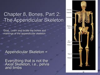
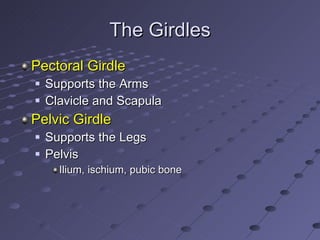
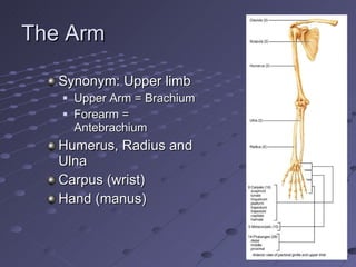
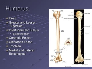
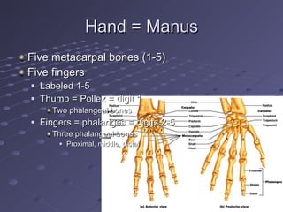
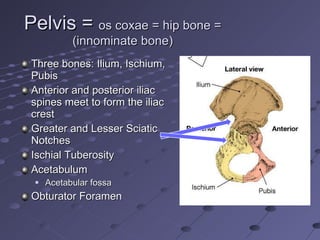
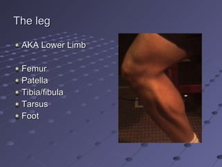
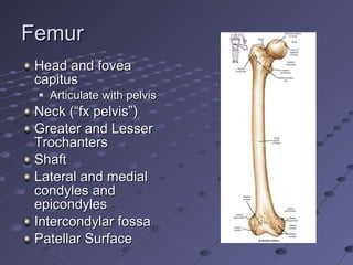
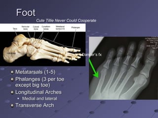
Chapter 8, Bones, Part   2:  The Appendicular Skeleton Appendicular Skeleton = Everything that is not the Axial Skeleton, i.e., pelvis and limbs Goal:  Learn and locate the bones and markings of the appendicular skeleton
The Girdles Pectoral Girdle Supports the Arms Clavicle and Scapula Pelvic Girdle Supports the Legs Pelvis Ilium, ischium, pubic bone
Clavicle  (collarbone) Manubrium to Acromion Frequently fractured
Scapula  (shoulder blade) Glenoid Spine Acromion Acromioclavicular joint Inferior and Superior Angles Origin of biceps brachii muscle: Coracoid Process Supraglenoid tubercle
The Arm Synonym: Upper limb Upper Arm = Brachium Forearm = Antebrachium Humerus, Radius and Ulna Carpus (wrist) Hand (manus)
Humerus Head Greater and Lesser Tubercles Intertubercular Sulcus Biceps tendon Coronoid Fossa Olecranon Fossa Trochlea Medial and Lateral Epicondyles
Radius  Ulna Head, neck, shaft Insertion of biceps brachii: Radial Tuberosity Radial Styloid Process Olecranon Trochlear notch Coronoid Process Ulnar Styloid Process Interosseous Membrane (between radius and ulna) Note how the two bones can cross “ Funny bone”
Carpus = Wrist Four Proximal Scaphoid, lunate, triquetrum, pisiform Four Distal Trapezium, trapezoid, capitate, hamate Scaphoid is frequently fractured
Hand = Manus Five metacarpal bones (1-5) Five fingers  Labeled 1-5 Thumb = Pollex = digit 1 Two phalangeal bones Fingers = phalanges = digits 2-5 Three phalangeal bones Proximal, middle, distal
The Girdles Pectoral Girdle Supports the Arms Clavicle and Scapula Pelvic Girdle Supports the Legs Pelvis (os coxae)
Pelvis =  os coxae = hip bone = (innominate bone) Three bones: Ilium, Ischium, Pubis Anterior and posterior iliac spines meet to form the iliac crest Greater and Lesser Sciatic Notches Ischial Tuberosity Acetabulum Acetabular fossa Obturator Foramen
More Pelvis Articular Surface for Articulation with Sacrum Difference between male and female Pelvic (or pubic) symphysis Fibrocartilage Stretches at childbirth (Relaxin)
The leg AKA Lower Limb Femur Patella Tibia/fibula Tarsus Foot
Femur Head and fovea capitus Articulate with pelvis  Neck (“fx pelvis”) Greater and Lesser Trochanters Shaft Lateral and medial condyles and epicondyles Intercondylar fossa Patellar Surface
Patella  = knee cap Sesamoid Bone Enclosed in the tendon of the quadriceps group of muscles “ Skyline” MRI of patella
Tibia  = shin bone Lateral and medial condyles Intercondylar eminence Tibial tuberosity Inferior articular surface Medial malleolus (= ankle bone) Interosseous Membrane
Fibula Head Shaft Lateral malleolus (= ankle bone) Not weight bearing Frequent fx Interosseous Membrane
Tarsus (7 bones)  Cute Tillie Never Could Cooperate   Navicular Cuboid Cunieform (3) Calcaneus Talus
Foot Metatarsals (1-5) Phalanges (3 per toe except big toe) Longitudinal Arches Medial and lateral Transverse Arch Dancer’s fx Cute Tillie Never Could Cooperate
Fractures  (a review) Bleeding Then clot Periosteal reaction Fibroblasts Osteoblasts Callus New bone “collar” Remodeling
“ Hip” fracture “ Grandma fell and broke her hip.” More accurately, “Grandma broke her femoral neck and then fell.” Sometimes the fx is at the intertrochanteric line Diabetes, hypertension, osteoporosis 25% die from complications in first year mostly related to immobility: Anesthesia Muscle Atrophy Pneumonia Decubitus ulcers Depression and disorientation
Douglas Iris

Chapter8 appendicularskeletonmarieb

  • 1.
    Chapter 8, Bones,Part 2: The Appendicular Skeleton Appendicular Skeleton = Everything that is not the Axial Skeleton, i.e., pelvis and limbs Goal: Learn and locate the bones and markings of the appendicular skeleton
  • 2.
    The Girdles PectoralGirdle Supports the Arms Clavicle and Scapula Pelvic Girdle Supports the Legs Pelvis Ilium, ischium, pubic bone
  • 3.
    Clavicle (collarbone)Manubrium to Acromion Frequently fractured
  • 4.
    Scapula (shoulderblade) Glenoid Spine Acromion Acromioclavicular joint Inferior and Superior Angles Origin of biceps brachii muscle: Coracoid Process Supraglenoid tubercle
  • 5.
    The Arm Synonym:Upper limb Upper Arm = Brachium Forearm = Antebrachium Humerus, Radius and Ulna Carpus (wrist) Hand (manus)
  • 6.
    Humerus Head Greaterand Lesser Tubercles Intertubercular Sulcus Biceps tendon Coronoid Fossa Olecranon Fossa Trochlea Medial and Lateral Epicondyles
  • 7.
    Radius UlnaHead, neck, shaft Insertion of biceps brachii: Radial Tuberosity Radial Styloid Process Olecranon Trochlear notch Coronoid Process Ulnar Styloid Process Interosseous Membrane (between radius and ulna) Note how the two bones can cross “ Funny bone”
  • 8.
    Carpus = WristFour Proximal Scaphoid, lunate, triquetrum, pisiform Four Distal Trapezium, trapezoid, capitate, hamate Scaphoid is frequently fractured
  • 9.
    Hand = ManusFive metacarpal bones (1-5) Five fingers Labeled 1-5 Thumb = Pollex = digit 1 Two phalangeal bones Fingers = phalanges = digits 2-5 Three phalangeal bones Proximal, middle, distal
  • 10.
    The Girdles PectoralGirdle Supports the Arms Clavicle and Scapula Pelvic Girdle Supports the Legs Pelvis (os coxae)
  • 11.
    Pelvis = os coxae = hip bone = (innominate bone) Three bones: Ilium, Ischium, Pubis Anterior and posterior iliac spines meet to form the iliac crest Greater and Lesser Sciatic Notches Ischial Tuberosity Acetabulum Acetabular fossa Obturator Foramen
  • 12.
    More Pelvis ArticularSurface for Articulation with Sacrum Difference between male and female Pelvic (or pubic) symphysis Fibrocartilage Stretches at childbirth (Relaxin)
  • 13.
    The leg AKALower Limb Femur Patella Tibia/fibula Tarsus Foot
  • 14.
    Femur Head andfovea capitus Articulate with pelvis Neck (“fx pelvis”) Greater and Lesser Trochanters Shaft Lateral and medial condyles and epicondyles Intercondylar fossa Patellar Surface
  • 15.
    Patella =knee cap Sesamoid Bone Enclosed in the tendon of the quadriceps group of muscles “ Skyline” MRI of patella
  • 16.
    Tibia =shin bone Lateral and medial condyles Intercondylar eminence Tibial tuberosity Inferior articular surface Medial malleolus (= ankle bone) Interosseous Membrane
  • 17.
    Fibula Head ShaftLateral malleolus (= ankle bone) Not weight bearing Frequent fx Interosseous Membrane
  • 18.
    Tarsus (7 bones) Cute Tillie Never Could Cooperate Navicular Cuboid Cunieform (3) Calcaneus Talus
  • 19.
    Foot Metatarsals (1-5)Phalanges (3 per toe except big toe) Longitudinal Arches Medial and lateral Transverse Arch Dancer’s fx Cute Tillie Never Could Cooperate
  • 20.
    Fractures (areview) Bleeding Then clot Periosteal reaction Fibroblasts Osteoblasts Callus New bone “collar” Remodeling
  • 21.
    “ Hip” fracture“ Grandma fell and broke her hip.” More accurately, “Grandma broke her femoral neck and then fell.” Sometimes the fx is at the intertrochanteric line Diabetes, hypertension, osteoporosis 25% die from complications in first year mostly related to immobility: Anesthesia Muscle Atrophy Pneumonia Decubitus ulcers Depression and disorientation
  • 22.