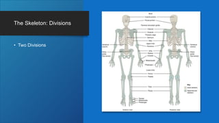
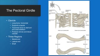
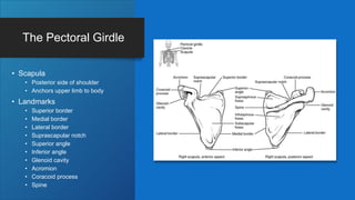
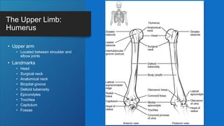
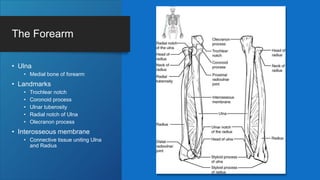
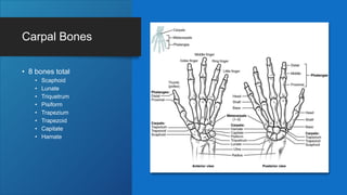
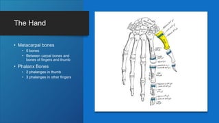
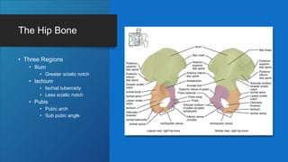
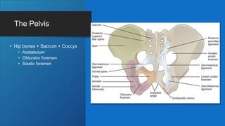
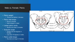
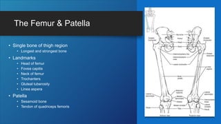
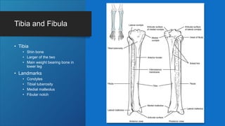
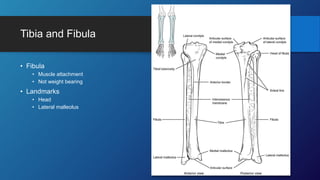
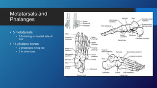
The Appendicular Skeleton
OpenStax Anatomy Chapter 8
The Pectoral Girdle: Objectives
DESCRIBE THE BONES THAT
FORM THE PECTORAL GIRDLE
LIST THE FUNCTIONS OF THE
PECTORAL GIRDLE
The Skeleton: Divisions
• Two Divisions
The Pectoral Girdle
• Clavicle
• Long bone, horizontal
• Supports scapula
• Transmits force to sternum
and axial skeleton
• Protects nerves and blood
vessels
• Three Regions
• Medial end
• Lateral end
• Shaft
The Pectoral Girdle
• Scapula
• Posterior side of shoulder
• Anchors upper limb to body
• Landmarks
• Superior border
• Medial border
• Lateral border
• Suprascapular notch
• Superior angle
• Inferior angle
• Glenoid cavity
• Acromion
• Coracoid process
• Spine
Bones of the Upper Limb: Objectives
Identify the divisions of the upper limb and describe the bones in
each region
List the bones and bony landmarks that articulate at each joint
of the upper limb
The Upper Limb:
Humerus
• Upper arm
• Located between shoulder and
elbow joints
• Landmarks
• Head
• Surgical neck
• Anatomical neck
• Bicipital groove
• Deltoid tuberosity
• Epicondyles
• Trochlea
• Capitulum
• Fossas
The Forearm
• Ulna
• Medial bone of forearm
• Landmarks
• Trochlear notch
• Coronoid process
• Ulnar tuberosity
• Radial notch of Ulna
• Olecranon process
• Interosseous membrane
• Connective tissue uniting Ulna
and Radius
The Forearm
• Radius
• Lateral bone of forearm
• Landmarks
• Head of radius
• Neck of radius
• Radial tuberosity
• Ulnar notch of the radius
• Styloid process of radius
• Interosseous membrane
• Connective tissue uniting Ulna
and Radius
Carpal Bones
• 8 bones total
• Scaphoid
• Lunate
• Triquetrum
• Pisiform
• Trapezium
• Trapezoid
• Capitate
• Hamate
Carpal Joints
• Scaphoid and Lunate articulate with radius
• Triquetrum articulates with fibrocartilaginous
pad
• Distal bones held together by ligaments
• Midcarpal joint
• Flexor retinaculum
• Spans top of area
• Maintains carpal grouping
• Carpal tunnel
The Hand
• Metacarpal bones
• 5 bones
• Between carpal bones and
bones of fingers and thumb
• Phalanx Bones
• 2 phalanges in thumb
• 3 phalanges in other fingers
The Pelvic
Girdle and
Pelvis:
Objectives
Define the pelvic girdle and
describe the bones and
ligaments of the pelvis
Explain the three regions of
the hip bone and identify
their bony landmarks
Describe the openings of the
pelvis and the boundaries of
the greater and lesser pelvis
The Hip Bone
• Three Regions
• Ilium
• Greater sciatic notch
• Ischium
• Ischial tuberosity
• Less sciatic notch
• Pubis
• Pubic arch
• Sub pubic angle
The Pelvis
• Hip bones + Sacrum + Coccyx
• Acetabulum
• Obturator foramen
• Sciatic foramen
Male vs. Female Pelvis
• Pelvic weight
• Thinner and lighter in females
• Pelvic inlet shape
• Round in female
• Heart shaped in males
• Lesser pelvic cavity
• Shorter and wider in females
• Subpubic angle
• Greater than 80 degrees in
females
• Less than 70 degrees in males
• Pelvic outlet shape
• Rounded and larger in females
Bones of the Lower Limb: Objectives
Identify the divisions of the lower limb and describe the bones of
each region
Describe the bones and bony landmarks that articulate at each
joint of the lower limb
The Femur & Patella
• Single bone of thigh region
• Longest and strongest bone
• Landmarks
• Head of femur
• Fovea capitis
• Neck of femur
• Trochanters
• Gluteal tuberosity
• Linea aspera
• Patella
• Sesamoid bone
• Tendon of quadriceps femoris
Tibia and Fibula
• Tibia
• Shin bone
• Larger of the two
• Main weight bearing bone in
lower leg
• Landmarks
• Condyles
• Tibial tuberosity
• Medial malleolus
• Fibular notch
Tibia and Fibula
• Fibula
• Muscle attachment
• Not weight bearing
• Landmarks
• Head
• Lateral malleolus
Tarsal Bones
• Talus
• Most superior bone
• Calcaneus
• Largest bone
• Cuboid
• Articulates with calcaneus
• Navicular
• Articulates with talus
• Cuneiform bones
• Medial , Lateral,
Intermmediate
Metatarsals and
Phalanges
• 5 metatarsals
• 1-5 starting on medial side of
foot
• 14 phalanx bones
• 2 phalanges in big toe
• 3 in other toes
Development of the Appendicular Skeleton:
Objectives
Describe the growth and development of the embryonic limb
buds
Discuss the appearance of primary and secondary ossification
centers
Limb Growth
• Limb Buds
• 4th week of development
• Upper and lower limbs
• Mesenchyme core covered with
ectoderm
• Apical ectodermal ridge
• Stimulates mesenchyme to
proliferate
• Outgrowth of limb
• 6th week
• Distal ends expand into paddle
shapes
• 7th week
• Upper limb rotates laterally 90
degrees
• Palm of hand faces anteriorly
Ossification of Bones
• Endochondral ossification
• All bones EXCEPT clavicle
• Mesenchyme within limb buds
differentiates into hyaline cartilage
• Primary ossification center forms
• Secondary ossification centers form
• Intramembranous ossification
• Clavicle ONLY
• Mesenchymal cells differentiate
directly into bone producing cells
• Complete at 25

OpenStax Anatomy and Physiology Chapter 8.pptx

  • 1.
  • 2.
    The Pectoral Girdle:Objectives DESCRIBE THE BONES THAT FORM THE PECTORAL GIRDLE LIST THE FUNCTIONS OF THE PECTORAL GIRDLE
  • 3.
  • 4.
    The Pectoral Girdle •Clavicle • Long bone, horizontal • Supports scapula • Transmits force to sternum and axial skeleton • Protects nerves and blood vessels • Three Regions • Medial end • Lateral end • Shaft
  • 5.
    The Pectoral Girdle •Scapula • Posterior side of shoulder • Anchors upper limb to body • Landmarks • Superior border • Medial border • Lateral border • Suprascapular notch • Superior angle • Inferior angle • Glenoid cavity • Acromion • Coracoid process • Spine
  • 6.
    Bones of theUpper Limb: Objectives Identify the divisions of the upper limb and describe the bones in each region List the bones and bony landmarks that articulate at each joint of the upper limb
  • 7.
    The Upper Limb: Humerus •Upper arm • Located between shoulder and elbow joints • Landmarks • Head • Surgical neck • Anatomical neck • Bicipital groove • Deltoid tuberosity • Epicondyles • Trochlea • Capitulum • Fossas
  • 8.
    The Forearm • Ulna •Medial bone of forearm • Landmarks • Trochlear notch • Coronoid process • Ulnar tuberosity • Radial notch of Ulna • Olecranon process • Interosseous membrane • Connective tissue uniting Ulna and Radius
  • 9.
    The Forearm • Radius •Lateral bone of forearm • Landmarks • Head of radius • Neck of radius • Radial tuberosity • Ulnar notch of the radius • Styloid process of radius • Interosseous membrane • Connective tissue uniting Ulna and Radius
  • 10.
    Carpal Bones • 8bones total • Scaphoid • Lunate • Triquetrum • Pisiform • Trapezium • Trapezoid • Capitate • Hamate
  • 11.
    Carpal Joints • Scaphoidand Lunate articulate with radius • Triquetrum articulates with fibrocartilaginous pad • Distal bones held together by ligaments • Midcarpal joint • Flexor retinaculum • Spans top of area • Maintains carpal grouping • Carpal tunnel
  • 12.
    The Hand • Metacarpalbones • 5 bones • Between carpal bones and bones of fingers and thumb • Phalanx Bones • 2 phalanges in thumb • 3 phalanges in other fingers
  • 13.
    The Pelvic Girdle and Pelvis: Objectives Definethe pelvic girdle and describe the bones and ligaments of the pelvis Explain the three regions of the hip bone and identify their bony landmarks Describe the openings of the pelvis and the boundaries of the greater and lesser pelvis
  • 14.
    The Hip Bone •Three Regions • Ilium • Greater sciatic notch • Ischium • Ischial tuberosity • Less sciatic notch • Pubis • Pubic arch • Sub pubic angle
  • 15.
    The Pelvis • Hipbones + Sacrum + Coccyx • Acetabulum • Obturator foramen • Sciatic foramen
  • 16.
    Male vs. FemalePelvis • Pelvic weight • Thinner and lighter in females • Pelvic inlet shape • Round in female • Heart shaped in males • Lesser pelvic cavity • Shorter and wider in females • Subpubic angle • Greater than 80 degrees in females • Less than 70 degrees in males • Pelvic outlet shape • Rounded and larger in females
  • 17.
    Bones of theLower Limb: Objectives Identify the divisions of the lower limb and describe the bones of each region Describe the bones and bony landmarks that articulate at each joint of the lower limb
  • 18.
    The Femur &Patella • Single bone of thigh region • Longest and strongest bone • Landmarks • Head of femur • Fovea capitis • Neck of femur • Trochanters • Gluteal tuberosity • Linea aspera • Patella • Sesamoid bone • Tendon of quadriceps femoris
  • 19.
    Tibia and Fibula •Tibia • Shin bone • Larger of the two • Main weight bearing bone in lower leg • Landmarks • Condyles • Tibial tuberosity • Medial malleolus • Fibular notch
  • 20.
    Tibia and Fibula •Fibula • Muscle attachment • Not weight bearing • Landmarks • Head • Lateral malleolus
  • 21.
    Tarsal Bones • Talus •Most superior bone • Calcaneus • Largest bone • Cuboid • Articulates with calcaneus • Navicular • Articulates with talus • Cuneiform bones • Medial , Lateral, Intermmediate
  • 22.
    Metatarsals and Phalanges • 5metatarsals • 1-5 starting on medial side of foot • 14 phalanx bones • 2 phalanges in big toe • 3 in other toes
  • 23.
    Development of theAppendicular Skeleton: Objectives Describe the growth and development of the embryonic limb buds Discuss the appearance of primary and secondary ossification centers
  • 24.
    Limb Growth • LimbBuds • 4th week of development • Upper and lower limbs • Mesenchyme core covered with ectoderm • Apical ectodermal ridge • Stimulates mesenchyme to proliferate • Outgrowth of limb • 6th week • Distal ends expand into paddle shapes • 7th week • Upper limb rotates laterally 90 degrees • Palm of hand faces anteriorly
  • 25.
    Ossification of Bones •Endochondral ossification • All bones EXCEPT clavicle • Mesenchyme within limb buds differentiates into hyaline cartilage • Primary ossification center forms • Secondary ossification centers form • Intramembranous ossification • Clavicle ONLY • Mesenchymal cells differentiate directly into bone producing cells • Complete at 25