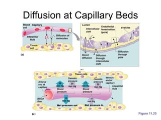
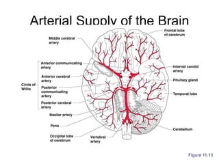
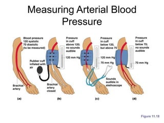
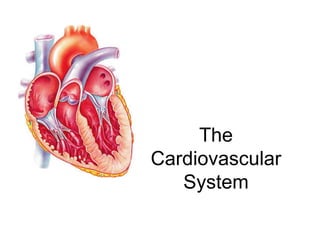
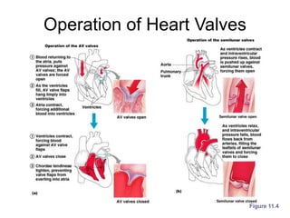
The
Cardiovascular
System
The Cardiovascular System
• A closed system of the heart and blood
vessels
– The heart pumps blood
– Blood vessels allow blood to circulate to all
parts of the body
• The function of the cardiovascular system
is to deliver oxygen and nutrients and to
remove carbon dioxide and other waste
products
The Heart
• Location
– Thorax between
the lungs
– Pointed apex
directed toward
left hip
• About the size of
your fist
The Heart: Coverings
• Pericardium – a double serous membrane
– Visceral pericardium - Next to heart
– Parietal pericardium - Outside layer
• Serous fluid fills the space between the
layers of pericardium
The Heart Wall: 3 layers
• Epicardium
• Outside layer
• This layer is the parietal pericardium
• Connective tissue layer
• Myocardium
• Middle layer
• Mostly cardiac muscle
• Endocardium
• Inner layer
• Endothelium
External Heart Anatomy
Figure 11.2a
The Heart: Chambers
• Right and left side act as
separate pumps
• Four chambers
– Atria
• Receiving chambers
– Right atrium
– Left atrium
– Ventricles
• Discharging chambers
– Right ventricle
– Left ventricle
Figure 11.2c
Blood
Circulation
Figure 11.3
The Heart: Valves
• Allow blood to flow in only one direction
• Four valves
– Atrioventricular valves – between atria and
ventricles
• Bicuspid valve (left)
• Tricuspid valve (right)
– Semilunar valves between ventricle and artery
• Pulmonary semilunar valve
• Aortic semilunar valve
The Heart: Valves
• Valves open as blood is pumped through
• Held in place by chordae tendineae (“heart
strings”)
• Close to prevent backflow
Operation of Heart Valves
Figure 11.4
The Heart:
Associated Great Vessels
• Aorta - leaves left ventricle
• Pulmonary arteries - leave right ventricle
• Vena cava - enters right atrium
• Pulmonary veins (four) - enter left atrium
Coronary Circulation
• Blood in the heart chambers does not
nourish the myocardium
• The heart has its own nourishing
circulatory system
– Coronary arteries
– Cardiac veins
– Blood empties into the right atrium via the
coronary sinus
The Heart: Conduction System
• Intrinsic conduction system
(nodal system)
– Heart muscle cells contract, without nerve
impulses, in a regular, continuous way
• Special tissue sets the pace
• Sinoatrial node (SA) - Pacemaker
• Atrioventricular node (AV)
• Atrioventricular bundle
• Bundle branches
• Purkinje fibers
The Heart’s Cardiac Cycle
• Atria contract simultaneously
• Atria relax, then ventricles contract
• Systole = contraction
• Diastole = relaxation
Heart Contractions
• Contraction is initiated by the sinoatrial node
• Sequential stimulation occurs at other
autorhythmic cells
Filling of Heart Chambers – the
Cardiac Cycle
Figure 11.6
The Heart: Cardiac Cycle
• Cardiac cycle – events of one complete
heart beat
– Mid-to-late diastole – blood flows into
ventricles
– Ventricular systole – blood pressure builds
before ventricle contracts, pushing out blood
– Early diastole – atria finish re-filling,
ventricular pressure is low
The Heart: Cardiac Output
• Cardiac output (CO)
– Amount of blood pumped by each side of the
heart in one minute
– CO = (heart rate [HR]) x (stroke volume [SV])
• Stroke volume
– Volume of blood pumped by each ventricle in
one contraction
Cardiac Output Regulation
Figure 11.7
Regulation of Heart Rate
• Stroke volume usually remains relatively
constant
– Starling’s law of the heart – the more that the
cardiac muscle is stretched, the stronger the
contraction
• Changing heart rate is the most common
way to change cardiac output
Regulation of Heart Rate
• Increased heart rate
– Sympathetic nervous system
• Activated in a Crisis
• Low blood pressure
– Hormones
• Epinephrine
• Thyroxine
– Exercise
– Decreased blood volume
Regulation of Heart Rate
• Decreased heart rate
– Parasympathetic nervous system
– High blood pressure or blood volume
– Decreased venous return
Blood Vessels: The Vascular
System
• Taking blood to the tissues and back
– Arteries
– Arterioles
– Capillaries
– Venules
– Veins
Figure 11.8a
The Vascular System
Figure 11.8b
Blood Vessels: Anatomy
• Three layers (tunics)
– Tunic intima:
• Endothelium
– Tunic media
• Smooth muscle
• Controlled by sympathetic nervous system
– Tunic externa
• Mostly fibrous connective tissue
Differences Between Blood
Vessel Types
• Walls of arteries are the thickest
• Lumens of veins are larger
• Skeletal muscle “milks” blood in veins
toward the heart
• Walls of capillaries are only one cell layer
thick to allow for exchanges between
blood and tissue
Movement of Blood Through
Vessels
• Most arterial blood is
pumped by the heart
• Veins use the milking
action of muscles to
help move blood
Figure 11.9
Capillary Beds
• Capillary beds
consist of two types
of vessels
– Vascular shunt –
directly connects an
arteriole to a venule
Figure 11.10
Capillary Beds
• True capillaries –
exchange vessels
• Oxygen and nutrients
cross to cells
• Carbon dioxide and
metabolic waste
products cross into
blood
Figure 11.10
Diffusion at Capillary Beds
Figure 11.20
Major Arteries of Systemic
Circulation
Figure 11.11
Major Veins of Systemic
Circulation
Figure 11.12
Arterial Supply of the Brain
Figure 11.13
Hepatic Portal Circulation
Figure 11.14
Circulation to the Fetus
Figure 11.15
Pulse
• Pulse – pressure
wave of blood
• Monitored at
“pressure points”
where pulse is
easily palpated
Figure 11.16
Blood Pressure
• Measurements by health professionals are
made on the pressure in large arteries
– Systolic – pressure at the peak of ventricular
contraction
– Diastolic – pressure when ventricles relax
• Pressure in blood vessels decreases as
the distance away from the heart
increases
Measuring Arterial Blood
Pressure
Figure 11.18
Blood Pressure: Effects of
Factors
• Neural factors
– Autonomic nervous system adjustments
(sympathetic division)
• Renal factors
– Regulation by altering blood volume
– Renin – hormonal control
Blood Pressure: Effects of
Factors
• Temperature
– Heat has a vasodilation effect
– Cold has a vasoconstricting effect
• Chemicals
– Various substances can cause increases or
decreases
• Diet
Variations in Blood Pressure
• Human normal range is variable
– Normal
• 140–110 mm Hg systolic
• 80–75 mm Hg diastolic
– Hypotension
• Low systolic (below 110 mm HG)
• Often associated with illness
– Hypertension
• High systolic (above 140 mm HG)
• Can be dangerous if it is chronic
Capillary Exchange
• Substances exchanged due to
concentration gradients
– Oxygen and nutrients leave the blood
– Carbon dioxide and other wastes leave the
cells
Capillary Exchange:
Mechanisms
• Direct diffusion across plasma membranes
• Endocytosis or exocytosis
• Some capillaries have gaps (intercellular
clefts)
– Plasma membrane not joined by tight
junctions
• Fenestrations of some capillaries
– Fenestrations = pores
Developmental Aspects of the
Cardiovascular System
• A simple “tube heart” develops in the
embryo and pumps by the fourth week
• The heart becomes a four-chambered
organ by the end of seven weeks
• Few structural changes occur after the
seventh week

Ch 11 cardiovascular system

  • 1.
  • 2.
    The Cardiovascular System •A closed system of the heart and blood vessels – The heart pumps blood – Blood vessels allow blood to circulate to all parts of the body • The function of the cardiovascular system is to deliver oxygen and nutrients and to remove carbon dioxide and other waste products
  • 3.
    The Heart • Location –Thorax between the lungs – Pointed apex directed toward left hip • About the size of your fist
  • 4.
    The Heart: Coverings •Pericardium – a double serous membrane – Visceral pericardium - Next to heart – Parietal pericardium - Outside layer • Serous fluid fills the space between the layers of pericardium
  • 5.
    The Heart Wall:3 layers • Epicardium • Outside layer • This layer is the parietal pericardium • Connective tissue layer • Myocardium • Middle layer • Mostly cardiac muscle • Endocardium • Inner layer • Endothelium
  • 6.
  • 7.
    The Heart: Chambers •Right and left side act as separate pumps • Four chambers – Atria • Receiving chambers – Right atrium – Left atrium – Ventricles • Discharging chambers – Right ventricle – Left ventricle Figure 11.2c
  • 8.
  • 9.
    The Heart: Valves •Allow blood to flow in only one direction • Four valves – Atrioventricular valves – between atria and ventricles • Bicuspid valve (left) • Tricuspid valve (right) – Semilunar valves between ventricle and artery • Pulmonary semilunar valve • Aortic semilunar valve
  • 10.
    The Heart: Valves •Valves open as blood is pumped through • Held in place by chordae tendineae (“heart strings”) • Close to prevent backflow
  • 11.
    Operation of HeartValves Figure 11.4
  • 12.
    The Heart: Associated GreatVessels • Aorta - leaves left ventricle • Pulmonary arteries - leave right ventricle • Vena cava - enters right atrium • Pulmonary veins (four) - enter left atrium
  • 13.
    Coronary Circulation • Bloodin the heart chambers does not nourish the myocardium • The heart has its own nourishing circulatory system – Coronary arteries – Cardiac veins – Blood empties into the right atrium via the coronary sinus
  • 14.
    The Heart: ConductionSystem • Intrinsic conduction system (nodal system) – Heart muscle cells contract, without nerve impulses, in a regular, continuous way • Special tissue sets the pace • Sinoatrial node (SA) - Pacemaker • Atrioventricular node (AV) • Atrioventricular bundle • Bundle branches • Purkinje fibers
  • 15.
    The Heart’s CardiacCycle • Atria contract simultaneously • Atria relax, then ventricles contract • Systole = contraction • Diastole = relaxation
  • 16.
    Heart Contractions • Contractionis initiated by the sinoatrial node • Sequential stimulation occurs at other autorhythmic cells
  • 17.
    Filling of HeartChambers – the Cardiac Cycle Figure 11.6
  • 18.
    The Heart: CardiacCycle • Cardiac cycle – events of one complete heart beat – Mid-to-late diastole – blood flows into ventricles – Ventricular systole – blood pressure builds before ventricle contracts, pushing out blood – Early diastole – atria finish re-filling, ventricular pressure is low
  • 19.
    The Heart: CardiacOutput • Cardiac output (CO) – Amount of blood pumped by each side of the heart in one minute – CO = (heart rate [HR]) x (stroke volume [SV]) • Stroke volume – Volume of blood pumped by each ventricle in one contraction
  • 20.
  • 21.
    Regulation of HeartRate • Stroke volume usually remains relatively constant – Starling’s law of the heart – the more that the cardiac muscle is stretched, the stronger the contraction • Changing heart rate is the most common way to change cardiac output
  • 22.
    Regulation of HeartRate • Increased heart rate – Sympathetic nervous system • Activated in a Crisis • Low blood pressure – Hormones • Epinephrine • Thyroxine – Exercise – Decreased blood volume
  • 23.
    Regulation of HeartRate • Decreased heart rate – Parasympathetic nervous system – High blood pressure or blood volume – Decreased venous return
  • 24.
    Blood Vessels: TheVascular System • Taking blood to the tissues and back – Arteries – Arterioles – Capillaries – Venules – Veins Figure 11.8a
  • 25.
  • 26.
    Blood Vessels: Anatomy •Three layers (tunics) – Tunic intima: • Endothelium – Tunic media • Smooth muscle • Controlled by sympathetic nervous system – Tunic externa • Mostly fibrous connective tissue
  • 27.
    Differences Between Blood VesselTypes • Walls of arteries are the thickest • Lumens of veins are larger • Skeletal muscle “milks” blood in veins toward the heart • Walls of capillaries are only one cell layer thick to allow for exchanges between blood and tissue
  • 28.
    Movement of BloodThrough Vessels • Most arterial blood is pumped by the heart • Veins use the milking action of muscles to help move blood Figure 11.9
  • 29.
    Capillary Beds • Capillarybeds consist of two types of vessels – Vascular shunt – directly connects an arteriole to a venule Figure 11.10
  • 30.
    Capillary Beds • Truecapillaries – exchange vessels • Oxygen and nutrients cross to cells • Carbon dioxide and metabolic waste products cross into blood Figure 11.10
  • 31.
    Diffusion at CapillaryBeds Figure 11.20
  • 32.
    Major Arteries ofSystemic Circulation Figure 11.11
  • 33.
    Major Veins ofSystemic Circulation Figure 11.12
  • 34.
    Arterial Supply ofthe Brain Figure 11.13
  • 35.
  • 36.
    Circulation to theFetus Figure 11.15
  • 37.
    Pulse • Pulse –pressure wave of blood • Monitored at “pressure points” where pulse is easily palpated Figure 11.16
  • 38.
    Blood Pressure • Measurementsby health professionals are made on the pressure in large arteries – Systolic – pressure at the peak of ventricular contraction – Diastolic – pressure when ventricles relax • Pressure in blood vessels decreases as the distance away from the heart increases
  • 39.
  • 40.
    Blood Pressure: Effectsof Factors • Neural factors – Autonomic nervous system adjustments (sympathetic division) • Renal factors – Regulation by altering blood volume – Renin – hormonal control
  • 41.
    Blood Pressure: Effectsof Factors • Temperature – Heat has a vasodilation effect – Cold has a vasoconstricting effect • Chemicals – Various substances can cause increases or decreases • Diet
  • 42.
    Variations in BloodPressure • Human normal range is variable – Normal • 140–110 mm Hg systolic • 80–75 mm Hg diastolic – Hypotension • Low systolic (below 110 mm HG) • Often associated with illness – Hypertension • High systolic (above 140 mm HG) • Can be dangerous if it is chronic
  • 43.
    Capillary Exchange • Substancesexchanged due to concentration gradients – Oxygen and nutrients leave the blood – Carbon dioxide and other wastes leave the cells
  • 44.
    Capillary Exchange: Mechanisms • Directdiffusion across plasma membranes • Endocytosis or exocytosis • Some capillaries have gaps (intercellular clefts) – Plasma membrane not joined by tight junctions • Fenestrations of some capillaries – Fenestrations = pores
  • 45.
    Developmental Aspects ofthe Cardiovascular System • A simple “tube heart” develops in the embryo and pumps by the fourth week • The heart becomes a four-chambered organ by the end of seven weeks • Few structural changes occur after the seventh week