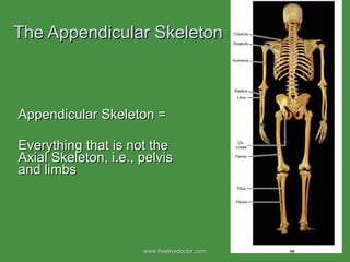
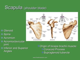
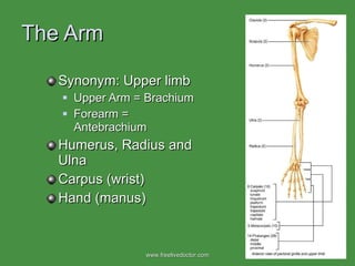
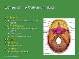
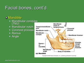
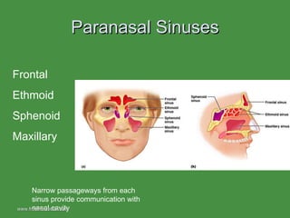
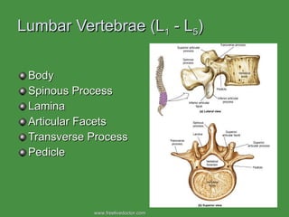
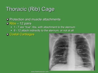
The Appendicular Skeleton  Appendicular Skeleton = Everything that is not the Axial Skeleton, i.e., pelvis and limbs www.freelivedoctor.com
Clavicle  (collarbone) Manubrium to Acromion Frequently fractured www.freelivedoctor.com
Scapula  (shoulder blade) Glenoid Spine Acromion Acromioclavicular joint Inferior and Superior Angles Origin of biceps brachii muscle: Coracoid Process Supraglenoid tubercle www.freelivedoctor.com
The Arm Synonym: Upper limb Upper Arm = Brachium Forearm = Antebrachium Humerus, Radius and Ulna Carpus (wrist) Hand (manus) www.freelivedoctor.com
Humerus Head Greater and Lesser Tubercles Intertubercular Sulcus Biceps tendon Coronoid Fossa Olecranon Fossa Trochlea Medial and Lateral Epicondyles www.freelivedoctor.com
Radius  Ulna Head, neck, shaft Insertion of biceps brachii: Radial Tuberosity Radial Styloid Process Olecranon Trochlear notch Coronoid Process Ulnar Styloid Process Interosseous Membrane (between radius and ulna) Note how the two bones can cross “ Funny bone” www.freelivedoctor.com
Carpus = Wrist Four Proximal Scaphoid, lunate, triquetrum, pisiform Four Distal Trapezium, trapezoid, capitate, hamate Scaphoid is frequently fractured www.freelivedoctor.com
Hand = Manus Five metacarpal bones (1-5) Five fingers  Labeled 1-5 Thumb = Pollex = digit 1 Two phalangeal bones Fingers = phalanges = digits 2-5 Three phalangeal bones Proximal, middle, distal www.freelivedoctor.com
The Girdles Pectoral Girdle Supports the Arms Clavicle and Scapula Pelvic Girdle Supports the Legs Pelvis (os coxae) www.freelivedoctor.com
Pelvis =  os coxae = hip bone Three bones: Ilium, Ischium, Pubis (innominate bone) Anterior and posterior iliac spines meet to form the iliac crest Greater and Lesser Sciatic Notches Ischial Tuberosity Acetabulum Acetabular fossa Obturator Foramen www.freelivedoctor.com
More Pelvis Articular Surface for Articulation with Sacrum Difference between male and female Pelvic (or pubic) symphysis Fibrocartilage Stretches at childbirth (Relaxin) www.freelivedoctor.com
The leg AKA Lower Limb Femur Patella Tibia/fibula Tarsus Foot www.freelivedoctor.com
Femur Head and fovea capitus Articulate with pelvis  Neck (“fx pelvis”) Greater and Lesser Trochanters Shaft Lateral and medial condyles and epicondyles Intercondylar fossa Patellar Surface www.freelivedoctor.com
Patella  = knee cap Sesamoid Bone Enclosed in the tendon of the quadriceps group of muscles “ Skyline” MRI of patella www.freelivedoctor.com
Tibia  = shin bone Lateral and medial condyles Intercondylar eminence Tibial tuberosity Inferior articular surface Medial malleolus (= ankle bone) Interosseous Membrane www.freelivedoctor.com
Fibula Head Shaft Lateral malleolus (= ankle bone) Not weight bearing Frequent fx Interosseous Membrane www.freelivedoctor.com
Tarsus (7 bones)  Cute Tillie Never Could Cooperate   Navicular Cuboid Cunieform (3) Calcaneus Talus www.freelivedoctor.com
Foot Metatarsals (1-5) Phalanges (3 per toe except big toe) Longitudinal Arches Medial and lateral Transverse Arch Dancer’s fx Cute Tillie Never Could Cooperate   www.freelivedoctor.com
Bones of the Skull www.freelivedoctor.com
Cranial Bones, AKA calvarium  vs.  Facial Bones www.freelivedoctor.com
Bones of the Calvarium Frontal  (forehead) Anterior fossa of base of skull Frontal Crests More prominent in male Frontal sinuses Parietal  (2) Sutures Occipital Foramen Magnum Occipital Condyles articulate with C 1 Occipital Crest www.freelivedoctor.com
Bones of the Calvarium, cont’d Temporal (4 parts):  Squamous (very thin), Mastoid (means breast-like) process Small sinus Petrous  Houses inner ear Acoustic meatus Jugular foramen Zygomatic process Styloid Process “ In one of these, the squamosal section of the temporal bone had been crushed, and another showed a jagged hole through the parietal bone.” The Bounty Trilogy, 1788 www.freelivedoctor.com
Bones of the Calvarium floor Sphenoid Sella turcica houses pituitary gland Ethmoid  (sieve)  Cribriform plate for olfactory nerve Crista galli Part of nasal septum Frontal Temporal Petrous part Occipital Foramen magnum www.freelivedoctor.com
Facial bones Maxilla (2) Alveolar margin for teeth Maxillary sinus Anterior portion of hard palate Palatine (2) Posterior aspect of hard palate Nasal (2) Inferior Nasal conchae (2) Will be covered in Respiratory System www.freelivedoctor.com
Facial bones, cont’d Zygomatic (2)  AKA cheek bones Zygomatic process of temporal bone Lacrimal (2) Lacrimal sac Vomer Part of nasal septum www.freelivedoctor.com
Facial bones, cont’d Mandible Mandibular condyles (TMJ) Mandibular notch Coronoid process Ramus Angle www.freelivedoctor.com
www.freelivedoctor.com
Sutures and Fontanels Fibrous Articulation, no movement, fused in adulthood Lambdoidal—betw occipital and parietal (mid) Saggital --- betw parietals Coronal– betw frontal and parietals Squamous – betw parietals and temporals Anterior and Posterior Fontanels “ Soft spot” of Newborn www.freelivedoctor.com
Paranasal Sinuses Frontal Ethmoid Sphenoid Maxillary Narrow passageways from each sinus provide communication with nasal cavity www.freelivedoctor.com
Hyoid Bone Inferior to the mandible Superior to the larynx Supports many cervical muscles Supports the larynx www.freelivedoctor.com
The Vertebral Column 7 Cervical 12 Thoracic 5 Lumbar 5 Sacral (fused) 4 Coccygeal (usually) Curvatures: Cervical, Thoracic, Lumbar, Sacral www.freelivedoctor.com
A “Typical” Vertebra Body and intervertebral disks Spinous Process Intervertebral Foramina Articular processes/facets Superior Inferior Vertebral (neural) Arch Pedicles and Laminae Vertebral Canal/foramen www.freelivedoctor.com
Cervical Vertebrae (C 1  – C  7 ) C 1  = Atlas (no body) C 1 -C 2  – site of rotation C 2  = Axis (dens) C 3  - C 6  are similar Note the  transverse foramina  in each cervical vertebra Vertebral Artery/Vein C 7  has the vertebra prominens C 1 www.freelivedoctor.com
Thoracic Vertebrae (T 1  - T 12 ) Large Spinous Processes Articulations for ribs, both superior and inferior www.freelivedoctor.com
Lumbar Vertebrae (L 1  - L 5 ) Body Spinous Process Lamina Articular Facets Transverse Process Pedicle www.freelivedoctor.com
Intervertebral Articulation www.freelivedoctor.com
Intervertebral Disk Nucleus Pulposus Annulus Fibrosus Fibrocartilage See Fig. 8-9 www.freelivedoctor.com
Damage to the Intervertebral Discs www.freelivedoctor.com
The Girdles Pectoral Girdle Supports the Arms Clavicle and Scapula Pelvic Girdle Supports the Legs Pelvis Ilium, ischium, pubic bone www.freelivedoctor.com
www.freelivedoctor.com
Sacrum (5 fused) Coccyx (3-5 fused) Sacroiliac joint Sacral Foramina www.freelivedoctor.com
Thoracic (Rib) Cage Protection and muscle attachments Ribs  – 12 pairs 1 - 7 are “true” ribs, with attachment to the sternum 8 - 12 attach indirectly to the sternum, or not at all Costal Cartilages www.freelivedoctor.com
Thoracic (Rib) Cage, cont’d Sternum Manubrium Clavicular Notch Body Xiphoid www.freelivedoctor.com

Appendicular&Axial Skeleton

  • 1.
    The Appendicular Skeleton Appendicular Skeleton = Everything that is not the Axial Skeleton, i.e., pelvis and limbs www.freelivedoctor.com
  • 2.
    Clavicle (collarbone)Manubrium to Acromion Frequently fractured www.freelivedoctor.com
  • 3.
    Scapula (shoulderblade) Glenoid Spine Acromion Acromioclavicular joint Inferior and Superior Angles Origin of biceps brachii muscle: Coracoid Process Supraglenoid tubercle www.freelivedoctor.com
  • 4.
    The Arm Synonym:Upper limb Upper Arm = Brachium Forearm = Antebrachium Humerus, Radius and Ulna Carpus (wrist) Hand (manus) www.freelivedoctor.com
  • 5.
    Humerus Head Greaterand Lesser Tubercles Intertubercular Sulcus Biceps tendon Coronoid Fossa Olecranon Fossa Trochlea Medial and Lateral Epicondyles www.freelivedoctor.com
  • 6.
    Radius UlnaHead, neck, shaft Insertion of biceps brachii: Radial Tuberosity Radial Styloid Process Olecranon Trochlear notch Coronoid Process Ulnar Styloid Process Interosseous Membrane (between radius and ulna) Note how the two bones can cross “ Funny bone” www.freelivedoctor.com
  • 7.
    Carpus = WristFour Proximal Scaphoid, lunate, triquetrum, pisiform Four Distal Trapezium, trapezoid, capitate, hamate Scaphoid is frequently fractured www.freelivedoctor.com
  • 8.
    Hand = ManusFive metacarpal bones (1-5) Five fingers Labeled 1-5 Thumb = Pollex = digit 1 Two phalangeal bones Fingers = phalanges = digits 2-5 Three phalangeal bones Proximal, middle, distal www.freelivedoctor.com
  • 9.
    The Girdles PectoralGirdle Supports the Arms Clavicle and Scapula Pelvic Girdle Supports the Legs Pelvis (os coxae) www.freelivedoctor.com
  • 10.
    Pelvis = os coxae = hip bone Three bones: Ilium, Ischium, Pubis (innominate bone) Anterior and posterior iliac spines meet to form the iliac crest Greater and Lesser Sciatic Notches Ischial Tuberosity Acetabulum Acetabular fossa Obturator Foramen www.freelivedoctor.com
  • 11.
    More Pelvis ArticularSurface for Articulation with Sacrum Difference between male and female Pelvic (or pubic) symphysis Fibrocartilage Stretches at childbirth (Relaxin) www.freelivedoctor.com
  • 12.
    The leg AKALower Limb Femur Patella Tibia/fibula Tarsus Foot www.freelivedoctor.com
  • 13.
    Femur Head andfovea capitus Articulate with pelvis Neck (“fx pelvis”) Greater and Lesser Trochanters Shaft Lateral and medial condyles and epicondyles Intercondylar fossa Patellar Surface www.freelivedoctor.com
  • 14.
    Patella =knee cap Sesamoid Bone Enclosed in the tendon of the quadriceps group of muscles “ Skyline” MRI of patella www.freelivedoctor.com
  • 15.
    Tibia =shin bone Lateral and medial condyles Intercondylar eminence Tibial tuberosity Inferior articular surface Medial malleolus (= ankle bone) Interosseous Membrane www.freelivedoctor.com
  • 16.
    Fibula Head ShaftLateral malleolus (= ankle bone) Not weight bearing Frequent fx Interosseous Membrane www.freelivedoctor.com
  • 17.
    Tarsus (7 bones) Cute Tillie Never Could Cooperate Navicular Cuboid Cunieform (3) Calcaneus Talus www.freelivedoctor.com
  • 18.
    Foot Metatarsals (1-5)Phalanges (3 per toe except big toe) Longitudinal Arches Medial and lateral Transverse Arch Dancer’s fx Cute Tillie Never Could Cooperate www.freelivedoctor.com
  • 19.
    Bones of theSkull www.freelivedoctor.com
  • 20.
    Bones of theSkull www.freelivedoctor.com
  • 21.
    Cranial Bones, AKAcalvarium vs. Facial Bones www.freelivedoctor.com
  • 22.
    Bones of theCalvarium Frontal (forehead) Anterior fossa of base of skull Frontal Crests More prominent in male Frontal sinuses Parietal (2) Sutures Occipital Foramen Magnum Occipital Condyles articulate with C 1 Occipital Crest www.freelivedoctor.com
  • 23.
    Bones of theCalvarium, cont’d Temporal (4 parts): Squamous (very thin), Mastoid (means breast-like) process Small sinus Petrous Houses inner ear Acoustic meatus Jugular foramen Zygomatic process Styloid Process “ In one of these, the squamosal section of the temporal bone had been crushed, and another showed a jagged hole through the parietal bone.” The Bounty Trilogy, 1788 www.freelivedoctor.com
  • 24.
    Bones of theCalvarium floor Sphenoid Sella turcica houses pituitary gland Ethmoid (sieve) Cribriform plate for olfactory nerve Crista galli Part of nasal septum Frontal Temporal Petrous part Occipital Foramen magnum www.freelivedoctor.com
  • 25.
    Facial bones Maxilla(2) Alveolar margin for teeth Maxillary sinus Anterior portion of hard palate Palatine (2) Posterior aspect of hard palate Nasal (2) Inferior Nasal conchae (2) Will be covered in Respiratory System www.freelivedoctor.com
  • 26.
    Facial bones, cont’dZygomatic (2) AKA cheek bones Zygomatic process of temporal bone Lacrimal (2) Lacrimal sac Vomer Part of nasal septum www.freelivedoctor.com
  • 27.
    Facial bones, cont’dMandible Mandibular condyles (TMJ) Mandibular notch Coronoid process Ramus Angle www.freelivedoctor.com
  • 28.
  • 29.
    Sutures and FontanelsFibrous Articulation, no movement, fused in adulthood Lambdoidal—betw occipital and parietal (mid) Saggital --- betw parietals Coronal– betw frontal and parietals Squamous – betw parietals and temporals Anterior and Posterior Fontanels “ Soft spot” of Newborn www.freelivedoctor.com
  • 30.
    Paranasal Sinuses FrontalEthmoid Sphenoid Maxillary Narrow passageways from each sinus provide communication with nasal cavity www.freelivedoctor.com
  • 31.
    Hyoid Bone Inferiorto the mandible Superior to the larynx Supports many cervical muscles Supports the larynx www.freelivedoctor.com
  • 32.
    The Vertebral Column7 Cervical 12 Thoracic 5 Lumbar 5 Sacral (fused) 4 Coccygeal (usually) Curvatures: Cervical, Thoracic, Lumbar, Sacral www.freelivedoctor.com
  • 33.
    A “Typical” VertebraBody and intervertebral disks Spinous Process Intervertebral Foramina Articular processes/facets Superior Inferior Vertebral (neural) Arch Pedicles and Laminae Vertebral Canal/foramen www.freelivedoctor.com
  • 34.
    Cervical Vertebrae (C1 – C 7 ) C 1 = Atlas (no body) C 1 -C 2 – site of rotation C 2 = Axis (dens) C 3 - C 6 are similar Note the transverse foramina in each cervical vertebra Vertebral Artery/Vein C 7 has the vertebra prominens C 1 www.freelivedoctor.com
  • 35.
    Thoracic Vertebrae (T1 - T 12 ) Large Spinous Processes Articulations for ribs, both superior and inferior www.freelivedoctor.com
  • 36.
    Lumbar Vertebrae (L1 - L 5 ) Body Spinous Process Lamina Articular Facets Transverse Process Pedicle www.freelivedoctor.com
  • 37.
  • 38.
    Intervertebral Disk NucleusPulposus Annulus Fibrosus Fibrocartilage See Fig. 8-9 www.freelivedoctor.com
  • 39.
    Damage to theIntervertebral Discs www.freelivedoctor.com
  • 40.
    The Girdles PectoralGirdle Supports the Arms Clavicle and Scapula Pelvic Girdle Supports the Legs Pelvis Ilium, ischium, pubic bone www.freelivedoctor.com
  • 41.
  • 42.
    Sacrum (5 fused)Coccyx (3-5 fused) Sacroiliac joint Sacral Foramina www.freelivedoctor.com
  • 43.
    Thoracic (Rib) CageProtection and muscle attachments Ribs – 12 pairs 1 - 7 are “true” ribs, with attachment to the sternum 8 - 12 attach indirectly to the sternum, or not at all Costal Cartilages www.freelivedoctor.com
  • 44.
    Thoracic (Rib) Cage,cont’d Sternum Manubrium Clavicular Notch Body Xiphoid www.freelivedoctor.com

Editor's Notes