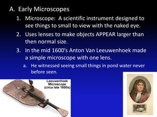
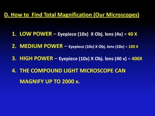
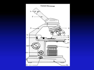
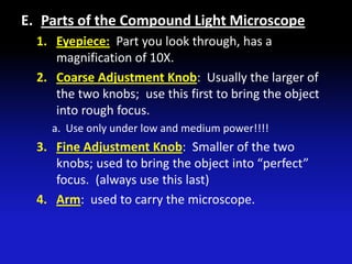
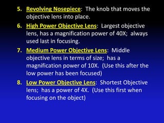
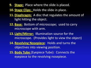
Chapter 2: Section 2
  (Viewing Cells)
   III. Viewing Cells
A. Early Microscopes
  1. Microscope: A scientific instrument designed to
     see things to small to view with the naked eye.
  2. Uses lenses to make objects APPEAR larger than
     then normal size.
  3. In the mid 1600’s Anton Van Leeuwenhoek made
     a simple microscope with one lens.
    a. He witnessed seeing small things in pond water never
       before seen.
B. Types of Lenses Used in Microscopes
  1. CONVEX LENS: lens that looks like a teardrop
     and magnifies.
    a. Used in microscopes and glasses for farsightedness.
  2. CONCAVE LENS: Lens that looks like an hourglass
     and bends light inward; good for looking at far
     away objects.
    a. Used in telescopes, binoculars, and glasses for
       nearsightedness.
Types of Lenses (diagram)
Convex vs Concave
C. Types of Microscopes
  1. Simple Microscope: has 1 lens, similar to a
     magnifying glass.
  2. Compound Light Microscope:
    a. Usually has 2 or 3 lenses inside of them; 1 in the
       eyepiece, one in the tube, and one in the objective.
    b. To find total magnification you must multiply the
       power of the lenses.
    c. Because there is an odd number of lenses the image
       will be upside down and reversed.
    d. This is the type we use in our school.
    e. Stereo Microscope: View through two eyes and
       produces a 3-D image.
3. ELECTRON MICROSOPES: Use a magnetic
   field in a vacuum to direct beams of electrons
   to view objects to mall to otherwise be seen;
   electron microscope images must be
   photographed or electronically produced.
  a. Scanning Electron Microscope (SEM): produces
     a 3-D image of the surface of a cell; magnifies
     around 200,000 X.
  b. Transmission Electron Microscope (TEM):
     Produces a 2-D image of a thinly sliced specimen;
     magnifies up to 1,000,000 X.
SEM – HUMAN HAIR
SEM - SPIDER
SEM - POLLEN
SEM – Fly’s HEAD
TEM – Fish Gills
TEM – Pollen Grains
TEM – Cell’s NUCLEUS
D. How to Find Total Magnification (Our Microscopes)


  1. LOW POWER – Eyepiece (10x) X Obj. lens (4x) = 40 X

  2. MEDIUM POWER – Eyepiece (10x) X Obj. lens (10x) = 100 X

  3. HIGH POWER – Eyepiece (10x) X Obj. lens (40 x) = 400X

  4. THE COMPOUND LIGHT MICROSCOPE CAN
     MAGNIFY UP TO 2000 x.
E. Parts of the Compound Light Microscope
  1. Eyepiece: Part you look through, has a
     magnification of 10X.
  2. Coarse Adjustment Knob: Usually the larger of
     the two knobs; use this first to bring the object
     into rough focus.
    a. Use only under low and medium power!!!!
  3. Fine Adjustment Knob: Smaller of the two
     knobs; used to bring the object into “perfect”
     focus. (always use this last)
  4. Arm: used to carry the microscope.
5. Revolving Nosepiece: The knob that moves the
   objective lens into place.
6. High Power Objective Lens: Largest objective
   lens, has a magnification power of 40X; always
   used last in focusing.
7. Medium Power Objective Lens: Middle
   objective lens in terms of size; has a
   magnification power of 10X. (Use this after the
   low power has been focused)
8. Low Power Objective Lens: Shortest Objective
   lens; has a power of 4X. (Use this first when
   focusing on the object)
9. Stage: Place where the slide is placed.
10. Stage Clips: Holds the slide in place.
11. Diaphragm: A disc that regulates the amount of
    light hitting the object.
12. Base: Bottom of microscope; used to carry
    microscope with arm.
13. Light/Mirror: Illumination source for the
    microscope. (Provides light to view the object)
14. Revolving Nosepiece: Holds and turns the
    objectives into viewing position.
15. Body Tube (Eyepiece Tube): Connects the
    eyepiece to the revolving nosepiece.
F. Other Tools for Microscope Use:
  1. Slides: Glass or plastic where specimen is placed.
  2. Coverslip: glass or plastic placed over the
     specimen to secure the object.
    a. Coverslip should be placed onto the slide on a 45
       degree angle and pushed down to force out air
       bubbles.
G. Rules of the Microscope
  1. Always carry the microscope holding the arm
     with one hand and supporting the base with the
     other hand.
  2. NEVER touch the lenses with your fingers!!!!
  3. The coarse adjustment know is used only when
     looking through the lowest power objective
     lens!!!
  4. The fine adjustment knob is used when the high
     power objective is in place
  5. Cover the microscope when you store it!!!
H. How to Use a Microscope
  1. Place the microscope on a flat surface that is clear of
     objects. The arm should be toward you.
  2. Look through the eyepiece. Adjust the diaphragm so
     light comes through the opening of the stage.
  3. Place a slide on the stage so the specimen is in the
     field of view. Hold it firmly in place by using the
     stage clips.
  4. Always focus with the coarse adjustment and low
     power objective first!!
  5. After the object is in focus on low power, turn the
     nosepiece until the high power objective is in place.
  6. Make sure the specimen is centered before changing
     powers!!
  7. USE ONLY THE FINE ADJUSTMENT TO FOCUS WITH
     THE HIGH POWER OBJECTIVE LENS!!!
I. Making a Wet Mount Slide
  1. Carefully place the item you want to look at in
     the center of a clean, glass slide. Make sure the
     sample is thin enough for light to pass through it.
  2. Use a dropper to place one or two drops of water
     on the sample.
  3. Hold a clean cover slip by the edges and place it
     at one edge of the water. Slowly lower the
     coverslip (45 degree anlge) onto the water until it
     lies flat.
  4. If you have too much water or a lot of air
     bubbles, touch the edge of a paper towel to the
     edge of the coverslip to draw off extra water and
     draw out unwanted air.
5. Sometimes Stain(Iodine) is used to make hidden
   cell pats visible.
  a. Other stains besides iodine are used in the science
     world.
J. History of The Cell Theory
   1. 1665 Robert Hooke was the
                                           Good looking Guy!!!!
      first person to see cells under
      the microscope.
     a. Hooke looked at a thin slice of
        cork and saw these empty little
        boxes which he called cells.
        (because they looked like little
        rooms)
2. In the 1830’s Matthias Schleiden studied plants
   and found they also had these little cells.
3. The same year Theodore Schwann discovered
   animals had cells!!!
4. Several years later Rudolf Virchow observed cells
   dividing and proposed that cells come from other
   cells that already exist.
5. These 4 Scientists are responsible for the ell
   theory.
K. The Cell Theory:
  1. All living organisms are made up of one or
     more cells.
    a. An organism can be on cell or many cells like most
       plants and animals.
  2. The cell is the basic unit of organization in
     organisms.
    a. Even in complex organisms the cell is the basic unit of
       structure and function.
  3. All cells come from other cells.
    a. Most cells can divide to form two new identical cells.

Chapter 2 section 2 (2011): Viewing Cells

  • 1.
    Chapter 2: Section2 (Viewing Cells) III. Viewing Cells
  • 2.
    A. Early Microscopes 1. Microscope: A scientific instrument designed to see things to small to view with the naked eye. 2. Uses lenses to make objects APPEAR larger than then normal size. 3. In the mid 1600’s Anton Van Leeuwenhoek made a simple microscope with one lens. a. He witnessed seeing small things in pond water never before seen.
  • 3.
    B. Types ofLenses Used in Microscopes 1. CONVEX LENS: lens that looks like a teardrop and magnifies. a. Used in microscopes and glasses for farsightedness. 2. CONCAVE LENS: Lens that looks like an hourglass and bends light inward; good for looking at far away objects. a. Used in telescopes, binoculars, and glasses for nearsightedness.
  • 4.
    Types of Lenses(diagram)
  • 5.
  • 6.
    C. Types ofMicroscopes 1. Simple Microscope: has 1 lens, similar to a magnifying glass. 2. Compound Light Microscope: a. Usually has 2 or 3 lenses inside of them; 1 in the eyepiece, one in the tube, and one in the objective. b. To find total magnification you must multiply the power of the lenses. c. Because there is an odd number of lenses the image will be upside down and reversed. d. This is the type we use in our school. e. Stereo Microscope: View through two eyes and produces a 3-D image.
  • 7.
    3. ELECTRON MICROSOPES:Use a magnetic field in a vacuum to direct beams of electrons to view objects to mall to otherwise be seen; electron microscope images must be photographed or electronically produced. a. Scanning Electron Microscope (SEM): produces a 3-D image of the surface of a cell; magnifies around 200,000 X. b. Transmission Electron Microscope (TEM): Produces a 2-D image of a thinly sliced specimen; magnifies up to 1,000,000 X.
  • 8.
  • 9.
  • 10.
  • 11.
  • 12.
  • 13.
  • 14.
  • 15.
    D. How toFind Total Magnification (Our Microscopes) 1. LOW POWER – Eyepiece (10x) X Obj. lens (4x) = 40 X 2. MEDIUM POWER – Eyepiece (10x) X Obj. lens (10x) = 100 X 3. HIGH POWER – Eyepiece (10x) X Obj. lens (40 x) = 400X 4. THE COMPOUND LIGHT MICROSCOPE CAN MAGNIFY UP TO 2000 x.
  • 17.
    E. Parts ofthe Compound Light Microscope 1. Eyepiece: Part you look through, has a magnification of 10X. 2. Coarse Adjustment Knob: Usually the larger of the two knobs; use this first to bring the object into rough focus. a. Use only under low and medium power!!!! 3. Fine Adjustment Knob: Smaller of the two knobs; used to bring the object into “perfect” focus. (always use this last) 4. Arm: used to carry the microscope.
  • 18.
    5. Revolving Nosepiece:The knob that moves the objective lens into place. 6. High Power Objective Lens: Largest objective lens, has a magnification power of 40X; always used last in focusing. 7. Medium Power Objective Lens: Middle objective lens in terms of size; has a magnification power of 10X. (Use this after the low power has been focused) 8. Low Power Objective Lens: Shortest Objective lens; has a power of 4X. (Use this first when focusing on the object)
  • 19.
    9. Stage: Placewhere the slide is placed. 10. Stage Clips: Holds the slide in place. 11. Diaphragm: A disc that regulates the amount of light hitting the object. 12. Base: Bottom of microscope; used to carry microscope with arm. 13. Light/Mirror: Illumination source for the microscope. (Provides light to view the object) 14. Revolving Nosepiece: Holds and turns the objectives into viewing position. 15. Body Tube (Eyepiece Tube): Connects the eyepiece to the revolving nosepiece.
  • 20.
    F. Other Toolsfor Microscope Use: 1. Slides: Glass or plastic where specimen is placed. 2. Coverslip: glass or plastic placed over the specimen to secure the object. a. Coverslip should be placed onto the slide on a 45 degree angle and pushed down to force out air bubbles.
  • 21.
    G. Rules ofthe Microscope 1. Always carry the microscope holding the arm with one hand and supporting the base with the other hand. 2. NEVER touch the lenses with your fingers!!!! 3. The coarse adjustment know is used only when looking through the lowest power objective lens!!! 4. The fine adjustment knob is used when the high power objective is in place 5. Cover the microscope when you store it!!!
  • 22.
    H. How toUse a Microscope 1. Place the microscope on a flat surface that is clear of objects. The arm should be toward you. 2. Look through the eyepiece. Adjust the diaphragm so light comes through the opening of the stage. 3. Place a slide on the stage so the specimen is in the field of view. Hold it firmly in place by using the stage clips. 4. Always focus with the coarse adjustment and low power objective first!! 5. After the object is in focus on low power, turn the nosepiece until the high power objective is in place. 6. Make sure the specimen is centered before changing powers!! 7. USE ONLY THE FINE ADJUSTMENT TO FOCUS WITH THE HIGH POWER OBJECTIVE LENS!!!
  • 23.
    I. Making aWet Mount Slide 1. Carefully place the item you want to look at in the center of a clean, glass slide. Make sure the sample is thin enough for light to pass through it. 2. Use a dropper to place one or two drops of water on the sample. 3. Hold a clean cover slip by the edges and place it at one edge of the water. Slowly lower the coverslip (45 degree anlge) onto the water until it lies flat. 4. If you have too much water or a lot of air bubbles, touch the edge of a paper towel to the edge of the coverslip to draw off extra water and draw out unwanted air.
  • 24.
    5. Sometimes Stain(Iodine)is used to make hidden cell pats visible. a. Other stains besides iodine are used in the science world.
  • 25.
    J. History ofThe Cell Theory 1. 1665 Robert Hooke was the Good looking Guy!!!! first person to see cells under the microscope. a. Hooke looked at a thin slice of cork and saw these empty little boxes which he called cells. (because they looked like little rooms)
  • 26.
    2. In the1830’s Matthias Schleiden studied plants and found they also had these little cells. 3. The same year Theodore Schwann discovered animals had cells!!! 4. Several years later Rudolf Virchow observed cells dividing and proposed that cells come from other cells that already exist. 5. These 4 Scientists are responsible for the ell theory.
  • 27.
    K. The CellTheory: 1. All living organisms are made up of one or more cells. a. An organism can be on cell or many cells like most plants and animals. 2. The cell is the basic unit of organization in organisms. a. Even in complex organisms the cell is the basic unit of structure and function. 3. All cells come from other cells. a. Most cells can divide to form two new identical cells.