SlideShare a Scribd company logo
Introduction to the
Microscope
• History
• Types
• Care
• Parts & functions
• Focusing
Circa 1000AD –
The first vision aid
was invented
(inventor unknown)
called a reading
stone. It was a glass sphere that
magnified when laid on top of reading
materials.
Circa 1284 –
Italian, Salvino D'Armate is credited
with inventing the first wearable eye
glasses.
1590 – Two Dutch eye glass makers,
Zaccharias Janssen and son Hans Janssen
experimented with multiple lenses
placed in a tube. The Janssens observed
that viewed objects in front of the tube
appeared greatly enlarged, creating both
the forerunner of the compound
microscope and the telescope.
1665 – English
physicist, Robert
Hooke looked at a
sliver of cork
through a
microscope lens and
noticed some
"pores" or "cells" in
it.
1674 – Anton van Leeuwenhoek built a
simple microscope with only one lens to
examine blood, yeast, insects and many
other tiny objects. Leeuwenhoek was the
first person to describe bacteria, and he
invented new methods for grinding and
polishing microscope lenses that allowed
for curvatures providing magnifications of
up to 270 diameters, the best available
lenses at that time.
18th century – Technical innovations
improved microscopes, leading to
microscopy becoming popular among
scientists. Lenses combining two types
of glass reduced the "chromatic effect"
the disturbing halos resulting from
differences in refraction of light.
1830 – Joseph Jackson Lister reduces
spherical aberration or the "chromatic
effect" by showing that several weak
lenses used together at certain distances
gave good magnification without
blurring the image. This was the
prototype for the compound microscope.
1872 – Ernst Abbe, then research
director of the Zeiss Optical Works,
wrote a mathematical formula called the
"Abbe Sine Condition". His formula
provided calculations that allowed for
the maximum resolution in microscopes
possible.
1903 – Richard
Zsigmondy developed
the ultramicroscope
that could study
objects below the
wavelength of light.
He won the Nobel
Prize in Chemistry in
1925.
1932 – Frits Zernike
invented the phase-
contrast microscope
that allowed for the
study of colorless and
transparent biological
materials for which he
won the Nobel Prize
in Physics in 1953.
1931 – Ernst Ruska co-invented the
electron microscope for which he won the
Nobel Prize in Physics in 1986. An
electron microscope depends on electrons
rather than light to view an object,
electrons are speeded up in a vacuum
until their wavelength is extremely short,
only one hundred-thousandth that of
white light. Electron microscopes make
1931 – Ernst
Ruska
it possible to
view objects
as small as
the diameter
of an atom.
1981 – Gerd Binnig and Heinrich Rohrer
invented the scanning tunneling
microscope that gives three-dimensional
images of objects down to the atomic
level. Binnig and Rohrer won the Nobel
Prize in Physics in 1986. The powerful
scanning tunneling microscope is the
strongest microscope to date.
•Compound Microscope
•Dissection Microscope
•Scanning Electron Microscope (SEM)
•Transmission Electron Microscope (TEM)
Compound microscopes are light
illuminated. The image seen with this type
of microscope is two dimensional. This
microscope is the most commonly used.
You can view individual cells, even living
ones. It has high magnification. However,
it has a low resolution.
Frog’s blood
1,000x
Paulownia Wood c.s.
200x
A dissection microscope is light
illuminated. The image that appears is
three dimensional. It is used for dissection
to get a better look at the
larger specimen. You cannot
see individual cells because
it has a low magnification.
(also called stereo
microscope)
Sunflower with moth
pupa in the stem
10x
Head of a moth pupa
60x
SEM use electron illumination. The
image is seen in 3-D. It has high
magnification and high resolution. The
specimen is coated in gold
and the electrons bounce
off to give you and exterior
view of the specimen. The
pictures are in black and
white.
cockroach antenna
pigeon blood
TEM is electron illuminated. This gives a
2-D view. Thin slices of specimen are
obtained. The electron beams pass
through this. It has
high magnification
and high resolution.
mitochondrion
bacillus bacteria
dividing
• Always carry with 2 hands
• Never touch the lenses with your fingers.
• Only use lens paper for cleaning
• Do not force knobs
• Keep objects clear of desk and cords
• When you are finished with your "scope",
rotate the nosepiece so that it's on the low
power objective, roll the stage down to
lowest level, rubber band the cord, then
replace the dust cover.
Objectives:
1. Identify the parts of the
microscope;
2. Describe what parts of the
microscope can do;
Materials:
• Microscope
• Activity Note book
• Learners’ Material
• Manila Paper
A. The Microscope, Its Parts and their
Functions
1. Get the microscope from its box or the
cabinet. Do this by grasping the curved
arm with one hand and supporting the
base with the other hand.
2. Carry it to your table or working place.
Remember to always use both hands
when carrying the microscope.
I. Activity, Analysis
3. Put the microscope down
gently on the laboratory table
with its arm facing you. Place
it about 7 centimeters away
from the edge of the table.
4. Wipe with tissue paper or
old t-shirt the metal parts of
the microscope.
Analysis 1. What are the
functions of the base and the
arm of the microscope?
5. Figure 1 shows
a light microscope
that most schools
have. Study and
use this to locate
different parts of
the microscope.
6. Look for the
revolving
nosepiece.
Note that
objectives are
attached it.
You should
know that
there are
lenses inside
the objectives.
Analysis 2. What have you
observed about the objectives?
Most schools have light microscopes
with three objectives. Others have four.
Usually, the shortest one marked 3x, 4x
or 5x is called the scanner. The low
power objective (LPO) is marked 10x or
12x while the high power objective
(HPO) is marked 40x, 43x or 60x. The
objectives magnify the object to be
observed to a certain size as indicated by
the 3x, 10x or 40x, etc. marks.
If the longest objective of
your microscope is marked 97x
or 100x or OIO or the word “oil”
on it, then it has an oil
immersion objective (OIO). This
objective is used to view bacteria,
very small protists and fungi. The
OIO requires the use of a special
oil such as quality cedarwood oil
or cargille’s immersion oil.
7. Find the coarse
adjustment. Slowly turn it
upwards, then downwards.
Analysis 3. What is
accomplished by turning the
coarse adjustment upwards?
downwards?
8. Looking from the side of the
microscope, raise the body tube.
Then, turn the revolving
nosepiece in any direction until
the LPO is back in position. You
will know an objective is in
position when it clicks. Note that
the revolving nosepiece makes
possible the changing from one
objective to another.
Analysis 4. What is the
other function of the
revolving nosepiece?
Analysis 5. Which part
connects the eyepiece to
the revolving nosepiece
with the objectives?
9. Locate the eyepiece. Notice
also that it is marked with a
number and an x. Know that the
eyepiece further magnifies the
image of the object that has been
magnified by the objective. If the
eyepiece is cloudy or dusty, wipe
it gently with a piece of lens
paper.
Use lens paper only in cleaning
the lenses of the eyepiece and
the objectives.
Remember
10. Look through the eyepiece. Do you
see anything?
11. Now, locate the mirror. Then,
position the microscope towards diffused
light from the windows or ceiling light.
Look through the eyepiece and with the
concave mirror (with depression) facing
up, move it until you see a bright circle of
light.
Never use direct sunlight as a light
source to view objects under the
microscope. Direct sunlight can
permanently damage the retina
of the eye.
Caution
The bright circle of light is called
the field of view of the microscope.
Adjust the position of the mirror so
that it is not glaring to the eyes.
Practice viewing through the
microscope using both eyes open.
This will reduce eyestrain.
Analysis 6. What are the two
functions of the eyepiece?
Analysis 7. Describe the
function of the mirror.
12. Locate the diaphragm.
While looking into the eyepiece,
rotate the diaphragm to the next
opening. Continue to do so until
the original opening you used is
back under the hole in the
stage.
Analysis 8. What do you
notice as you change the
diaphragm openings?
Analaysis 9. What can you
infer as to the function of
the diaphragm?
13. Find the
inclination joint.
Analysis 10. What parts of the
microscope are being connected
by the inclination joint?
14. Grasp the arm and slowly
pull it towards you. Sit down
and try looking through the
eyepiece.
Tilting of the microscope allows
one to do observations while seating
down. This is however, only done
when materials observed do not
contain liquids like water.
Remember
Ocular lens
Body Tube
Revolving Nosepiece
Arm
Objective Lens
Stage
Stage
Clips Coarse adjustment knob
Fine adjustment knob
Base
Diaphragm
Light
Ocular lens
magnifies; where you
look through to see the
image of your specimen.
They are usually 10X or
15X power. Our
microscopes have an ocular
lens power of 10x.
arm
supports the tube and
connects it to the
base
stage
the flat platform
where you place
your slides
coarse adjustment knob
moves stage (or body
tube) up and down
fine adjustment knob
small, round knob on
the side of the
microscope used to
fine-tune the focus of
your specimen
after using the coarse
adjustment knob
base
the bottom of the
microscope, used for
support
body tube
connects the eyepiece
to the objective
lenses
revolving nosepiece
the part that holds two
or more objective lenses
and can be rotated to
easily change power
objective lens
Adds to the magnification
Usually you will find 3 or
4 objective lenses on a
microscope. They almost
always consist of 4X, 10X,
40X and 100X
powers. When coupled
with a 10X (most common)
objective lenses
eyepiece lens, we get total
magnifications of 40X (4X
times 10X), 100X , 400X
and 1000X.
The shortest
lens is the lowest power, the
longest one is the lens with
the greatest power. Lenses
are color coded.
objective lenses
The high power objective
lenses are retractable (i.e.
40XR). This means that if
they hit a slide, the end of
the lens will
push in (spring loaded)
thereby protecting the lens
and the slide.
stage clips
Stage clips hold the slides in
place. If your microscope
has a mechanical stage, you
will be able to move the
slide around by turning two
knobs. One
moves it left and right, the
other moves it up and down.
diaphragm
controls the amount of light
going through the specimen
Many microscopes have a
rotating disk under the
stage. This diaphragm has
different sized holes and is
used to vary the intensity
and size of the cone of light
diaphragm
that is projected upward into
the slide. There is no set rule
regarding which setting to use
for a particular power. Rather,
the setting is a function of the
transparency of the specimen,
the degree of contrast
you desire and the particular
objective lens in use.
light
makes the specimen
easier to see
The proper way to focus a microscope is
to start with the lowest power objective
lens first and while looking from the
side, crank the lens down as close to the
specimen as possible without touching
it. Now, look through the eyepiece lens
and focus upward only until the image
is sharp. If you can't get it in focus,
repeat the process again.
Once the image is sharp with the low
power lens, you should be able to
simply click in the next power lens and
do minor adjustments with the focus
knob. If your microscope has a fine
focus adjustment, turning it a bit should
be all that's necessary. Continue with
subsequent objective lenses and fine
focus each time.
Rotate to 40x objective, locate desired
portion of specimen in the center of the
field. Refocus very carefully so that the
specimen is focused as sharply as
possible. (Do not
alter focus for the
Following steps )
Partially rotate so that 40x and 100x
objectives straddle the specimen.
Place a small drop of oil on the slide in
the center of the lighted area. (Take care
not to dribble on the stage.)
Put the small drop
of oil directly over
the area of the
specimen to be
Examined.
Rotate so that the 100x oil immersion
objective touches the oil and clicks into
place.
Focus only with fine focus. Hopefully,
the specimen will come into focus easily.
Do not change focus dramatically.
Clean up!: When you have finished for
the day, wipe the 100x oil immersion
objective carefully with lens paper to
remove all oil. Wipe oil from the slide
thoroughly with a Kimwipe. Cleanse
stage should any oil have spilled on it.
Recap the immersion oil container
securely, replace in drawer.
Q1. What are the functions
of the base and the arm of
the microscope? The base
provides support to the
microscope. The arm on the
other hand supports the
body tube and it is where
the microscope is held.
Q2. What have you observed
about the objectives?
Answers can be: they are of
different lengths, they are
marked with numbers
followed by x, some may say:
there are three or four
objectives attached to the
revolving nosepiece.
Q3. What is accomplished
by turning the coarse
adjustment upwards?
Downwards? Turning the
coarse adjustment upwards
and downwards raises and
lowers the body tube with
the objectives respectively. It
also focuses or brings out the
object to be observed.
Q4. What is the other
function of the revolving
nosepiece? It facilitates
the changing of objectives.
Q5. Which connects the
eyepiece to the revolving
nosepiece with the
objectives? Body tube.
Q6. What are the two
functions of the eyepiece? It is
where you look through in the
microscope. It also magnifies
the image of the object that
has been magnified by the
objective.
Q7. Describe the function of
the mirror. It reflects light up
to the diaphragm, object to be
observed and lenses.
Q8. What do you notice as you
change the diaphragm openings?
The size of the openings differ.
The amount of light reflected also
changes in that the bigger the
opening, the greater is the amount
of light reflected.
Q9. What can you infer as to the
function of the diaphragm? The
diaphragm regulates the amount
of light reflected to the object to be
viewed.
Q10. What parts of the
microscope are being
connected by the inclination
joint? The arm and the base of
the microscope.
Q11. What does this
movement do? It allows one to
tilt the microscope so viewing
is possible while seated.
Microscope.ppt
Microscope.ppt

More Related Content

Similar to Microscope.ppt

Types of miscroscope, by kk sahu
Types of miscroscope, by kk sahu Types of miscroscope, by kk sahu
Types of miscroscope, by kk sahu
KAUSHAL SAHU
 
Chapter 1 Microscopy
Chapter 1 MicroscopyChapter 1 Microscopy
Chapter 1 Microscopy
Bulacan State University
 
Grade 7 - Lesson 1 - Microscope and Its Functions
Grade 7 - Lesson 1 - Microscope and Its FunctionsGrade 7 - Lesson 1 - Microscope and Its Functions
Grade 7 - Lesson 1 - Microscope and Its Functions
OrtegaSyrineMay
 
The microscope
The microscopeThe microscope
The microscope
Anisa Farah
 
SCIENCE-7-QUARTER-2-LAS (1).pdf
SCIENCE-7-QUARTER-2-LAS (1).pdfSCIENCE-7-QUARTER-2-LAS (1).pdf
SCIENCE-7-QUARTER-2-LAS (1).pdf
Harry James Thompson
 
What in the world
What in the worldWhat in the world
What in the world
Sally Inglis
 
Microscopy use this.ppt
Microscopy use this.pptMicroscopy use this.ppt
Microscopy use this.ppt
ShantanuDharekar
 
Microscope & Microscopy.pptxvjvgbngn bjjfhbxjb
Microscope & Microscopy.pptxvjvgbngn bjjfhbxjbMicroscope & Microscopy.pptxvjvgbngn bjjfhbxjb
Microscope & Microscopy.pptxvjvgbngn bjjfhbxjb
AmirAmeerAli
 
1.0 - The Light Miscroscope (2).pptx0ppt
1.0 - The Light Miscroscope (2).pptx0ppt1.0 - The Light Miscroscope (2).pptx0ppt
1.0 - The Light Miscroscope (2).pptx0ppt
stephenopokuasante
 
1.0 - The Light Miscroscope.ppt microscopy
1.0 - The Light Miscroscope.ppt microscopy1.0 - The Light Miscroscope.ppt microscopy
1.0 - The Light Miscroscope.ppt microscopy
stephenopokuasante
 
Compound microscope parts
Compound microscope partsCompound microscope parts
Compound microscope parts
amscope
 
How to use microscope (basic biology) unm
How to use microscope (basic biology) unmHow to use microscope (basic biology) unm
How to use microscope (basic biology) unm
Jeny Hardiah
 
Microscope
MicroscopeMicroscope
Microscope
SalalKhan5
 
Microscope.pptx
Microscope.pptxMicroscope.pptx
Microscope.pptx
NinaAngela2
 
Microscope.pptx
Microscope.pptxMicroscope.pptx
Microscope.pptx
NinaAngela2
 
microscope.ppt
microscope.pptmicroscope.ppt
microscope.ppt
SusanaMolinaAlvarez
 
Microscope
MicroscopeMicroscope
Microscope
Anup Bajracharya
 
اجهزة مختبرية -م2.pptx
اجهزة مختبرية -م2.pptxاجهزة مختبرية -م2.pptx
اجهزة مختبرية -م2.pptx
saraso888
 
Microscopy
MicroscopyMicroscopy
Microscopy
YhanzieCapilitan
 

Similar to Microscope.ppt (20)

Types of miscroscope, by kk sahu
Types of miscroscope, by kk sahu Types of miscroscope, by kk sahu
Types of miscroscope, by kk sahu
 
Chapter 1 Microscopy
Chapter 1 MicroscopyChapter 1 Microscopy
Chapter 1 Microscopy
 
Grade 7 - Lesson 1 - Microscope and Its Functions
Grade 7 - Lesson 1 - Microscope and Its FunctionsGrade 7 - Lesson 1 - Microscope and Its Functions
Grade 7 - Lesson 1 - Microscope and Its Functions
 
The microscope
The microscopeThe microscope
The microscope
 
SCIENCE-7-QUARTER-2-LAS (1).pdf
SCIENCE-7-QUARTER-2-LAS (1).pdfSCIENCE-7-QUARTER-2-LAS (1).pdf
SCIENCE-7-QUARTER-2-LAS (1).pdf
 
What in the world
What in the worldWhat in the world
What in the world
 
Microscopy use this.ppt
Microscopy use this.pptMicroscopy use this.ppt
Microscopy use this.ppt
 
Microscope & Microscopy.pptxvjvgbngn bjjfhbxjb
Microscope & Microscopy.pptxvjvgbngn bjjfhbxjbMicroscope & Microscopy.pptxvjvgbngn bjjfhbxjb
Microscope & Microscopy.pptxvjvgbngn bjjfhbxjb
 
1.0 - The Light Miscroscope (2).pptx0ppt
1.0 - The Light Miscroscope (2).pptx0ppt1.0 - The Light Miscroscope (2).pptx0ppt
1.0 - The Light Miscroscope (2).pptx0ppt
 
1.0 - The Light Miscroscope.ppt microscopy
1.0 - The Light Miscroscope.ppt microscopy1.0 - The Light Miscroscope.ppt microscopy
1.0 - The Light Miscroscope.ppt microscopy
 
Compound microscope parts
Compound microscope partsCompound microscope parts
Compound microscope parts
 
How to use microscope (basic biology) unm
How to use microscope (basic biology) unmHow to use microscope (basic biology) unm
How to use microscope (basic biology) unm
 
Microscope
MicroscopeMicroscope
Microscope
 
Microscope.pptx
Microscope.pptxMicroscope.pptx
Microscope.pptx
 
Microscope.pptx
Microscope.pptxMicroscope.pptx
Microscope.pptx
 
microscope.ppt
microscope.pptmicroscope.ppt
microscope.ppt
 
The microscope
The microscopeThe microscope
The microscope
 
Microscope
MicroscopeMicroscope
Microscope
 
اجهزة مختبرية -م2.pptx
اجهزة مختبرية -م2.pptxاجهزة مختبرية -م2.pptx
اجهزة مختبرية -م2.pptx
 
Microscopy
MicroscopyMicroscopy
Microscopy
 

More from LunaLedezma3

Kwento ng Walang-Panginoon-Grade-10 .docx
Kwento ng Walang-Panginoon-Grade-10 .docxKwento ng Walang-Panginoon-Grade-10 .docx
Kwento ng Walang-Panginoon-Grade-10 .docx
LunaLedezma3
 
mapeh-ten-all-ppt-3rd-qtr_compress .pptx
mapeh-ten-all-ppt-3rd-qtr_compress .pptxmapeh-ten-all-ppt-3rd-qtr_compress .pptx
mapeh-ten-all-ppt-3rd-qtr_compress .pptx
LunaLedezma3
 
LOCATING-PLACES-USING-COORDINATES-NEW.ppt
LOCATING-PLACES-USING-COORDINATES-NEW.pptLOCATING-PLACES-USING-COORDINATES-NEW.ppt
LOCATING-PLACES-USING-COORDINATES-NEW.ppt
LunaLedezma3
 
HEALTH 3RD QUARTER - MAPEH GRADE 10.pptx
HEALTH 3RD QUARTER - MAPEH GRADE 10.pptxHEALTH 3RD QUARTER - MAPEH GRADE 10.pptx
HEALTH 3RD QUARTER - MAPEH GRADE 10.pptx
LunaLedezma3
 
QUIZ IN ARTS AND P.E. (3RD QUARTER) MAPEH 10.pptx
QUIZ IN ARTS AND P.E. (3RD QUARTER) MAPEH 10.pptxQUIZ IN ARTS AND P.E. (3RD QUARTER) MAPEH 10.pptx
QUIZ IN ARTS AND P.E. (3RD QUARTER) MAPEH 10.pptx
LunaLedezma3
 
QUIZ IN MUSIC (3RD QUARTER) MUSIC 10.pptx
QUIZ IN MUSIC (3RD QUARTER) MUSIC 10.pptxQUIZ IN MUSIC (3RD QUARTER) MUSIC 10.pptx
QUIZ IN MUSIC (3RD QUARTER) MUSIC 10.pptx
LunaLedezma3
 
HEAT-TRANSFER science 7 third quarter.pptx
HEAT-TRANSFER science 7 third quarter.pptxHEAT-TRANSFER science 7 third quarter.pptx
HEAT-TRANSFER science 7 third quarter.pptx
LunaLedezma3
 
Ang Kalupi ni Benjamin Pascual (CUF) .pptx
Ang Kalupi ni Benjamin Pascual (CUF) .pptxAng Kalupi ni Benjamin Pascual (CUF) .pptx
Ang Kalupi ni Benjamin Pascual (CUF) .pptx
LunaLedezma3
 
Grade-8-Quarter 1_ESP-Curriculum Guide.docx
Grade-8-Quarter 1_ESP-Curriculum Guide.docxGrade-8-Quarter 1_ESP-Curriculum Guide.docx
Grade-8-Quarter 1_ESP-Curriculum Guide.docx
LunaLedezma3
 
Grade 7- Science (3rd Quarter) Waves.pptx
Grade 7- Science (3rd Quarter) Waves.pptxGrade 7- Science (3rd Quarter) Waves.pptx
Grade 7- Science (3rd Quarter) Waves.pptx
LunaLedezma3
 
PHYSICAL EDUCATION GRADE 10 3RD QUARTER.pptx
PHYSICAL EDUCATION GRADE 10 3RD QUARTER.pptxPHYSICAL EDUCATION GRADE 10 3RD QUARTER.pptx
PHYSICAL EDUCATION GRADE 10 3RD QUARTER.pptx
LunaLedezma3
 
GRADE SEVEN VALUES EDUCATION FOR FRIDAY
GRADE SEVEN VALUES EDUCATION  FOR FRIDAYGRADE SEVEN VALUES EDUCATION  FOR FRIDAY
GRADE SEVEN VALUES EDUCATION FOR FRIDAY
LunaLedezma3
 
Speed and Velocity Science_Grade - Seven
Speed and Velocity Science_Grade - SevenSpeed and Velocity Science_Grade - Seven
Speed and Velocity Science_Grade - Seven
LunaLedezma3
 
end behavior.....pdf
end behavior.....pdfend behavior.....pdf
end behavior.....pdf
LunaLedezma3
 
Acids-and-bases-ppt.ppt
Acids-and-bases-ppt.pptAcids-and-bases-ppt.ppt
Acids-and-bases-ppt.ppt
LunaLedezma3
 
ACCT1_Users+of+Accounting.pptx
ACCT1_Users+of+Accounting.pptxACCT1_Users+of+Accounting.pptx
ACCT1_Users+of+Accounting.pptx
LunaLedezma3
 
Cell Reproduction.ppt
Cell Reproduction.pptCell Reproduction.ppt
Cell Reproduction.ppt
LunaLedezma3
 
LESSON-1-Afro-Latin-American-and-Popular-Music.pptx
LESSON-1-Afro-Latin-American-and-Popular-Music.pptxLESSON-1-Afro-Latin-American-and-Popular-Music.pptx
LESSON-1-Afro-Latin-American-and-Popular-Music.pptx
LunaLedezma3
 
end behavior.....pptx
end behavior.....pptxend behavior.....pptx
end behavior.....pptx
LunaLedezma3
 
377434531-Rubric-Photo-Contest.docx
377434531-Rubric-Photo-Contest.docx377434531-Rubric-Photo-Contest.docx
377434531-Rubric-Photo-Contest.docx
LunaLedezma3
 

More from LunaLedezma3 (20)

Kwento ng Walang-Panginoon-Grade-10 .docx
Kwento ng Walang-Panginoon-Grade-10 .docxKwento ng Walang-Panginoon-Grade-10 .docx
Kwento ng Walang-Panginoon-Grade-10 .docx
 
mapeh-ten-all-ppt-3rd-qtr_compress .pptx
mapeh-ten-all-ppt-3rd-qtr_compress .pptxmapeh-ten-all-ppt-3rd-qtr_compress .pptx
mapeh-ten-all-ppt-3rd-qtr_compress .pptx
 
LOCATING-PLACES-USING-COORDINATES-NEW.ppt
LOCATING-PLACES-USING-COORDINATES-NEW.pptLOCATING-PLACES-USING-COORDINATES-NEW.ppt
LOCATING-PLACES-USING-COORDINATES-NEW.ppt
 
HEALTH 3RD QUARTER - MAPEH GRADE 10.pptx
HEALTH 3RD QUARTER - MAPEH GRADE 10.pptxHEALTH 3RD QUARTER - MAPEH GRADE 10.pptx
HEALTH 3RD QUARTER - MAPEH GRADE 10.pptx
 
QUIZ IN ARTS AND P.E. (3RD QUARTER) MAPEH 10.pptx
QUIZ IN ARTS AND P.E. (3RD QUARTER) MAPEH 10.pptxQUIZ IN ARTS AND P.E. (3RD QUARTER) MAPEH 10.pptx
QUIZ IN ARTS AND P.E. (3RD QUARTER) MAPEH 10.pptx
 
QUIZ IN MUSIC (3RD QUARTER) MUSIC 10.pptx
QUIZ IN MUSIC (3RD QUARTER) MUSIC 10.pptxQUIZ IN MUSIC (3RD QUARTER) MUSIC 10.pptx
QUIZ IN MUSIC (3RD QUARTER) MUSIC 10.pptx
 
HEAT-TRANSFER science 7 third quarter.pptx
HEAT-TRANSFER science 7 third quarter.pptxHEAT-TRANSFER science 7 third quarter.pptx
HEAT-TRANSFER science 7 third quarter.pptx
 
Ang Kalupi ni Benjamin Pascual (CUF) .pptx
Ang Kalupi ni Benjamin Pascual (CUF) .pptxAng Kalupi ni Benjamin Pascual (CUF) .pptx
Ang Kalupi ni Benjamin Pascual (CUF) .pptx
 
Grade-8-Quarter 1_ESP-Curriculum Guide.docx
Grade-8-Quarter 1_ESP-Curriculum Guide.docxGrade-8-Quarter 1_ESP-Curriculum Guide.docx
Grade-8-Quarter 1_ESP-Curriculum Guide.docx
 
Grade 7- Science (3rd Quarter) Waves.pptx
Grade 7- Science (3rd Quarter) Waves.pptxGrade 7- Science (3rd Quarter) Waves.pptx
Grade 7- Science (3rd Quarter) Waves.pptx
 
PHYSICAL EDUCATION GRADE 10 3RD QUARTER.pptx
PHYSICAL EDUCATION GRADE 10 3RD QUARTER.pptxPHYSICAL EDUCATION GRADE 10 3RD QUARTER.pptx
PHYSICAL EDUCATION GRADE 10 3RD QUARTER.pptx
 
GRADE SEVEN VALUES EDUCATION FOR FRIDAY
GRADE SEVEN VALUES EDUCATION  FOR FRIDAYGRADE SEVEN VALUES EDUCATION  FOR FRIDAY
GRADE SEVEN VALUES EDUCATION FOR FRIDAY
 
Speed and Velocity Science_Grade - Seven
Speed and Velocity Science_Grade - SevenSpeed and Velocity Science_Grade - Seven
Speed and Velocity Science_Grade - Seven
 
end behavior.....pdf
end behavior.....pdfend behavior.....pdf
end behavior.....pdf
 
Acids-and-bases-ppt.ppt
Acids-and-bases-ppt.pptAcids-and-bases-ppt.ppt
Acids-and-bases-ppt.ppt
 
ACCT1_Users+of+Accounting.pptx
ACCT1_Users+of+Accounting.pptxACCT1_Users+of+Accounting.pptx
ACCT1_Users+of+Accounting.pptx
 
Cell Reproduction.ppt
Cell Reproduction.pptCell Reproduction.ppt
Cell Reproduction.ppt
 
LESSON-1-Afro-Latin-American-and-Popular-Music.pptx
LESSON-1-Afro-Latin-American-and-Popular-Music.pptxLESSON-1-Afro-Latin-American-and-Popular-Music.pptx
LESSON-1-Afro-Latin-American-and-Popular-Music.pptx
 
end behavior.....pptx
end behavior.....pptxend behavior.....pptx
end behavior.....pptx
 
377434531-Rubric-Photo-Contest.docx
377434531-Rubric-Photo-Contest.docx377434531-Rubric-Photo-Contest.docx
377434531-Rubric-Photo-Contest.docx
 

Recently uploaded

Model Attribute Check Company Auto Property
Model Attribute  Check Company Auto PropertyModel Attribute  Check Company Auto Property
Model Attribute Check Company Auto Property
Celine George
 
Digital Tools and AI for Teaching Learning and Research
Digital Tools and AI for Teaching Learning and ResearchDigital Tools and AI for Teaching Learning and Research
Digital Tools and AI for Teaching Learning and Research
Vikramjit Singh
 
Honest Reviews of Tim Han LMA Course Program.pptx
Honest Reviews of Tim Han LMA Course Program.pptxHonest Reviews of Tim Han LMA Course Program.pptx
Honest Reviews of Tim Han LMA Course Program.pptx
timhan337
 
"Protectable subject matters, Protection in biotechnology, Protection of othe...
"Protectable subject matters, Protection in biotechnology, Protection of othe..."Protectable subject matters, Protection in biotechnology, Protection of othe...
"Protectable subject matters, Protection in biotechnology, Protection of othe...
SACHIN R KONDAGURI
 
Guidance_and_Counselling.pdf B.Ed. 4th Semester
Guidance_and_Counselling.pdf B.Ed. 4th SemesterGuidance_and_Counselling.pdf B.Ed. 4th Semester
Guidance_and_Counselling.pdf B.Ed. 4th Semester
Atul Kumar Singh
 
678020731-Sumas-y-Restas-Para-Colorear.pdf
678020731-Sumas-y-Restas-Para-Colorear.pdf678020731-Sumas-y-Restas-Para-Colorear.pdf
678020731-Sumas-y-Restas-Para-Colorear.pdf
CarlosHernanMontoyab2
 
Welcome to TechSoup New Member Orientation and Q&A (May 2024).pdf
Welcome to TechSoup   New Member Orientation and Q&A (May 2024).pdfWelcome to TechSoup   New Member Orientation and Q&A (May 2024).pdf
Welcome to TechSoup New Member Orientation and Q&A (May 2024).pdf
TechSoup
 
Lapbook sobre os Regimes Totalitários.pdf
Lapbook sobre os Regimes Totalitários.pdfLapbook sobre os Regimes Totalitários.pdf
Lapbook sobre os Regimes Totalitários.pdf
Jean Carlos Nunes Paixão
 
Acetabularia Information For Class 9 .docx
Acetabularia Information For Class 9  .docxAcetabularia Information For Class 9  .docx
Acetabularia Information For Class 9 .docx
vaibhavrinwa19
 
Synthetic Fiber Construction in lab .pptx
Synthetic Fiber Construction in lab .pptxSynthetic Fiber Construction in lab .pptx
Synthetic Fiber Construction in lab .pptx
Pavel ( NSTU)
 
Sha'Carri Richardson Presentation 202345
Sha'Carri Richardson Presentation 202345Sha'Carri Richardson Presentation 202345
Sha'Carri Richardson Presentation 202345
beazzy04
 
Phrasal Verbs.XXXXXXXXXXXXXXXXXXXXXXXXXX
Phrasal Verbs.XXXXXXXXXXXXXXXXXXXXXXXXXXPhrasal Verbs.XXXXXXXXXXXXXXXXXXXXXXXXXX
Phrasal Verbs.XXXXXXXXXXXXXXXXXXXXXXXXXX
MIRIAMSALINAS13
 
Biological Screening of Herbal Drugs in detailed.
Biological Screening of Herbal Drugs in detailed.Biological Screening of Herbal Drugs in detailed.
Biological Screening of Herbal Drugs in detailed.
Ashokrao Mane college of Pharmacy Peth-Vadgaon
 
Additional Benefits for Employee Website.pdf
Additional Benefits for Employee Website.pdfAdditional Benefits for Employee Website.pdf
Additional Benefits for Employee Website.pdf
joachimlavalley1
 
Unit 8 - Information and Communication Technology (Paper I).pdf
Unit 8 - Information and Communication Technology (Paper I).pdfUnit 8 - Information and Communication Technology (Paper I).pdf
Unit 8 - Information and Communication Technology (Paper I).pdf
Thiyagu K
 
The basics of sentences session 5pptx.pptx
The basics of sentences session 5pptx.pptxThe basics of sentences session 5pptx.pptx
The basics of sentences session 5pptx.pptx
heathfieldcps1
 
Instructions for Submissions thorugh G- Classroom.pptx
Instructions for Submissions thorugh G- Classroom.pptxInstructions for Submissions thorugh G- Classroom.pptx
Instructions for Submissions thorugh G- Classroom.pptx
Jheel Barad
 
special B.ed 2nd year old paper_20240531.pdf
special B.ed 2nd year old paper_20240531.pdfspecial B.ed 2nd year old paper_20240531.pdf
special B.ed 2nd year old paper_20240531.pdf
Special education needs
 
The French Revolution Class 9 Study Material pdf free download
The French Revolution Class 9 Study Material pdf free downloadThe French Revolution Class 9 Study Material pdf free download
The French Revolution Class 9 Study Material pdf free download
Vivekanand Anglo Vedic Academy
 
Palestine last event orientationfvgnh .pptx
Palestine last event orientationfvgnh .pptxPalestine last event orientationfvgnh .pptx
Palestine last event orientationfvgnh .pptx
RaedMohamed3
 

Recently uploaded (20)

Model Attribute Check Company Auto Property
Model Attribute  Check Company Auto PropertyModel Attribute  Check Company Auto Property
Model Attribute Check Company Auto Property
 
Digital Tools and AI for Teaching Learning and Research
Digital Tools and AI for Teaching Learning and ResearchDigital Tools and AI for Teaching Learning and Research
Digital Tools and AI for Teaching Learning and Research
 
Honest Reviews of Tim Han LMA Course Program.pptx
Honest Reviews of Tim Han LMA Course Program.pptxHonest Reviews of Tim Han LMA Course Program.pptx
Honest Reviews of Tim Han LMA Course Program.pptx
 
"Protectable subject matters, Protection in biotechnology, Protection of othe...
"Protectable subject matters, Protection in biotechnology, Protection of othe..."Protectable subject matters, Protection in biotechnology, Protection of othe...
"Protectable subject matters, Protection in biotechnology, Protection of othe...
 
Guidance_and_Counselling.pdf B.Ed. 4th Semester
Guidance_and_Counselling.pdf B.Ed. 4th SemesterGuidance_and_Counselling.pdf B.Ed. 4th Semester
Guidance_and_Counselling.pdf B.Ed. 4th Semester
 
678020731-Sumas-y-Restas-Para-Colorear.pdf
678020731-Sumas-y-Restas-Para-Colorear.pdf678020731-Sumas-y-Restas-Para-Colorear.pdf
678020731-Sumas-y-Restas-Para-Colorear.pdf
 
Welcome to TechSoup New Member Orientation and Q&A (May 2024).pdf
Welcome to TechSoup   New Member Orientation and Q&A (May 2024).pdfWelcome to TechSoup   New Member Orientation and Q&A (May 2024).pdf
Welcome to TechSoup New Member Orientation and Q&A (May 2024).pdf
 
Lapbook sobre os Regimes Totalitários.pdf
Lapbook sobre os Regimes Totalitários.pdfLapbook sobre os Regimes Totalitários.pdf
Lapbook sobre os Regimes Totalitários.pdf
 
Acetabularia Information For Class 9 .docx
Acetabularia Information For Class 9  .docxAcetabularia Information For Class 9  .docx
Acetabularia Information For Class 9 .docx
 
Synthetic Fiber Construction in lab .pptx
Synthetic Fiber Construction in lab .pptxSynthetic Fiber Construction in lab .pptx
Synthetic Fiber Construction in lab .pptx
 
Sha'Carri Richardson Presentation 202345
Sha'Carri Richardson Presentation 202345Sha'Carri Richardson Presentation 202345
Sha'Carri Richardson Presentation 202345
 
Phrasal Verbs.XXXXXXXXXXXXXXXXXXXXXXXXXX
Phrasal Verbs.XXXXXXXXXXXXXXXXXXXXXXXXXXPhrasal Verbs.XXXXXXXXXXXXXXXXXXXXXXXXXX
Phrasal Verbs.XXXXXXXXXXXXXXXXXXXXXXXXXX
 
Biological Screening of Herbal Drugs in detailed.
Biological Screening of Herbal Drugs in detailed.Biological Screening of Herbal Drugs in detailed.
Biological Screening of Herbal Drugs in detailed.
 
Additional Benefits for Employee Website.pdf
Additional Benefits for Employee Website.pdfAdditional Benefits for Employee Website.pdf
Additional Benefits for Employee Website.pdf
 
Unit 8 - Information and Communication Technology (Paper I).pdf
Unit 8 - Information and Communication Technology (Paper I).pdfUnit 8 - Information and Communication Technology (Paper I).pdf
Unit 8 - Information and Communication Technology (Paper I).pdf
 
The basics of sentences session 5pptx.pptx
The basics of sentences session 5pptx.pptxThe basics of sentences session 5pptx.pptx
The basics of sentences session 5pptx.pptx
 
Instructions for Submissions thorugh G- Classroom.pptx
Instructions for Submissions thorugh G- Classroom.pptxInstructions for Submissions thorugh G- Classroom.pptx
Instructions for Submissions thorugh G- Classroom.pptx
 
special B.ed 2nd year old paper_20240531.pdf
special B.ed 2nd year old paper_20240531.pdfspecial B.ed 2nd year old paper_20240531.pdf
special B.ed 2nd year old paper_20240531.pdf
 
The French Revolution Class 9 Study Material pdf free download
The French Revolution Class 9 Study Material pdf free downloadThe French Revolution Class 9 Study Material pdf free download
The French Revolution Class 9 Study Material pdf free download
 
Palestine last event orientationfvgnh .pptx
Palestine last event orientationfvgnh .pptxPalestine last event orientationfvgnh .pptx
Palestine last event orientationfvgnh .pptx
 

Microscope.ppt

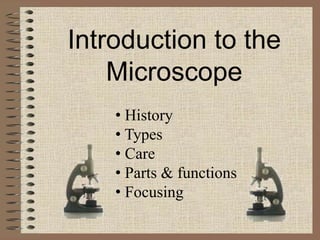
  • 1. Introduction to the Microscope • History • Types • Care • Parts & functions • Focusing
  • 2. Circa 1000AD – The first vision aid was invented (inventor unknown) called a reading stone. It was a glass sphere that magnified when laid on top of reading materials.
  • 3. Circa 1284 – Italian, Salvino D'Armate is credited with inventing the first wearable eye glasses.
  • 4. 1590 – Two Dutch eye glass makers, Zaccharias Janssen and son Hans Janssen experimented with multiple lenses placed in a tube. The Janssens observed that viewed objects in front of the tube appeared greatly enlarged, creating both the forerunner of the compound microscope and the telescope.
  • 5. 1665 – English physicist, Robert Hooke looked at a sliver of cork through a microscope lens and noticed some "pores" or "cells" in it.
  • 6. 1674 – Anton van Leeuwenhoek built a simple microscope with only one lens to examine blood, yeast, insects and many other tiny objects. Leeuwenhoek was the first person to describe bacteria, and he invented new methods for grinding and polishing microscope lenses that allowed for curvatures providing magnifications of up to 270 diameters, the best available lenses at that time.
  • 7. 18th century – Technical innovations improved microscopes, leading to microscopy becoming popular among scientists. Lenses combining two types of glass reduced the "chromatic effect" the disturbing halos resulting from differences in refraction of light.
  • 8. 1830 – Joseph Jackson Lister reduces spherical aberration or the "chromatic effect" by showing that several weak lenses used together at certain distances gave good magnification without blurring the image. This was the prototype for the compound microscope.
  • 9. 1872 – Ernst Abbe, then research director of the Zeiss Optical Works, wrote a mathematical formula called the "Abbe Sine Condition". His formula provided calculations that allowed for the maximum resolution in microscopes possible.
  • 10. 1903 – Richard Zsigmondy developed the ultramicroscope that could study objects below the wavelength of light. He won the Nobel Prize in Chemistry in 1925.
  • 11. 1932 – Frits Zernike invented the phase- contrast microscope that allowed for the study of colorless and transparent biological materials for which he won the Nobel Prize in Physics in 1953.
  • 12. 1931 – Ernst Ruska co-invented the electron microscope for which he won the Nobel Prize in Physics in 1986. An electron microscope depends on electrons rather than light to view an object, electrons are speeded up in a vacuum until their wavelength is extremely short, only one hundred-thousandth that of white light. Electron microscopes make
  • 13. 1931 – Ernst Ruska it possible to view objects as small as the diameter of an atom.
  • 14. 1981 – Gerd Binnig and Heinrich Rohrer invented the scanning tunneling microscope that gives three-dimensional images of objects down to the atomic level. Binnig and Rohrer won the Nobel Prize in Physics in 1986. The powerful scanning tunneling microscope is the strongest microscope to date.
  • 15. •Compound Microscope •Dissection Microscope •Scanning Electron Microscope (SEM) •Transmission Electron Microscope (TEM)
  • 16. Compound microscopes are light illuminated. The image seen with this type of microscope is two dimensional. This microscope is the most commonly used. You can view individual cells, even living ones. It has high magnification. However, it has a low resolution.
  • 18. A dissection microscope is light illuminated. The image that appears is three dimensional. It is used for dissection to get a better look at the larger specimen. You cannot see individual cells because it has a low magnification. (also called stereo microscope)
  • 19. Sunflower with moth pupa in the stem 10x Head of a moth pupa 60x
  • 20. SEM use electron illumination. The image is seen in 3-D. It has high magnification and high resolution. The specimen is coated in gold and the electrons bounce off to give you and exterior view of the specimen. The pictures are in black and white.
  • 22. TEM is electron illuminated. This gives a 2-D view. Thin slices of specimen are obtained. The electron beams pass through this. It has high magnification and high resolution.
  • 24. • Always carry with 2 hands • Never touch the lenses with your fingers. • Only use lens paper for cleaning • Do not force knobs • Keep objects clear of desk and cords • When you are finished with your "scope", rotate the nosepiece so that it's on the low power objective, roll the stage down to lowest level, rubber band the cord, then replace the dust cover.
  • 25. Objectives: 1. Identify the parts of the microscope; 2. Describe what parts of the microscope can do;
  • 26. Materials: • Microscope • Activity Note book • Learners’ Material • Manila Paper
  • 27. A. The Microscope, Its Parts and their Functions 1. Get the microscope from its box or the cabinet. Do this by grasping the curved arm with one hand and supporting the base with the other hand. 2. Carry it to your table or working place. Remember to always use both hands when carrying the microscope. I. Activity, Analysis
  • 28. 3. Put the microscope down gently on the laboratory table with its arm facing you. Place it about 7 centimeters away from the edge of the table. 4. Wipe with tissue paper or old t-shirt the metal parts of the microscope.
  • 29. Analysis 1. What are the functions of the base and the arm of the microscope? 5. Figure 1 shows a light microscope that most schools have. Study and use this to locate different parts of the microscope.
  • 30. 6. Look for the revolving nosepiece. Note that objectives are attached it. You should know that there are lenses inside the objectives.
  • 31.
  • 32. Analysis 2. What have you observed about the objectives? Most schools have light microscopes with three objectives. Others have four. Usually, the shortest one marked 3x, 4x or 5x is called the scanner. The low power objective (LPO) is marked 10x or 12x while the high power objective (HPO) is marked 40x, 43x or 60x. The objectives magnify the object to be observed to a certain size as indicated by the 3x, 10x or 40x, etc. marks.
  • 33. If the longest objective of your microscope is marked 97x or 100x or OIO or the word “oil” on it, then it has an oil immersion objective (OIO). This objective is used to view bacteria, very small protists and fungi. The OIO requires the use of a special oil such as quality cedarwood oil or cargille’s immersion oil.
  • 34. 7. Find the coarse adjustment. Slowly turn it upwards, then downwards. Analysis 3. What is accomplished by turning the coarse adjustment upwards? downwards?
  • 35. 8. Looking from the side of the microscope, raise the body tube. Then, turn the revolving nosepiece in any direction until the LPO is back in position. You will know an objective is in position when it clicks. Note that the revolving nosepiece makes possible the changing from one objective to another.
  • 36. Analysis 4. What is the other function of the revolving nosepiece? Analysis 5. Which part connects the eyepiece to the revolving nosepiece with the objectives?
  • 37. 9. Locate the eyepiece. Notice also that it is marked with a number and an x. Know that the eyepiece further magnifies the image of the object that has been magnified by the objective. If the eyepiece is cloudy or dusty, wipe it gently with a piece of lens paper.
  • 38. Use lens paper only in cleaning the lenses of the eyepiece and the objectives. Remember 10. Look through the eyepiece. Do you see anything? 11. Now, locate the mirror. Then, position the microscope towards diffused light from the windows or ceiling light. Look through the eyepiece and with the concave mirror (with depression) facing up, move it until you see a bright circle of light.
  • 39. Never use direct sunlight as a light source to view objects under the microscope. Direct sunlight can permanently damage the retina of the eye. Caution The bright circle of light is called the field of view of the microscope. Adjust the position of the mirror so that it is not glaring to the eyes. Practice viewing through the microscope using both eyes open. This will reduce eyestrain.
  • 40. Analysis 6. What are the two functions of the eyepiece? Analysis 7. Describe the function of the mirror. 12. Locate the diaphragm. While looking into the eyepiece, rotate the diaphragm to the next opening. Continue to do so until the original opening you used is back under the hole in the stage.
  • 41. Analysis 8. What do you notice as you change the diaphragm openings? Analaysis 9. What can you infer as to the function of the diaphragm? 13. Find the inclination joint.
  • 42. Analysis 10. What parts of the microscope are being connected by the inclination joint? 14. Grasp the arm and slowly pull it towards you. Sit down and try looking through the eyepiece. Tilting of the microscope allows one to do observations while seating down. This is however, only done when materials observed do not contain liquids like water. Remember
  • 43.
  • 44. Ocular lens Body Tube Revolving Nosepiece Arm Objective Lens Stage Stage Clips Coarse adjustment knob Fine adjustment knob Base Diaphragm Light
  • 45. Ocular lens magnifies; where you look through to see the image of your specimen. They are usually 10X or 15X power. Our microscopes have an ocular lens power of 10x.
  • 46. arm supports the tube and connects it to the base
  • 47. stage the flat platform where you place your slides
  • 48. coarse adjustment knob moves stage (or body tube) up and down
  • 49. fine adjustment knob small, round knob on the side of the microscope used to fine-tune the focus of your specimen after using the coarse adjustment knob
  • 50. base the bottom of the microscope, used for support
  • 51. body tube connects the eyepiece to the objective lenses
  • 52. revolving nosepiece the part that holds two or more objective lenses and can be rotated to easily change power
  • 53. objective lens Adds to the magnification Usually you will find 3 or 4 objective lenses on a microscope. They almost always consist of 4X, 10X, 40X and 100X powers. When coupled with a 10X (most common)
  • 54. objective lenses eyepiece lens, we get total magnifications of 40X (4X times 10X), 100X , 400X and 1000X. The shortest lens is the lowest power, the longest one is the lens with the greatest power. Lenses are color coded.
  • 55. objective lenses The high power objective lenses are retractable (i.e. 40XR). This means that if they hit a slide, the end of the lens will push in (spring loaded) thereby protecting the lens and the slide.
  • 56. stage clips Stage clips hold the slides in place. If your microscope has a mechanical stage, you will be able to move the slide around by turning two knobs. One moves it left and right, the other moves it up and down.
  • 57. diaphragm controls the amount of light going through the specimen Many microscopes have a rotating disk under the stage. This diaphragm has different sized holes and is used to vary the intensity and size of the cone of light
  • 58. diaphragm that is projected upward into the slide. There is no set rule regarding which setting to use for a particular power. Rather, the setting is a function of the transparency of the specimen, the degree of contrast you desire and the particular objective lens in use.
  • 60. The proper way to focus a microscope is to start with the lowest power objective lens first and while looking from the side, crank the lens down as close to the specimen as possible without touching it. Now, look through the eyepiece lens and focus upward only until the image is sharp. If you can't get it in focus, repeat the process again.
  • 61. Once the image is sharp with the low power lens, you should be able to simply click in the next power lens and do minor adjustments with the focus knob. If your microscope has a fine focus adjustment, turning it a bit should be all that's necessary. Continue with subsequent objective lenses and fine focus each time.
  • 62. Rotate to 40x objective, locate desired portion of specimen in the center of the field. Refocus very carefully so that the specimen is focused as sharply as possible. (Do not alter focus for the Following steps )
  • 63. Partially rotate so that 40x and 100x objectives straddle the specimen.
  • 64. Place a small drop of oil on the slide in the center of the lighted area. (Take care not to dribble on the stage.) Put the small drop of oil directly over the area of the specimen to be Examined.
  • 65. Rotate so that the 100x oil immersion objective touches the oil and clicks into place.
  • 66. Focus only with fine focus. Hopefully, the specimen will come into focus easily. Do not change focus dramatically.
  • 67. Clean up!: When you have finished for the day, wipe the 100x oil immersion objective carefully with lens paper to remove all oil. Wipe oil from the slide thoroughly with a Kimwipe. Cleanse stage should any oil have spilled on it. Recap the immersion oil container securely, replace in drawer.
  • 68. Q1. What are the functions of the base and the arm of the microscope? The base provides support to the microscope. The arm on the other hand supports the body tube and it is where the microscope is held.
  • 69. Q2. What have you observed about the objectives? Answers can be: they are of different lengths, they are marked with numbers followed by x, some may say: there are three or four objectives attached to the revolving nosepiece.
  • 70. Q3. What is accomplished by turning the coarse adjustment upwards? Downwards? Turning the coarse adjustment upwards and downwards raises and lowers the body tube with the objectives respectively. It also focuses or brings out the object to be observed.
  • 71. Q4. What is the other function of the revolving nosepiece? It facilitates the changing of objectives. Q5. Which connects the eyepiece to the revolving nosepiece with the objectives? Body tube.
  • 72. Q6. What are the two functions of the eyepiece? It is where you look through in the microscope. It also magnifies the image of the object that has been magnified by the objective. Q7. Describe the function of the mirror. It reflects light up to the diaphragm, object to be observed and lenses.
  • 73. Q8. What do you notice as you change the diaphragm openings? The size of the openings differ. The amount of light reflected also changes in that the bigger the opening, the greater is the amount of light reflected. Q9. What can you infer as to the function of the diaphragm? The diaphragm regulates the amount of light reflected to the object to be viewed.
  • 74. Q10. What parts of the microscope are being connected by the inclination joint? The arm and the base of the microscope. Q11. What does this movement do? It allows one to tilt the microscope so viewing is possible while seated.