Download as PDF, PPTX
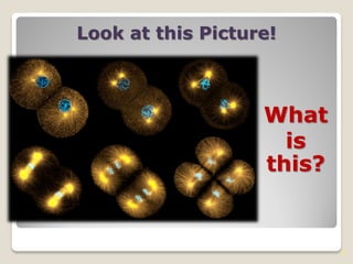


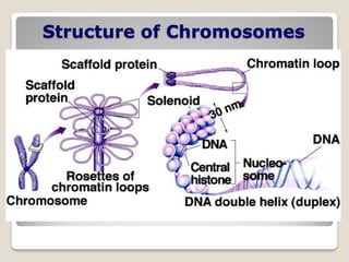


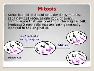
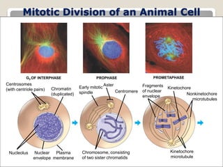


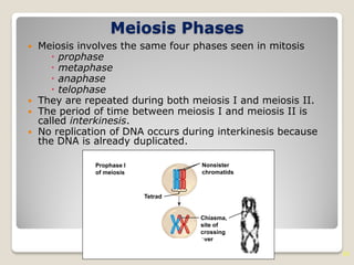


The document discusses the cell cycle and how cells divide through mitosis and meiosis. It provides details on the following: 1) The cell cycle consists of interphase and the mitotic phase. Interphase includes the G1, S, and G2 phases where the cell grows and duplicates its DNA. 2) Mitosis and meiosis are types of cell division. Mitosis produces two identical daughter cells through chromosome duplication and separation. Meiosis reduces the chromosome number by half to produce gametes. 3) Chromosomes duplicate and separate through different phases - prophase, metaphase, anaphase, and telophase. Sister chromatids separate in anaphase to move into two daughter cells