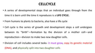
CELLCYCLE
• A series of developmental steps that an individual goes through from the
time it is born until the time it reproduces is a LIFE CYCLE.
• From humans to plants to bacteria, also have a life cycle
• Cell cycle is the series of growth and development steps a cell undergoes
between its “birth”—formation by the division of a mother cell—and
reproduction—division to make two new daughter cells.
• Division of cell includes several tasks- it must grow, copy its genetic material
(DNA), and physically split into two daughter cells
• In eukaryotic cells, or cells with a nucleus, the stages of the cell cycle are
divided into two major phases: INTERPHASE and the MITOTIC (M) PHASE.
• During interphase, the cell grows and makes a copy of its DNA.
• During the mitotic (M) phase, the cell separates its DNA into two sets and
divides its cytoplasm, forming two new cells.
INTERPHASE - preparation of cell division happens in 3 steps.
1. G1PHASE
2. S PHASE
3. G2PHASE
G1PHASE: Also called FIRST GAP PHASE
The cell grows physically larger, copies organelles, and makes the molecular
building blocks it will need in later steps.
S PHASE: The cell synthesizes a complete copy of the DNA in its nucleus.
It also duplicates a microtubule-organizing structure called the centrosome.
The centrosomes help separate DNA during M phase.
G2PHASE: SECOND GAP PHASE
The cell grows more, makes proteins and organelles, and begins to
reorganize its contents in preparation for mitosis.
G2phase ends when mitosis begins.
• TheG1, S, and G2PHASE together are known as interphase.
• The prefix inter- means between, reflecting that interphase takes place
between one mitotic (M) phase and the next.
MITOSIS
• Mitosis is a type of cell division in which one cell (the mother) divides to produce
two new cells (the daughters) that are genetically identical to itself.
• Mitosis is the part of the division process in which the DNA of the cell's nucleus is
split into two equal sets of chromosomes.
Cell division under light microscope
• Mitosis was first discovered in plant cells by STRASBURGER (1875)
• Later, W.FLEMMING (1879) discovered in animal cells.
• The mitosis was coined by FLEMMING (1882).
• The great majority of the cell divisions that happen in your body involve
mitosis
• During development and growth, mitosis populates an organism’s body with
cells, and throughout an organism’s life, it replaces old, worn-out cells with
new ones
OCCURRENCE :
The cells undergoes mitosis are called MITOCYTES
Plants – meristematic cells.
Animal – stem cells, germinal epithelium and embryonic cells.
 It occurs during regeneration. Root tips are best example to study mitosis
REASONS FOR MITOSIS
1. Growth
2. Repair / Healing
3. Asexual reproduction
CHARACTERSTICS OF MITOSIS:
 A diploid cell will gives rise to a diploid cell
 Chromosomes remains same
 The DNA remains identically same
 One cell (2N) gives rise to two cells (2N)
oDIPLOID CELLS: A cell with two sets of chromosomes.
All body cells are diploid ( somatic cells)
oHAPLOID CELLS: A cell with single set of chromosomes
All gamets ( sperm cells, eggs ) [ germ line cells]
• SOMATIC CELLS : A cells whose genes will not pass on to next generation
• GERM CELLS : A cell that is destined to form a gamet ( sperm, egg ) .
A cell whose genes will pass on to next generation
MITOSIS IS THE DIVISION OF SOMATIC CELLS
DEPENDING ON CELL TYPE:
Mitosis can take a few minutes or a few days
• Muscle cells
• Nerve cells
• Skin cells
• Digestive tract cell.
• Glomerular cells
• Mitosis consists of four basic phases: prophase, metaphase,
anaphase, and telophase.
• These phases occur in strict sequential order, and cytokinesis - the
process of dividing the cell contents to make two new cells - starts in
anaphase or telophase.
• Before the beginning of mitosis the cell is in interphase (late G2 phase) and has
already copied its DNA, so the chromosomes in the nucleus each consist of two
connected copies, called sister chromatids.
CENTROSOMES:
• Functions as the major microtubule-organizing center to ensure that
chromosome segregation during cell division.
• Forming the bipolar mitotic spindle required to accurately divide genetic
material between daughter cells during cell division
• As mitosis is initiated, the two centrosomes move apart to form the poles of a
bipolar mitotic spindle
• In early prophase, the cell starts to break down some structures and build
others
• This setting the stage for division of the chromosomes.
The chromosomes start to condense
The mitotic spindle begins to form
The spindle is a structure made of microtubules, strong fibers that are part
of the cell’s “skeleton.” Its main role is to organize the chromosomes.
The nucleolus (or nucleoli, plural), a part of the nucleus where ribosomes
are made, disappears. This is a sign that the nucleus is getting ready to break
down.
EARLY
PROPHASE
• In late prophase (sometimes also called prometaphase), the mitotic
spindle begins to capture and organize the chromosomes.
• The chromosomes become even more condensed
• The nuclear envelope breaks down, releasing the chromosomes.
• The mitotic spindle grows more, and some of the microtubules start to
“capture” chromosomes.
LATE PROPHASE
(PROMETAPHASE)
•Microtubules can bind to chromosomes
at the kinetochore, a patch of protein
found on the centromere of each sister
chromatid.
•Microtubules that bind a chromosome
are called kinetochore microtubules.
•Microtubules that don’t bind to
kinetochores can grab on to microtubules
from the opposite pole, stabilizing the
spindle.
• More microtubules extend from each
centrosome towards the edge of the cell,
forming a structure called the aster.
• In metaphase, the spindle has captured all the chromosomes and lined them
up at the middle of the cell, ready to divide.
• All the chromosomes align at the metaphase plate
• At this stage, the two kinetochores of each chromosome should be attached to
microtubules from opposite spindle poles.
• Before proceeding to anaphase, the cell will check to make sure that all the
chromosomes are at the metaphase plate with their kinetochores correctly
attached to microtubules. This is called the spindle checkpoint and helps
ensure that the sister chromatids will split evenly between the two daughter
cells when they separate in the next step.
METAPHASE
• In anaphase, the sister chromatids separate from each other and are
pulled towards opposite ends of the cell.
• The protein “glue” that holds the sister chromatids together is broken
down, allowing them to separate. Each is now its own chromosome. The
chromosomes of each pair are pulled towards opposite ends of the cell.
• Microtubules not attached to chromosomes elongate and push apart,
separating the poles and making the cell longer.
ANAPHASE
• In telophase, the cell is nearly done dividing, and it starts to re-establish its
normal structures as cytokinesis (division of the cell contents) takes place.
• The mitotic spindle is broken down into its building blocks.
• Two new nuclei form, one for each set of chromosomes. Nuclear membranes
and nucleoli reappear.
• The chromosomes begin to decondense and return to their “stringy” form.
TELOPHASE
• Cytokinesis, the division of the cytoplasm to form two new cells, overlaps
with the final stages of mitosis
• In animal cells, cytokinesis is contractile, pinching the cell in two like a coin
purse with a drawstring.
• The “drawstring” is a band of filaments made of a protein called actin, and
the pinch crease is known as the cleavage furrow.
• Plant cells can’t be divided like this because they have a cell wall and are
too stiff. Instead, a structure called the cell plate forms down the middle of
the cell, splitting it into two daughter cells separated by a new wall.
CYTOKINESIS
 When cytokinesis finishes, we end up
with two new cells, each with a
complete set of chromosomes identical
to those of the mother cell.
 The daughter cells can now begin
their own cellular “lives,”
•Every somatic cells in our body has 46 chromosomes
•Every gamet cells has 23 chromosomes
MEIOISIS
MONOSOMY 22
MONOSOMY 18
MONOSOMY 21
TRISOMY 18
TRISOMY 21
MITOSIS PPT.pptx

MITOSIS PPT.pptx

  • 1.
    CELLCYCLE • A seriesof developmental steps that an individual goes through from the time it is born until the time it reproduces is a LIFE CYCLE. • From humans to plants to bacteria, also have a life cycle • Cell cycle is the series of growth and development steps a cell undergoes between its “birth”—formation by the division of a mother cell—and reproduction—division to make two new daughter cells. • Division of cell includes several tasks- it must grow, copy its genetic material (DNA), and physically split into two daughter cells
  • 2.
    • In eukaryoticcells, or cells with a nucleus, the stages of the cell cycle are divided into two major phases: INTERPHASE and the MITOTIC (M) PHASE. • During interphase, the cell grows and makes a copy of its DNA. • During the mitotic (M) phase, the cell separates its DNA into two sets and divides its cytoplasm, forming two new cells. INTERPHASE - preparation of cell division happens in 3 steps. 1. G1PHASE 2. S PHASE 3. G2PHASE
  • 3.
    G1PHASE: Also calledFIRST GAP PHASE The cell grows physically larger, copies organelles, and makes the molecular building blocks it will need in later steps. S PHASE: The cell synthesizes a complete copy of the DNA in its nucleus. It also duplicates a microtubule-organizing structure called the centrosome. The centrosomes help separate DNA during M phase. G2PHASE: SECOND GAP PHASE The cell grows more, makes proteins and organelles, and begins to reorganize its contents in preparation for mitosis. G2phase ends when mitosis begins.
  • 4.
    • TheG1, S,and G2PHASE together are known as interphase. • The prefix inter- means between, reflecting that interphase takes place between one mitotic (M) phase and the next.
  • 5.
    MITOSIS • Mitosis isa type of cell division in which one cell (the mother) divides to produce two new cells (the daughters) that are genetically identical to itself. • Mitosis is the part of the division process in which the DNA of the cell's nucleus is split into two equal sets of chromosomes. Cell division under light microscope
  • 6.
    • Mitosis wasfirst discovered in plant cells by STRASBURGER (1875) • Later, W.FLEMMING (1879) discovered in animal cells. • The mitosis was coined by FLEMMING (1882). • The great majority of the cell divisions that happen in your body involve mitosis • During development and growth, mitosis populates an organism’s body with cells, and throughout an organism’s life, it replaces old, worn-out cells with new ones
  • 7.
    OCCURRENCE : The cellsundergoes mitosis are called MITOCYTES Plants – meristematic cells. Animal – stem cells, germinal epithelium and embryonic cells.  It occurs during regeneration. Root tips are best example to study mitosis REASONS FOR MITOSIS 1. Growth 2. Repair / Healing 3. Asexual reproduction
  • 8.
    CHARACTERSTICS OF MITOSIS: A diploid cell will gives rise to a diploid cell  Chromosomes remains same  The DNA remains identically same  One cell (2N) gives rise to two cells (2N) oDIPLOID CELLS: A cell with two sets of chromosomes. All body cells are diploid ( somatic cells) oHAPLOID CELLS: A cell with single set of chromosomes All gamets ( sperm cells, eggs ) [ germ line cells]
  • 9.
    • SOMATIC CELLS: A cells whose genes will not pass on to next generation • GERM CELLS : A cell that is destined to form a gamet ( sperm, egg ) . A cell whose genes will pass on to next generation MITOSIS IS THE DIVISION OF SOMATIC CELLS
  • 10.
    DEPENDING ON CELLTYPE: Mitosis can take a few minutes or a few days • Muscle cells • Nerve cells • Skin cells • Digestive tract cell. • Glomerular cells
  • 11.
    • Mitosis consistsof four basic phases: prophase, metaphase, anaphase, and telophase. • These phases occur in strict sequential order, and cytokinesis - the process of dividing the cell contents to make two new cells - starts in anaphase or telophase.
  • 12.
    • Before thebeginning of mitosis the cell is in interphase (late G2 phase) and has already copied its DNA, so the chromosomes in the nucleus each consist of two connected copies, called sister chromatids.
  • 13.
    CENTROSOMES: • Functions asthe major microtubule-organizing center to ensure that chromosome segregation during cell division. • Forming the bipolar mitotic spindle required to accurately divide genetic material between daughter cells during cell division • As mitosis is initiated, the two centrosomes move apart to form the poles of a bipolar mitotic spindle
  • 15.
    • In earlyprophase, the cell starts to break down some structures and build others • This setting the stage for division of the chromosomes. The chromosomes start to condense The mitotic spindle begins to form The spindle is a structure made of microtubules, strong fibers that are part of the cell’s “skeleton.” Its main role is to organize the chromosomes. The nucleolus (or nucleoli, plural), a part of the nucleus where ribosomes are made, disappears. This is a sign that the nucleus is getting ready to break down. EARLY PROPHASE
  • 17.
    • In lateprophase (sometimes also called prometaphase), the mitotic spindle begins to capture and organize the chromosomes. • The chromosomes become even more condensed • The nuclear envelope breaks down, releasing the chromosomes. • The mitotic spindle grows more, and some of the microtubules start to “capture” chromosomes. LATE PROPHASE (PROMETAPHASE)
  • 18.
    •Microtubules can bindto chromosomes at the kinetochore, a patch of protein found on the centromere of each sister chromatid. •Microtubules that bind a chromosome are called kinetochore microtubules. •Microtubules that don’t bind to kinetochores can grab on to microtubules from the opposite pole, stabilizing the spindle. • More microtubules extend from each centrosome towards the edge of the cell, forming a structure called the aster.
  • 19.
    • In metaphase,the spindle has captured all the chromosomes and lined them up at the middle of the cell, ready to divide. • All the chromosomes align at the metaphase plate • At this stage, the two kinetochores of each chromosome should be attached to microtubules from opposite spindle poles. • Before proceeding to anaphase, the cell will check to make sure that all the chromosomes are at the metaphase plate with their kinetochores correctly attached to microtubules. This is called the spindle checkpoint and helps ensure that the sister chromatids will split evenly between the two daughter cells when they separate in the next step. METAPHASE
  • 21.
    • In anaphase,the sister chromatids separate from each other and are pulled towards opposite ends of the cell. • The protein “glue” that holds the sister chromatids together is broken down, allowing them to separate. Each is now its own chromosome. The chromosomes of each pair are pulled towards opposite ends of the cell. • Microtubules not attached to chromosomes elongate and push apart, separating the poles and making the cell longer. ANAPHASE
  • 23.
    • In telophase,the cell is nearly done dividing, and it starts to re-establish its normal structures as cytokinesis (division of the cell contents) takes place. • The mitotic spindle is broken down into its building blocks. • Two new nuclei form, one for each set of chromosomes. Nuclear membranes and nucleoli reappear. • The chromosomes begin to decondense and return to their “stringy” form. TELOPHASE
  • 25.
    • Cytokinesis, thedivision of the cytoplasm to form two new cells, overlaps with the final stages of mitosis • In animal cells, cytokinesis is contractile, pinching the cell in two like a coin purse with a drawstring. • The “drawstring” is a band of filaments made of a protein called actin, and the pinch crease is known as the cleavage furrow. • Plant cells can’t be divided like this because they have a cell wall and are too stiff. Instead, a structure called the cell plate forms down the middle of the cell, splitting it into two daughter cells separated by a new wall. CYTOKINESIS
  • 26.
     When cytokinesisfinishes, we end up with two new cells, each with a complete set of chromosomes identical to those of the mother cell.  The daughter cells can now begin their own cellular “lives,”
  • 27.
    •Every somatic cellsin our body has 46 chromosomes •Every gamet cells has 23 chromosomes
  • 28.
  • 52.
  • 53.
  • 55.