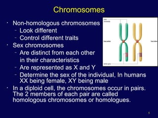
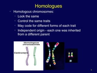
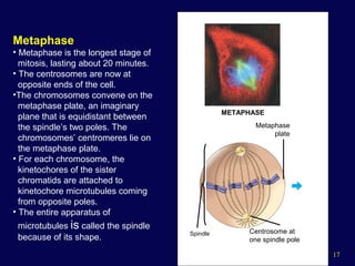
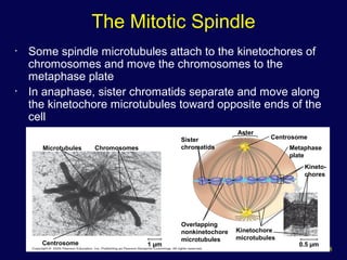
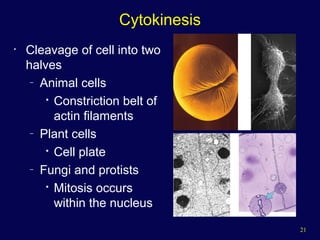
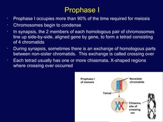
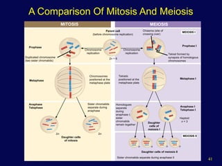
1
The Cell Cycle and How Cells Divide
2
Phases of the Cell Cycle
• The cell cycle consists of
– Interphase – normal cell activity
– The mitotic phase – cell division
INTERPHASE
Growth
G 1
(DNA synthesis)
Growth
G2
CellDivsion
3
Functions of Cell Division
20 µm100 µm 200 µm
(a) Reproduction. An amoeba,
a single-celled eukaryote, is
dividing into two cells. Each
new cell will be an individual
organism (LM).
(b) Growth and development.
This micrograph shows a
sand dollar embryo shortly after
the fertilized egg divided, forming
two cells (LM).
(c) Tissue renewal. These dividing
bone marrow cells (arrow) will
give rise to new blood cells (LM).
4
Cell Division
• An integral part of the cell cycle
• Mitosis results in genetically identical daughter
cells
• Cells duplicate their genetic material
– Before they divide, ensuring that each daughter
cell receives an exact copy of the genetic
material, DNA
5
Chromosomes
• Non-homologous chromosomes
– Look different
– Control different traits
• Sex chromosomes
– Are distinct from each other
in their characteristics
– Are represented as X and Y
– Determine the sex of the individual, In humans
XX being female, XY being male
• In a diploid cell, the chromosomes occur in pairs.
The 2 members of each pair are called
homologous chromosomes or homologues.
6
Homologues
• Homologous chromosomes:
• Look the same
• Control the same traits
• May code for different forms of each trait
• Independent origin - each one was inherited
from a different parent
7
Chromosome Duplication
0.5 µm
Chromosome
duplication
(including DNA
synthesis)
Centromere
Separation
of sister
chromatids
Sister
chromatids
Centrometers Sister chromatids
A eukaryotic cell has multiple
chromosomes, one of which is
represented here. Before
duplication, each chromosome
has a single DNA molecule.
Once duplicated, a chromosome
consists of two sister chromatids
connected at the centromere. Each
chromatid contains a copy of the
DNA molecule.
Mechanical processes separate
the sister chromatids into two
chromosomes and distribute
them to two daughter cells.
• In preparation for cell division, DNA is replicated and the chromosomes condense
• Each duplicated chromosome has two sister chromatids, which separate during cell
division
8
• Because of duplication, each condensed chromosome
consists of 2 identical chromatids joined by a centromere.
• Each duplicated chromosome contains 2 identical DNA
molecules (unless a mutation occurred), one in each
chromatid:
Chromosome Duplication
Copyright © The McGraw-Hill Companies, Inc. Permission required for reproduction or display.
Two unduplicated
chromosomes
Centromere
Sister
chromatids
Sister
chromatids
Duplication
Non-sister
chromatids
Two duplicated chromosomes
9
Structure of Chromosomes
• The centromere is a constricted region of the chromosome containing a
specific DNA sequence, to which is bound 2 discs of protein called
kinetochores.
• Kinetochores serve as points of attachment for microtubules that move
the chromosomes during cell division:
Copyright © The McGraw-Hill Companies, Inc. Permission required for reproduction or display.
Metaphase chromosome
Kinetochore
Kinetochore
microtubules
Centromere
region of
chromosome
Sister Chromatids
10
Phases of the Cell Cycle
• Interphase
– G1 - primary growth
– S - genome replicated
– G2 - secondary growth
• M - mitosis
• C - cytokinesis
11
Interphase
• G1 - Cells undergo majority of growth
• S - Each chromosome replicates (Synthesizes) to
produce sister chromatids
– Attached at centromere
– Contains attachment site (kinetochore)
• G2 - Chromosomes condense - Assemble
machinery for division such as centrioles
12
Mitosis

Some haploid & diploid cells divide by mitosis.

Each new cell receives one copy of every
chromosome that was present in the original cell.

Produces 2 new cells that are both genetically
identical to the original cell.
DNA duplication
during interphase
Mitosis
Diploid Cell
13
Mitotic Division of an Animal Cell
G2 OF INTERPHASE PROPHASE PROMETAPHASE
Centrosomes
(with centriole pairs) Chromatin
(duplicated)
Early mitotic
spindle
Aster
Centromere
Fragments
of nuclear
envelope
Kinetochore
Nucleolus Nuclear
envelope
Plasma
membrane
Chromosome, consisting
of two sister chromatids
Kinetochore
microtubule
Nonkinetochore
microtubules
14
METAPHASE ANAPHASE TELOPHASE AND CYTOKINESIS
Spindle
Metaphase
plate Nucleolus
forming
Cleavage
furrow
Nuclear
envelope
formingCentrosome at
one spindle pole
Daughter
chromosomes
Mitotic Division of an Animal Cell
15
G2 of Interphase
• A nuclear envelope bounds
the nucleus.
• The nucleus contains one or
more nucleoli (singular,
nucleolus).
• Two centrosomes have
formed by replication of a
single centrosome.
• In animal cells, each
centrosome features two
centrioles.
• Chromosomes, duplicated
during S phase, cannot be
seen individually because
they have not yet condensed.
The light micrographs show dividing lung cells
from a newt, which has 22 chromosomes in
its somatic cells (chromosomes appear blue,
microtubules green, intermediate filaments
red). For simplicity, the drawings show only
four chromosomes.
G2 OF INTERPHASE
Centrosomes
(with centriole pairs) Chromatin
(duplicated)
Nucleolus Nuclear
envelope
Plasma
membrane
16
Prophase
• The chromatin fibers become
more tightly coiled, condensing
into discrete chromosomes
observable with a light
microscope.
• The nucleoli disappear.
• Each duplicated chromosome
appears as two identical sister
chromatids joined together.
• The mitotic spindle begins to form.
It is composed of the centrosomes
and the microtubules that extend
from them. The radial arrays of
shorter microtubules that extend
from the centrosomes are called
asters (“stars”).
• The centrosomes move away from
each other, apparently propelled
by the lengthening microtubules
between them.
PROPHASE
Early mitotic
spindle
Aster
Centromere
Chromosome, consisting
of two sister chromatids
17
Metaphase
• Metaphase is the longest stage of
mitosis, lasting about 20 minutes.
• The centrosomes are now at
opposite ends of the cell.
•The chromosomes convene on the
metaphase plate, an imaginary
plane that is equidistant between
the spindle’s two poles. The
chromosomes’ centromeres lie on
the metaphase plate.
• For each chromosome, the
kinetochores of the sister
chromatids are attached to
kinetochore microtubules coming
from opposite poles.
• The entire apparatus of
microtubules is called the spindle
because of its shape.
METAPHASE
Spindle
Metaphase
plate
Centrosome at
one spindle pole
18
• Some spindle microtubules attach to the kinetochores of
chromosomes and move the chromosomes to the
metaphase plate
• In anaphase, sister chromatids separate and move along
the kinetochore microtubules toward opposite ends of the
cell
Microtubules Chromosomes
Sister
chromatids
Aster
Centrosome
Metaphase
plate
Kineto-
chores
Kinetochore
microtubules
0.5 µm
Overlapping
nonkinetochore
microtubules
1 µmCentrosome
The Mitotic Spindle
19
Anaphase
• Anaphase is the shortest stage of
mitosis, lasting only a few minutes.
• Anaphase begins when the two sister
chromatids of each pair suddenly part.
Each chromatid thus becomes a full-
fledged chromosome.
• The two liberated chromosomes begin
moving toward opposite ends of the cell,
as their kinetochore microtubules
shorten. Because these microtubules are
attached at the centromere region, the
chromosomes move centromere first (at
about 1 µm/min).
• The cell elongates as the
nonkinetochore microtubules lengthen.
• By the end of anaphase, the two ends of
the cell have equivalent—and
complete—collections of chromosomes.
ANAPHASE
Daughter
chromosomes
20
Telophase
• Two daughter nuclei begin to
form in the cell.
• Nuclear envelopes arise from
the fragments of the parent
cell’s nuclear envelope and
other portions of the
endomembrane system.
• The chromosomes become
less condensed.
• Mitosis, the division of one
nucleus into two genetically
identical nuclei, is now
complete.
TELOPHASE AND CYTOKINESIS
Nucleolus
forming
Cleavage
furrow
Nuclear
envelope
forming
21
Cytokinesis
• Cleavage of cell into two
halves
– Animal cells

Constriction belt of
actin filaments
– Plant cells

Cell plate
– Fungi and protists

Mitosis occurs
within the nucleus
22
Cytokinesis In Animal And Plant Cells
Daughter cells
Cleavage furrow
Contractile ring of
microfilaments
Daughter cells
100 µm
1 µmVesicles
forming
cell plate
Wall of
patent cell Cell plate
New cell wall
(a) Cleavage of an animal cell (SEM) (b) Cell plate formation in a plant cell (SEM)
23
24
Meiosis and Sexual Life Cycles
• Living organisms are distinguished by their ability to
reproduce their own kind
• Heredity
– Is the transmission of traits from one generation to the
next
• Variation
– Shows that offspring differ somewhat in appearance
from parents and siblings
25
Sex Cells - Gametes
• Unlike somatic cells, sperm and egg cells
are haploid cells, containing only one set of
chromosomes
• At sexual maturity the ovaries and testes
produce haploid gametes by meiosis
26
Sexual Reproduction - The Human Life Cycle
• During fertilization,
sperm and ovum fuse
forming a diploid
zygote
• The zygote develops
into an adult organism
Haploid (n)
Diploid (2n)
Haploid gametes (n = 23)
Ovum (n)
Sperm
Cell (n)
MEIOSIS FERTILIZATION
Ovary Testis Diploid
zygote
(2n = 46)
Mitosis and
development
Multicellular diploid
adults (2n = 46)
27
Meiosis
• Reduces the chromosome number such that
each daughter
• Cell has a haploid set of chromosomes
• Ensures that the next generation will have:
– Diploid number of chromosome
– Exchange of genetic information
(combination of traits
– that differs from that of either parent)
28
Meiosis
• Only diploid cells can divide by meiosis.
• Prior to meiosis I, DNA replication occurs.
• During meiosis, there will be two nuclear divisions, and the result will be
four haploid nuclei.
• No replication of DNA occurs between meiosis I and meiosis II.
29
Meiosis
• Meiosis reduces the
number of chromosome
sets from diploid to
haploid
• Meiosis takes place in
two sets of divisions
– Meiosis I reduces the
number of chromosomes
from diploid to haploid
– Meiosis II produces four
haploid daughter cells
Figure 13.7
Interphase
Homologous pair
of chromosomes
in diploid parent cell
Chromosomes
replicate
Homologous pair of replicated chromosomes
Sister
chromatids Diploid cell with
replicated
chromosomes
1
2
Homologous
chromosomes
separate
Haploid cells with
replicated chromosomes
Sister chromatids
separate
Haploid cells with unreplicated chromosomes
Meiosis I
Meiosis II
30
Meiosis Phases
• Meiosis involves the same four phases seen in
mitosis

prophase

metaphase

anaphase

telophase
• They are repeated during both meiosis I and
meiosis II.
• The period of time between meiosis I and meiosis
II is called interkinesis.
• No replication of DNA occurs during interkinesis
because the DNA is already duplicated.
31
Prophase I
• Prophase I occupies more than 90% of the time required for meiosis
• Chromosomes begin to condense
• In synapsis, the 2 members of each homologous pair of chromosomes
line up side-by-side, aligned gene by gene, to form a tetrad consisting
of 4 chromatids
• During synapsis, sometimes there is an exchange of homologous parts
between non-sister chromatids. This exchange is called crossing over
• Each tetrad usually has one or more chiasmata, X-shaped regions
where crossing over occurred
Prophase I
of meiosis
Tetrad
Nonsister
chromatids
Chiasma,
site of
crossing
over
32
Metaphase I
• At metaphase I, tetrads line up at the metaphase plate, with one
chromosome facing each pole
• Microtubules from one pole are attached to the kinetochore of one
chromosome of each tetrad
• Microtubules from the other pole are attached to the kinetochore of the
other chromosome
Sister
chromatids
Chiasmata
Spindle
Centromere
(with kinetochore)
Metaphase
plate
Homologous
chromosomes
separate
Sister chromatids
remain attached
Microtubule
attached to
kinetochore
Tetrad
PROPHASE I METAPHASE I ANAPHASE I
Homologous chromosomes
(red and blue) pair and
exchange segments; 2n = 6
Pairs of homologous
chromosomes split up
Tetrads line up
33
Anaphase I
• In anaphase I, pairs of homologous chromosomes separate
• One chromosome moves toward each pole, guided by the
spindle apparatus
• Sister chromatids remain attached at the centromere and
move as one unit toward the pole
Sister
chromatids
Chiasmata
Spindle
Centromere
(with kinetochore)
Metaphase
plate
Homologous
chromosomes
separate
Sister chromatids
remain attached
Microtubule
attached to
kinetochore
Tetrad
PROPHASE I METAPHASE I ANAPHASE I
Homologous chromosomes
(red and blue) pair and
exchange segments; 2n = 6
Pairs of homologous
chromosomes split up
Tetrads line up
34
Telophase I and Cytokinesis
• In the beginning of telophase I, each half of the
cell has a haploid set of chromosomes; each
chromosome still consists of two sister chromatids
• Cytokinesis usually occurs simultaneously, forming
two haploid daughter cells
• In animal cells, a cleavage furrow forms; in plant
cells, a cell plate forms
• No chromosome replication occurs between the
end of meiosis I and the beginning of meiosis II
because the chromosomes are already replicated
35
Prophase II
• Meiosis II is very similar to mitosis
• In prophase II, a spindle apparatus forms
• In late prophase II, chromosomes (each still composed of
two chromatids) move toward the metaphase plate
Cleavage
furrow
PROPHASE II METAPHASE II ANAPHASE II
TELOPHASE I AND
CYTOKINESIS
TELOPHASE II AND
CYTOKINESIS
Sister chromatids
separate
Haploid daughter cells
forming
36
Metaphase II
• At metaphase II, the sister chromatids are at the metaphase plate
• Because of crossing over in meiosis I, the two sister chromatids of each
chromosome are no longer genetically identical
• The kinetochores of sister chromatids attach to microtubules extending
from opposite poles
Cleavage
furrow
PROPHASE II METAPHASE II ANAPHASE II
TELOPHASE I AND
CYTOKINESIS
TELOPHASE II AND
CYTOKINESIS
Sister chromatids
separate
Haploid daughter cells
forming
37
Anaphase II
• At anaphase II, the sister chromatids separate
• The sister chromatids of each chromosome now move as
two newly individual chromosomes toward opposite poles
Cleavage
furrow
PROPHASE II METAPHASE II ANAPHASE II
TELOPHASE I AND
CYTOKINESIS
TELOPHASE II AND
CYTOKINESIS
Sister chromatids
separate
Haploid daughter cells
forming
38
Telophase II and Cytokinesis
• In telophase II, the chromosomes arrive at opposite poles
• Nuclei form, and the chromosomes begin decondensing
• Cytokinesis separates the cytoplasm
• At the end of meiosis, there are four daughter cells, each with a
haploid set of unreplicated chromosomes
• Each daughter cell is genetically distinct from the others and from the
parent cell
Cleavage
furrow
PROPHASE II METAPHASE II ANAPHASE II
TELOPHASE I AND
CYTOKINESIS
TELOPHASE II AND
CYTOKINESIS
Sister chromatids
separate
Haploid daughter cells
forming
39
A Comparison of Mitosis and Meiosis
• Mitosis conserves the number of chromosome
sets, producing cells that are genetically identical
to the parent cell
• Meiosis reduces the number of chromosomes sets
from two (diploid) to one (haploid), producing cells
that differ genetically from each other and from the
parent cell
• The mechanism for separating sister chromatids is
virtually identical in meiosis II and mitosis
40
• Three events are unique to meiosis, and all three
occur in meiosis l:
– Synapsis and crossing over in prophase I:
Homologous chromosomes physically connect and
exchange genetic information
– At the metaphase plate, there are paired homologous
chromosomes (tetrads), instead of individual replicated
chromosomes
– At anaphase I of meiosis, homologous pairs move
toward opposite poles of the cell. In anaphase II of
meiosis, the sister chromatids separate
A Comparison of Mitosis and Meiosis
41
MITOSIS MEIOSIS
Prophase
Duplicated chromosome
(two sister chromatids)
Chromosome
replication
Chromosome
replication
Parent cell
(before chromosome replication)
Chiasma (site of
crossing over)
MEIOSIS I
Prophase I
Tetrad formed by
synapsis of homologous
chromosomes
Metaphase
Chromosomes
positioned at the
metaphase plate
Tetrads
positioned at the
metaphase plate
Metaphase I
Anaphase I
Telophase I
Haploid
n = 3
MEIOSIS II
Daughter
cells of
meiosis I
Homologues
separate
during
anaphase I;
sister
chromatids
remain together
Daughter cells of meiosis II
n n n n
Sister chromatids separate during anaphase II
Anaphase
Telophase
Sister chromatids
separate during
anaphase
2n 2n
Daughter cells
of mitosis
2n = 6
A Comparison Of Mitosis And Meiosis
42
Comparison
• Meiosis
• DNA duplication
followed by 2 cell
divisions
• Sysnapsis
• Crossing-over
• One diploid cell
produces 4
haploid cells
• Each new cell
has a unique
combination of
genes
• Mitosis
• Homologous
chromosomes do not
pair up
• No genetic exchange
between homologous
chromosomes
• One diploid cell
produces 2 diploid
cells or one haploid
cell produces 2
haploid cells
• New cells are
genetically identical to
original cell (except for
mutation)
43
Sexual Reproduction - The Human Life Cycle
• During fertilization,
sperm and ovum fuse
forming a diploid
zygote
• The zygote develops
into an adult organism
Haploid (n)
Diploid (2n)
Haploid gametes (n = 23)
Ovum (n)
Sperm
Cell (n)
MEIOSIS FERTILIZATION
Ovary Testis Diploid
zygote
(2n = 46)
Mitosis and
development
Multicellular diploid
adults (2n = 46)
44
Regulation of cell cycle
• Each cyclin is associated with
a particular phase, transition,
or set of phases in the cell
cycle and
• helps drive the events of that
phase or period. For instance;
– M cyclin promotes the
events of M phase, such as
nuclear envelope breakdown
and chromosome
condensation
Most important core cell cycle regulators: proteins called cyclins,
enzymes called Cdks, and an enzyme complex called the APC/C
Cyclins are among the most important core cell cycle regulators
- Cyclins are a group of related proteins and
- four basic types found in humans and most other eukaryotes
–
- G1 cyclin, G1/S cyclins, S cyclins, and M cyclins​
45
Cyclin-dependent kinases
• To drive the cell cycle forward, a
cyclin must activate or inactivate
different target proteins inside of
the cell
• In cell cycle regulation Cyclins work
with a family of enzymes called
the cyclin-dependent kinases (Cdks)
• Binding of a cyclin activates inactive
Cdks
• Activation makes a functional
enzyme and allowing it to modify
target proteins
46
Cdks
• Cdks are kinases –phosphorylate specific target proteins
• The attached phosphate group acts like a switch, making
the target protein more or less active
• Cyclin attaching to a Cdk exert two important effects:
– activates the Cdk as a kinase
– directs the Cdk to a specific set of target proteins
• G1/S cyclins send Cdks to S phase targets (e.g., promoting
DNA replication)
M cyclins send Cdks to M
phase targets
(e.g., making the nuclear
membrane break down)
also be negatively regulated by
phosphorylation of other sites
47
Maturation-promoting factor (MPF)
• Example of cyclins and Cdks working together to
control cell cycle transitions
• Researchers found that cells in M phase contained an
unknown factor that could force frog egg cells (stuck
in G2) to enter M phase
• This mystery molecule was called MPF
• Discovered in the 1980s to be a Cdk bound to its M
cyclin partner
• M cyclin builds up as cell division progresses to G2/M
• Trigger M phase
• Activation signal is the intactness of DNA
48
Action of MPF complex
• The MPF complexes add phosphate tags to several
different proteins in the nuclear envelope
• Resulting in its breakdown (a key event of early M phase)
and
• Activate targets that promote chromosome condensation
and other M phase events
• Trigger nuclear envelope breakdown
49
The anaphase-promoting
complex/cyclosome (APC/C)
• MPF also triggers its own destruction by activating the anaphase-promoting
complex/cyclosome(APC/C)
• A protein complex that causes M cyclins to be destroyed starting in anaphase
• The destruction of M cyclins pushes the cell out of mitosis
• Allow the new daughter cells to enter G1
• The APC/C also causes destruction of the proteins that hold the sister chromatids
together, allowing them to separate in anaphase and move to opposite poles of
the cell.
50
Action of APC/C
• The APC/C is an enzyme
• It adds a small protein tag called ubiquitin (Ub)
• When a target is tagged with ubiquitin, it is sent to
the proteasome
• Thought as the recycle bin of the cell, and destroyed
• Eg. The APC/C attaches a ubiquitin tag to M cyclins
• causing them to be chopped up by the proteasome and
• Allow the newly forming daughter cells to enter G1
• The APC/C also uses ubiquitin tagging to trigger the
separation of sister chromatids during mitosis
• If the APC/C gets the right signals at metaphase, it sets
off a chain of events that destroys cohesin, the protein
glue that holds sister chromatids together
51
APC/C
• The APC/C first adds a
ubiquitin tag to a protein called
securin and
• Send it for recycling
• Securin normally binds to, and
inactivates, a protein called
separase
• When securin is sent for
recycling, separase becomes
active and be functional
• Separase chops up the cohesin
that holds sister chromatids
together, allowing them to
separate
52
Checkpoints and regulators
• Cdks, cyclins, and the APC/C are direct regulators of cell
cycle transitions
• They respond to signals from inside and outside the cell
• These signals determine whether the cell moves forward
in the cell cycle
• Positive cues, like growth factors may increase activity of
Cdks and cyclins
• Negative ones, like DNA damage decrease or block activity
• Cells deal with DNA damage by;
• Fixing it if possible and/or by
• Preventing cell division
• A protein called p53, a famous tumor suppressor gene
involved in DNA damage response
53
Cell cycle check point in DNA damage
• p53 works on multiple levels to ensure that cells do not pass on their
damaged DNA through cell division
First, it stops the cell cycle at the
G1checkpoint by triggering
production of Cdk inhibitor (CKI)
proteins
The CKI proteins bind to Cdk-cyclin
complexes and block their activity
p53's second job is to activate DNA
repair enzymes
If DNA damage is not fixable, p53
will play its third and final role:
triggering programmed cell death so
damaged DNA is not passed on

Celldivision

  • 1.
    1 The Cell Cycleand How Cells Divide
  • 2.
    2 Phases of theCell Cycle • The cell cycle consists of – Interphase – normal cell activity – The mitotic phase – cell division INTERPHASE Growth G 1 (DNA synthesis) Growth G2 CellDivsion
  • 3.
    3 Functions of CellDivision 20 µm100 µm 200 µm (a) Reproduction. An amoeba, a single-celled eukaryote, is dividing into two cells. Each new cell will be an individual organism (LM). (b) Growth and development. This micrograph shows a sand dollar embryo shortly after the fertilized egg divided, forming two cells (LM). (c) Tissue renewal. These dividing bone marrow cells (arrow) will give rise to new blood cells (LM).
  • 4.
    4 Cell Division • Anintegral part of the cell cycle • Mitosis results in genetically identical daughter cells • Cells duplicate their genetic material – Before they divide, ensuring that each daughter cell receives an exact copy of the genetic material, DNA
  • 5.
    5 Chromosomes • Non-homologous chromosomes –Look different – Control different traits • Sex chromosomes – Are distinct from each other in their characteristics – Are represented as X and Y – Determine the sex of the individual, In humans XX being female, XY being male • In a diploid cell, the chromosomes occur in pairs. The 2 members of each pair are called homologous chromosomes or homologues.
  • 6.
    6 Homologues • Homologous chromosomes: •Look the same • Control the same traits • May code for different forms of each trait • Independent origin - each one was inherited from a different parent
  • 7.
    7 Chromosome Duplication 0.5 µm Chromosome duplication (includingDNA synthesis) Centromere Separation of sister chromatids Sister chromatids Centrometers Sister chromatids A eukaryotic cell has multiple chromosomes, one of which is represented here. Before duplication, each chromosome has a single DNA molecule. Once duplicated, a chromosome consists of two sister chromatids connected at the centromere. Each chromatid contains a copy of the DNA molecule. Mechanical processes separate the sister chromatids into two chromosomes and distribute them to two daughter cells. • In preparation for cell division, DNA is replicated and the chromosomes condense • Each duplicated chromosome has two sister chromatids, which separate during cell division
  • 8.
    8 • Because ofduplication, each condensed chromosome consists of 2 identical chromatids joined by a centromere. • Each duplicated chromosome contains 2 identical DNA molecules (unless a mutation occurred), one in each chromatid: Chromosome Duplication Copyright © The McGraw-Hill Companies, Inc. Permission required for reproduction or display. Two unduplicated chromosomes Centromere Sister chromatids Sister chromatids Duplication Non-sister chromatids Two duplicated chromosomes
  • 9.
    9 Structure of Chromosomes •The centromere is a constricted region of the chromosome containing a specific DNA sequence, to which is bound 2 discs of protein called kinetochores. • Kinetochores serve as points of attachment for microtubules that move the chromosomes during cell division: Copyright © The McGraw-Hill Companies, Inc. Permission required for reproduction or display. Metaphase chromosome Kinetochore Kinetochore microtubules Centromere region of chromosome Sister Chromatids
  • 10.
    10 Phases of theCell Cycle • Interphase – G1 - primary growth – S - genome replicated – G2 - secondary growth • M - mitosis • C - cytokinesis
  • 11.
    11 Interphase • G1 -Cells undergo majority of growth • S - Each chromosome replicates (Synthesizes) to produce sister chromatids – Attached at centromere – Contains attachment site (kinetochore) • G2 - Chromosomes condense - Assemble machinery for division such as centrioles
  • 12.
    12 Mitosis  Some haploid &diploid cells divide by mitosis.  Each new cell receives one copy of every chromosome that was present in the original cell.  Produces 2 new cells that are both genetically identical to the original cell. DNA duplication during interphase Mitosis Diploid Cell
  • 13.
    13 Mitotic Division ofan Animal Cell G2 OF INTERPHASE PROPHASE PROMETAPHASE Centrosomes (with centriole pairs) Chromatin (duplicated) Early mitotic spindle Aster Centromere Fragments of nuclear envelope Kinetochore Nucleolus Nuclear envelope Plasma membrane Chromosome, consisting of two sister chromatids Kinetochore microtubule Nonkinetochore microtubules
  • 14.
    14 METAPHASE ANAPHASE TELOPHASEAND CYTOKINESIS Spindle Metaphase plate Nucleolus forming Cleavage furrow Nuclear envelope formingCentrosome at one spindle pole Daughter chromosomes Mitotic Division of an Animal Cell
  • 15.
    15 G2 of Interphase •A nuclear envelope bounds the nucleus. • The nucleus contains one or more nucleoli (singular, nucleolus). • Two centrosomes have formed by replication of a single centrosome. • In animal cells, each centrosome features two centrioles. • Chromosomes, duplicated during S phase, cannot be seen individually because they have not yet condensed. The light micrographs show dividing lung cells from a newt, which has 22 chromosomes in its somatic cells (chromosomes appear blue, microtubules green, intermediate filaments red). For simplicity, the drawings show only four chromosomes. G2 OF INTERPHASE Centrosomes (with centriole pairs) Chromatin (duplicated) Nucleolus Nuclear envelope Plasma membrane
  • 16.
    16 Prophase • The chromatinfibers become more tightly coiled, condensing into discrete chromosomes observable with a light microscope. • The nucleoli disappear. • Each duplicated chromosome appears as two identical sister chromatids joined together. • The mitotic spindle begins to form. It is composed of the centrosomes and the microtubules that extend from them. The radial arrays of shorter microtubules that extend from the centrosomes are called asters (“stars”). • The centrosomes move away from each other, apparently propelled by the lengthening microtubules between them. PROPHASE Early mitotic spindle Aster Centromere Chromosome, consisting of two sister chromatids
  • 17.
    17 Metaphase • Metaphase isthe longest stage of mitosis, lasting about 20 minutes. • The centrosomes are now at opposite ends of the cell. •The chromosomes convene on the metaphase plate, an imaginary plane that is equidistant between the spindle’s two poles. The chromosomes’ centromeres lie on the metaphase plate. • For each chromosome, the kinetochores of the sister chromatids are attached to kinetochore microtubules coming from opposite poles. • The entire apparatus of microtubules is called the spindle because of its shape. METAPHASE Spindle Metaphase plate Centrosome at one spindle pole
  • 18.
    18 • Some spindlemicrotubules attach to the kinetochores of chromosomes and move the chromosomes to the metaphase plate • In anaphase, sister chromatids separate and move along the kinetochore microtubules toward opposite ends of the cell Microtubules Chromosomes Sister chromatids Aster Centrosome Metaphase plate Kineto- chores Kinetochore microtubules 0.5 µm Overlapping nonkinetochore microtubules 1 µmCentrosome The Mitotic Spindle
  • 19.
    19 Anaphase • Anaphase isthe shortest stage of mitosis, lasting only a few minutes. • Anaphase begins when the two sister chromatids of each pair suddenly part. Each chromatid thus becomes a full- fledged chromosome. • The two liberated chromosomes begin moving toward opposite ends of the cell, as their kinetochore microtubules shorten. Because these microtubules are attached at the centromere region, the chromosomes move centromere first (at about 1 µm/min). • The cell elongates as the nonkinetochore microtubules lengthen. • By the end of anaphase, the two ends of the cell have equivalent—and complete—collections of chromosomes. ANAPHASE Daughter chromosomes
  • 20.
    20 Telophase • Two daughternuclei begin to form in the cell. • Nuclear envelopes arise from the fragments of the parent cell’s nuclear envelope and other portions of the endomembrane system. • The chromosomes become less condensed. • Mitosis, the division of one nucleus into two genetically identical nuclei, is now complete. TELOPHASE AND CYTOKINESIS Nucleolus forming Cleavage furrow Nuclear envelope forming
  • 21.
    21 Cytokinesis • Cleavage ofcell into two halves – Animal cells  Constriction belt of actin filaments – Plant cells  Cell plate – Fungi and protists  Mitosis occurs within the nucleus
  • 22.
    22 Cytokinesis In AnimalAnd Plant Cells Daughter cells Cleavage furrow Contractile ring of microfilaments Daughter cells 100 µm 1 µmVesicles forming cell plate Wall of patent cell Cell plate New cell wall (a) Cleavage of an animal cell (SEM) (b) Cell plate formation in a plant cell (SEM)
  • 23.
  • 24.
    24 Meiosis and SexualLife Cycles • Living organisms are distinguished by their ability to reproduce their own kind • Heredity – Is the transmission of traits from one generation to the next • Variation – Shows that offspring differ somewhat in appearance from parents and siblings
  • 25.
    25 Sex Cells -Gametes • Unlike somatic cells, sperm and egg cells are haploid cells, containing only one set of chromosomes • At sexual maturity the ovaries and testes produce haploid gametes by meiosis
  • 26.
    26 Sexual Reproduction -The Human Life Cycle • During fertilization, sperm and ovum fuse forming a diploid zygote • The zygote develops into an adult organism Haploid (n) Diploid (2n) Haploid gametes (n = 23) Ovum (n) Sperm Cell (n) MEIOSIS FERTILIZATION Ovary Testis Diploid zygote (2n = 46) Mitosis and development Multicellular diploid adults (2n = 46)
  • 27.
    27 Meiosis • Reduces thechromosome number such that each daughter • Cell has a haploid set of chromosomes • Ensures that the next generation will have: – Diploid number of chromosome – Exchange of genetic information (combination of traits – that differs from that of either parent)
  • 28.
    28 Meiosis • Only diploidcells can divide by meiosis. • Prior to meiosis I, DNA replication occurs. • During meiosis, there will be two nuclear divisions, and the result will be four haploid nuclei. • No replication of DNA occurs between meiosis I and meiosis II.
  • 29.
    29 Meiosis • Meiosis reducesthe number of chromosome sets from diploid to haploid • Meiosis takes place in two sets of divisions – Meiosis I reduces the number of chromosomes from diploid to haploid – Meiosis II produces four haploid daughter cells Figure 13.7 Interphase Homologous pair of chromosomes in diploid parent cell Chromosomes replicate Homologous pair of replicated chromosomes Sister chromatids Diploid cell with replicated chromosomes 1 2 Homologous chromosomes separate Haploid cells with replicated chromosomes Sister chromatids separate Haploid cells with unreplicated chromosomes Meiosis I Meiosis II
  • 30.
    30 Meiosis Phases • Meiosisinvolves the same four phases seen in mitosis  prophase  metaphase  anaphase  telophase • They are repeated during both meiosis I and meiosis II. • The period of time between meiosis I and meiosis II is called interkinesis. • No replication of DNA occurs during interkinesis because the DNA is already duplicated.
  • 31.
    31 Prophase I • ProphaseI occupies more than 90% of the time required for meiosis • Chromosomes begin to condense • In synapsis, the 2 members of each homologous pair of chromosomes line up side-by-side, aligned gene by gene, to form a tetrad consisting of 4 chromatids • During synapsis, sometimes there is an exchange of homologous parts between non-sister chromatids. This exchange is called crossing over • Each tetrad usually has one or more chiasmata, X-shaped regions where crossing over occurred Prophase I of meiosis Tetrad Nonsister chromatids Chiasma, site of crossing over
  • 32.
    32 Metaphase I • Atmetaphase I, tetrads line up at the metaphase plate, with one chromosome facing each pole • Microtubules from one pole are attached to the kinetochore of one chromosome of each tetrad • Microtubules from the other pole are attached to the kinetochore of the other chromosome Sister chromatids Chiasmata Spindle Centromere (with kinetochore) Metaphase plate Homologous chromosomes separate Sister chromatids remain attached Microtubule attached to kinetochore Tetrad PROPHASE I METAPHASE I ANAPHASE I Homologous chromosomes (red and blue) pair and exchange segments; 2n = 6 Pairs of homologous chromosomes split up Tetrads line up
  • 33.
    33 Anaphase I • Inanaphase I, pairs of homologous chromosomes separate • One chromosome moves toward each pole, guided by the spindle apparatus • Sister chromatids remain attached at the centromere and move as one unit toward the pole Sister chromatids Chiasmata Spindle Centromere (with kinetochore) Metaphase plate Homologous chromosomes separate Sister chromatids remain attached Microtubule attached to kinetochore Tetrad PROPHASE I METAPHASE I ANAPHASE I Homologous chromosomes (red and blue) pair and exchange segments; 2n = 6 Pairs of homologous chromosomes split up Tetrads line up
  • 34.
    34 Telophase I andCytokinesis • In the beginning of telophase I, each half of the cell has a haploid set of chromosomes; each chromosome still consists of two sister chromatids • Cytokinesis usually occurs simultaneously, forming two haploid daughter cells • In animal cells, a cleavage furrow forms; in plant cells, a cell plate forms • No chromosome replication occurs between the end of meiosis I and the beginning of meiosis II because the chromosomes are already replicated
  • 35.
    35 Prophase II • MeiosisII is very similar to mitosis • In prophase II, a spindle apparatus forms • In late prophase II, chromosomes (each still composed of two chromatids) move toward the metaphase plate Cleavage furrow PROPHASE II METAPHASE II ANAPHASE II TELOPHASE I AND CYTOKINESIS TELOPHASE II AND CYTOKINESIS Sister chromatids separate Haploid daughter cells forming
  • 36.
    36 Metaphase II • Atmetaphase II, the sister chromatids are at the metaphase plate • Because of crossing over in meiosis I, the two sister chromatids of each chromosome are no longer genetically identical • The kinetochores of sister chromatids attach to microtubules extending from opposite poles Cleavage furrow PROPHASE II METAPHASE II ANAPHASE II TELOPHASE I AND CYTOKINESIS TELOPHASE II AND CYTOKINESIS Sister chromatids separate Haploid daughter cells forming
  • 37.
    37 Anaphase II • Atanaphase II, the sister chromatids separate • The sister chromatids of each chromosome now move as two newly individual chromosomes toward opposite poles Cleavage furrow PROPHASE II METAPHASE II ANAPHASE II TELOPHASE I AND CYTOKINESIS TELOPHASE II AND CYTOKINESIS Sister chromatids separate Haploid daughter cells forming
  • 38.
    38 Telophase II andCytokinesis • In telophase II, the chromosomes arrive at opposite poles • Nuclei form, and the chromosomes begin decondensing • Cytokinesis separates the cytoplasm • At the end of meiosis, there are four daughter cells, each with a haploid set of unreplicated chromosomes • Each daughter cell is genetically distinct from the others and from the parent cell Cleavage furrow PROPHASE II METAPHASE II ANAPHASE II TELOPHASE I AND CYTOKINESIS TELOPHASE II AND CYTOKINESIS Sister chromatids separate Haploid daughter cells forming
  • 39.
    39 A Comparison ofMitosis and Meiosis • Mitosis conserves the number of chromosome sets, producing cells that are genetically identical to the parent cell • Meiosis reduces the number of chromosomes sets from two (diploid) to one (haploid), producing cells that differ genetically from each other and from the parent cell • The mechanism for separating sister chromatids is virtually identical in meiosis II and mitosis
  • 40.
    40 • Three eventsare unique to meiosis, and all three occur in meiosis l: – Synapsis and crossing over in prophase I: Homologous chromosomes physically connect and exchange genetic information – At the metaphase plate, there are paired homologous chromosomes (tetrads), instead of individual replicated chromosomes – At anaphase I of meiosis, homologous pairs move toward opposite poles of the cell. In anaphase II of meiosis, the sister chromatids separate A Comparison of Mitosis and Meiosis
  • 41.
    41 MITOSIS MEIOSIS Prophase Duplicated chromosome (twosister chromatids) Chromosome replication Chromosome replication Parent cell (before chromosome replication) Chiasma (site of crossing over) MEIOSIS I Prophase I Tetrad formed by synapsis of homologous chromosomes Metaphase Chromosomes positioned at the metaphase plate Tetrads positioned at the metaphase plate Metaphase I Anaphase I Telophase I Haploid n = 3 MEIOSIS II Daughter cells of meiosis I Homologues separate during anaphase I; sister chromatids remain together Daughter cells of meiosis II n n n n Sister chromatids separate during anaphase II Anaphase Telophase Sister chromatids separate during anaphase 2n 2n Daughter cells of mitosis 2n = 6 A Comparison Of Mitosis And Meiosis
  • 42.
    42 Comparison • Meiosis • DNAduplication followed by 2 cell divisions • Sysnapsis • Crossing-over • One diploid cell produces 4 haploid cells • Each new cell has a unique combination of genes • Mitosis • Homologous chromosomes do not pair up • No genetic exchange between homologous chromosomes • One diploid cell produces 2 diploid cells or one haploid cell produces 2 haploid cells • New cells are genetically identical to original cell (except for mutation)
  • 43.
    43 Sexual Reproduction -The Human Life Cycle • During fertilization, sperm and ovum fuse forming a diploid zygote • The zygote develops into an adult organism Haploid (n) Diploid (2n) Haploid gametes (n = 23) Ovum (n) Sperm Cell (n) MEIOSIS FERTILIZATION Ovary Testis Diploid zygote (2n = 46) Mitosis and development Multicellular diploid adults (2n = 46)
  • 44.
    44 Regulation of cellcycle • Each cyclin is associated with a particular phase, transition, or set of phases in the cell cycle and • helps drive the events of that phase or period. For instance; – M cyclin promotes the events of M phase, such as nuclear envelope breakdown and chromosome condensation Most important core cell cycle regulators: proteins called cyclins, enzymes called Cdks, and an enzyme complex called the APC/C Cyclins are among the most important core cell cycle regulators - Cyclins are a group of related proteins and - four basic types found in humans and most other eukaryotes – - G1 cyclin, G1/S cyclins, S cyclins, and M cyclins​
  • 45.
    45 Cyclin-dependent kinases • Todrive the cell cycle forward, a cyclin must activate or inactivate different target proteins inside of the cell • In cell cycle regulation Cyclins work with a family of enzymes called the cyclin-dependent kinases (Cdks) • Binding of a cyclin activates inactive Cdks • Activation makes a functional enzyme and allowing it to modify target proteins
  • 46.
    46 Cdks • Cdks arekinases –phosphorylate specific target proteins • The attached phosphate group acts like a switch, making the target protein more or less active • Cyclin attaching to a Cdk exert two important effects: – activates the Cdk as a kinase – directs the Cdk to a specific set of target proteins • G1/S cyclins send Cdks to S phase targets (e.g., promoting DNA replication) M cyclins send Cdks to M phase targets (e.g., making the nuclear membrane break down) also be negatively regulated by phosphorylation of other sites
  • 47.
    47 Maturation-promoting factor (MPF) •Example of cyclins and Cdks working together to control cell cycle transitions • Researchers found that cells in M phase contained an unknown factor that could force frog egg cells (stuck in G2) to enter M phase • This mystery molecule was called MPF • Discovered in the 1980s to be a Cdk bound to its M cyclin partner • M cyclin builds up as cell division progresses to G2/M • Trigger M phase • Activation signal is the intactness of DNA
  • 48.
    48 Action of MPFcomplex • The MPF complexes add phosphate tags to several different proteins in the nuclear envelope • Resulting in its breakdown (a key event of early M phase) and • Activate targets that promote chromosome condensation and other M phase events • Trigger nuclear envelope breakdown
  • 49.
    49 The anaphase-promoting complex/cyclosome (APC/C) •MPF also triggers its own destruction by activating the anaphase-promoting complex/cyclosome(APC/C) • A protein complex that causes M cyclins to be destroyed starting in anaphase • The destruction of M cyclins pushes the cell out of mitosis • Allow the new daughter cells to enter G1 • The APC/C also causes destruction of the proteins that hold the sister chromatids together, allowing them to separate in anaphase and move to opposite poles of the cell.
  • 50.
    50 Action of APC/C •The APC/C is an enzyme • It adds a small protein tag called ubiquitin (Ub) • When a target is tagged with ubiquitin, it is sent to the proteasome • Thought as the recycle bin of the cell, and destroyed • Eg. The APC/C attaches a ubiquitin tag to M cyclins • causing them to be chopped up by the proteasome and • Allow the newly forming daughter cells to enter G1 • The APC/C also uses ubiquitin tagging to trigger the separation of sister chromatids during mitosis • If the APC/C gets the right signals at metaphase, it sets off a chain of events that destroys cohesin, the protein glue that holds sister chromatids together
  • 51.
    51 APC/C • The APC/Cfirst adds a ubiquitin tag to a protein called securin and • Send it for recycling • Securin normally binds to, and inactivates, a protein called separase • When securin is sent for recycling, separase becomes active and be functional • Separase chops up the cohesin that holds sister chromatids together, allowing them to separate
  • 52.
    52 Checkpoints and regulators •Cdks, cyclins, and the APC/C are direct regulators of cell cycle transitions • They respond to signals from inside and outside the cell • These signals determine whether the cell moves forward in the cell cycle • Positive cues, like growth factors may increase activity of Cdks and cyclins • Negative ones, like DNA damage decrease or block activity • Cells deal with DNA damage by; • Fixing it if possible and/or by • Preventing cell division • A protein called p53, a famous tumor suppressor gene involved in DNA damage response
  • 53.
    53 Cell cycle checkpoint in DNA damage • p53 works on multiple levels to ensure that cells do not pass on their damaged DNA through cell division First, it stops the cell cycle at the G1checkpoint by triggering production of Cdk inhibitor (CKI) proteins The CKI proteins bind to Cdk-cyclin complexes and block their activity p53's second job is to activate DNA repair enzymes If DNA damage is not fixable, p53 will play its third and final role: triggering programmed cell death so damaged DNA is not passed on