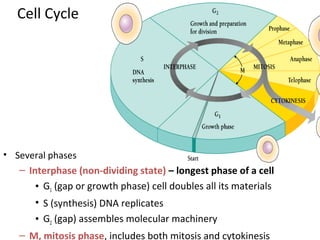
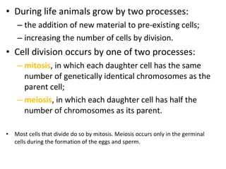
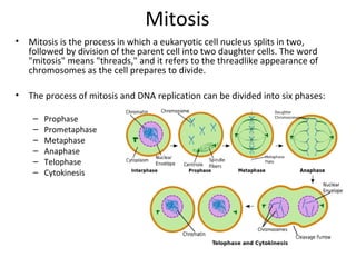
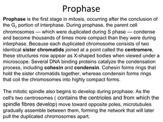
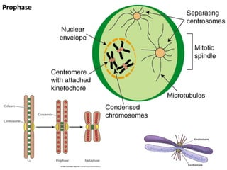
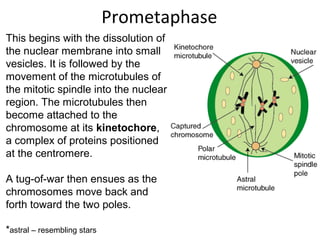
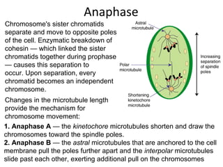
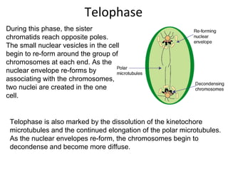
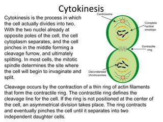
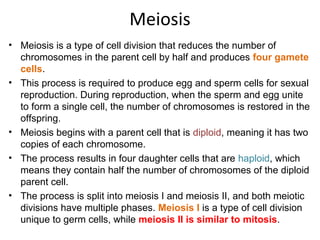
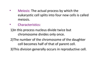
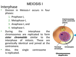
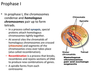
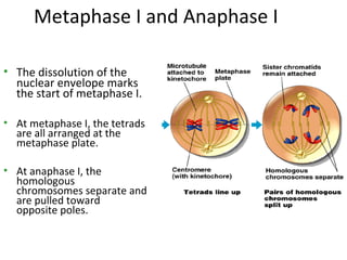
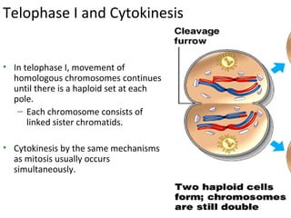
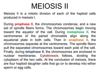
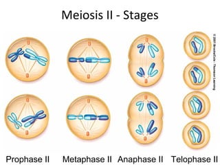
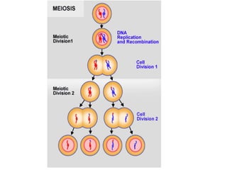
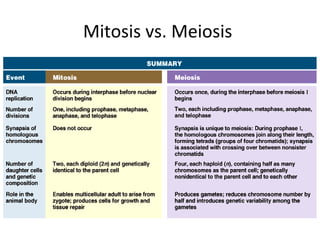
Cell Division
Md. Saiful Islam
Dept. of Pharmaceutical Sciences
North South University
Facebook Group: Pharmacy Universe
Youtube Channel: Pharmacy Universe
The study of the cell cycle focuses on mechanisms that
regulate the timing and frequency of DNA duplication and cell
division. As a biological concept, the cell cycle is defined as the
period between successive divisions of a cell. During this
period, the contents of the cell must be accurately replicated.
The cell cycle is regulated by cyclins and cyclin-dependent kinases.
How long is one cell cycle?
Depends. Eg. Skin cells every 24 hours. Some bacteria every 2
hours. Some cells every 3 months. Cancer cells very short. Nerve
cells never.
Programmed cell death:
Each cell type will only do so many cell cycles then die. (Apoptosis)
The cell cycle
Cell Cycle
• Several phases
– Interphase (non-dividing state) – longest phase of a cell
• G1 (gap or growth phase) cell doubles all its materials
• S (synthesis) DNA replicates
• G2 (gap) assembles molecular machinery
– M, mitosis phase, includes both mitosis and cytokinesis
• During life animals grow by two processes:
– the addition of new material to pre-existing cells;
– increasing the number of cells by division.
• Cell division occurs by one of two processes:
– mitosis, in which each daughter cell has the same
number of genetically identical chromosomes as the
parent cell;
– meiosis, in which each daughter cell has half the
number of chromosomes as its parent.
• Most cells that divide do so by mitosis. Meiosis occurs only in the germinal
cells during the formation of the eggs and sperm.
MITOSIS
Mitosis
• Mitosis is the process in which a eukaryotic cell nucleus splits in two,
followed by division of the parent cell into two daughter cells. The word
"mitosis" means "threads," and it refers to the threadlike appearance of
chromosomes as the cell prepares to divide.
• The process of mitosis and DNA replication can be divided into six phases:
– Prophase
– Prometaphase
– Metaphase
– Anaphase
– Telophase
– Cytokinesis
Prophase
Prophase is the first stage in mitosis, occurring after the conclusion of
the G2 portion of interphase. During prophase, the parent cell
chromosomes — which were duplicated during S phase — condense
and become thousands of times more compact than they were during
interphase. Because each duplicated chromosome consists of two
identical sister chromatids joined at a point called the centromere,
these structures now appear as X-shaped bodies when viewed under a
microscope. Several DNA binding proteins catalyze the condensation
process, including cohesin and condensin. Cohesin forms rings that
hold the sister chromatids together, whereas condensin forms rings
that coil the chromosomes into highly compact forms.
The mitotic spindle also begins to develop during prophase. As the
cell's two centrosomes ( contains the centrioles and from which the
spindle fibres develop) move toward opposite poles, microtubules
gradually assemble between them, forming the network that will later
pull the duplicated chromosomes apart.
Prophase
Prometaphase
This begins with the dissolution of
the nuclear membrane into small
vesicles. It is followed by the
movement of the microtubules of
the mitotic spindle into the nuclear
region. The microtubules then
become attached to the
chromosome at its kinetochore,
a complex of proteins positioned
at the centromere.
A tug-of-war then ensues as the
chromosomes move back and
forth toward the two poles.
*astral – resembling stars
Metaphase
During metaphase (longest stage
of mitosis) the chromosomes align
along the cell equator and form a
disc called metaphase plate.
At this point, the tension within the
cell becomes balanced, and the
chromosomes no longer move
back and forth. In addition, the
spindle is now complete, and three
groups of spindle microtubules are
apparent:
1. Kinetochore microtubules attach the chromosomes to the spindle
pole. 2. Interpolar microtubules extend from the spindle pole across
the equator, almost to the opposite spindle pole; and 3. astral
microtubules extend from the spindle pole to the cell membrane.
Anaphase
Chromosome's sister chromatids
separate and move to opposite poles
of the cell. Enzymatic breakdown of
cohesin — which linked the sister
chromatids together during prophase
— causes this separation to
occur. Upon separation, every
chromatid becomes an independent
chromosome.
Changes in the microtubule length
provide the mechanism for
chromosome movement:
1. Anaphase A — the kinetochore microtubules shorten and draw the
chromosomes toward the spindle poles.
2. Anaphase B — the astral microtubules that are anchored to the cell
membrane pull the poles further apart and the interpolar microtubules
slide past each other, exerting additional pull on the chromosomes
Telophase
During this phase, the sister
chromatids reach opposite poles.
The small nuclear vesicles in the cell
begin to re-form around the group of
chromosomes at each end. As the
nuclear envelope re-forms by
associating with the chromosomes,
two nuclei are created in the one
cell.
Telophase is also marked by the dissolution of the kinetochore
microtubules and the continued elongation of the polar microtubules.
As the nuclear envelopes re-form, the chromosomes begin to
decondense and become more diffuse.
Cytokinesis
Cytokinesis is the process in which
the cell actually divides into two.
With the two nuclei already at
opposite poles of the cell, the cell
cytoplasm separates, and the cell
pinches in the middle forming a
cleavage furrow, and ultimately
splitting. In most cells, the mitotic
spindle determines the site where
the cell will begin to invaginate and
split.
Cleavage occurs by the contraction of a thin ring of actin filaments
that form the contractile ring. The contractile ring defines the
cleavage line for the cell. If the ring is not positioned at the center of
the cell, an asymmetrical division takes place. The ring contracts
and eventually pinches the cell until it separates into two
independent daughter cells.
RESULT OF MITOSIS
• Two daughter nuclei
• Each with same chromosome
number as parent cell
• Chromosomes in unduplicated
form
Cell Cycle Regulation
• Basic regulation depends on cyclins (which are cyclically
synthesized and degraded) and enzymes known as cyclin-
dependant kinases.
• Cell cycle Checkpoints – points where the cell monitors the
previous steps before allowing the cycle to proceed further.
Inhibitory Signals. There are a few including:-
- DNA damage checkpoint (G1/S) – stops DNA replication till
damage is repaired or initiates apoptosis.
- DNA replication checkpoint (G2/M) – prevents entry into
mitosis before all DNA is properly replicated and no
replication forks are left.
- Mitotic Spindle attachment checkpoint – prevents entry
into Anaphase before all chromosome centromeres are
bipolarly attached to the mitotic spindle.
MEIOSIS
Meiosis
• Meiosis is a type of cell division that reduces the number of
chromosomes in the parent cell by half and produces four gamete
cells.
• This process is required to produce egg and sperm cells for sexual
reproduction. During reproduction, when the sperm and egg unite
to form a single cell, the number of chromosomes is restored in the
offspring.
• Meiosis begins with a parent cell that is diploid, meaning it has two
copies of each chromosome.
• The process results in four daughter cells that are haploid, which
means they contain half the number of chromosomes of the diploid
parent cell.
• The process is split into meiosis I and meiosis II, and both meiotic
divisions have multiple phases. Meiosis I is a type of cell division
unique to germ cells, while meiosis II is similar to mitosis.
• Meiosis: The actual process by which the
eukaryotic cell splits into four new cells is called
meiosis.
• Characteristics:
1)In this process nucleus divide twice but
chromosome divides only once.
2)The number of the chromosome of the daughter
cell becomes half of that of parent cell.
3)This division generally occurs in reproductive cell.
Interphase
• Division in Meiosis-I occurs in four
phases:
1. Prophase-I,
2. Metaphase-I,
3. Anaphase-I, and
4. telophase-I.
• During the interphase the
chromosomes are replicated to form
sister chromatids similar to the
interphase of mitosis. These are
genetically identical and joined at the
centromere.
• Also, the single centrosome
is replicated.
MEIOSIS I
Prophase I
• In prophase I, the chromosomes
condense and homologous
chromosomes pair up to form
tetrads.
– In a process called synapsis, special
proteins attach homologous
chromosomes tightly together.
– At several sites the chromatids of
homologous chromosomes are crossed
(chiasmata) and segments of the
chromosomes cross over takes place
(also called recombination).
– Recombination is a process that breaks,
recombines and rejoins sections of DNA
to produce new combinations of genes.
– A spindle forms from each
centrosome
Metaphase I and Anaphase I
• The dissolution of the
nuclear envelope marks
the start of metaphase I.
• At metaphase I, the tetrads
are all arranged at the
metaphase plate.
• At anaphase I, the
homologous
chromosomes separate and
are pulled toward
opposite poles.
Telophase I and Cytokinesis
• In telophase I, movement of
homologous chromosomes continues
until there is a haploid set at each
pole.
– Each chromosome consists of
linked sister chromatids.
• Cytokinesis by the same mechanisms
as mitosis usually occurs
simultaneously.
During prophase II, the chromosomes condense, and a new
set of spindle fibers forms. The chromosomes begin moving
toward the equator of the cell. During metaphase II, the
centromeres of the paired chromatids align along the
equatorial plate in both cells. Then in anaphase II, the
chromosomes separate at the centromeres. The spindle fibers
pull the separated chromosomes toward each pole of the cell.
Finally, during telophase II, the chromosomes are enclosed in
nuclear membranes. Cytokinesis follows, dividing the
cytoplasm of the two cells. At the conclusion of meiosis, there
are four haploid daughter cells that go on to develop into either
sperm or egg cells.
Meiosis II is a mitotic division of each of the haploid cells
produced in meiosis I.
MEIOSIS II
Meiosis II - Stages
Prophase II Metaphase II Anaphase II Telophase II
Mitosis vs. Meiosis
THANK YOU

Cell division/Cell Cycle/ DNA duplication

  • 1.
    Cell Division Md. SaifulIslam Dept. of Pharmaceutical Sciences North South University Facebook Group: Pharmacy Universe Youtube Channel: Pharmacy Universe
  • 2.
    The study ofthe cell cycle focuses on mechanisms that regulate the timing and frequency of DNA duplication and cell division. As a biological concept, the cell cycle is defined as the period between successive divisions of a cell. During this period, the contents of the cell must be accurately replicated. The cell cycle is regulated by cyclins and cyclin-dependent kinases. How long is one cell cycle? Depends. Eg. Skin cells every 24 hours. Some bacteria every 2 hours. Some cells every 3 months. Cancer cells very short. Nerve cells never. Programmed cell death: Each cell type will only do so many cell cycles then die. (Apoptosis) The cell cycle
  • 3.
    Cell Cycle • Severalphases – Interphase (non-dividing state) – longest phase of a cell • G1 (gap or growth phase) cell doubles all its materials • S (synthesis) DNA replicates • G2 (gap) assembles molecular machinery – M, mitosis phase, includes both mitosis and cytokinesis
  • 4.
    • During lifeanimals grow by two processes: – the addition of new material to pre-existing cells; – increasing the number of cells by division. • Cell division occurs by one of two processes: – mitosis, in which each daughter cell has the same number of genetically identical chromosomes as the parent cell; – meiosis, in which each daughter cell has half the number of chromosomes as its parent. • Most cells that divide do so by mitosis. Meiosis occurs only in the germinal cells during the formation of the eggs and sperm.
  • 5.
  • 6.
    Mitosis • Mitosis isthe process in which a eukaryotic cell nucleus splits in two, followed by division of the parent cell into two daughter cells. The word "mitosis" means "threads," and it refers to the threadlike appearance of chromosomes as the cell prepares to divide. • The process of mitosis and DNA replication can be divided into six phases: – Prophase – Prometaphase – Metaphase – Anaphase – Telophase – Cytokinesis
  • 7.
    Prophase Prophase is thefirst stage in mitosis, occurring after the conclusion of the G2 portion of interphase. During prophase, the parent cell chromosomes — which were duplicated during S phase — condense and become thousands of times more compact than they were during interphase. Because each duplicated chromosome consists of two identical sister chromatids joined at a point called the centromere, these structures now appear as X-shaped bodies when viewed under a microscope. Several DNA binding proteins catalyze the condensation process, including cohesin and condensin. Cohesin forms rings that hold the sister chromatids together, whereas condensin forms rings that coil the chromosomes into highly compact forms. The mitotic spindle also begins to develop during prophase. As the cell's two centrosomes ( contains the centrioles and from which the spindle fibres develop) move toward opposite poles, microtubules gradually assemble between them, forming the network that will later pull the duplicated chromosomes apart.
  • 8.
  • 9.
    Prometaphase This begins withthe dissolution of the nuclear membrane into small vesicles. It is followed by the movement of the microtubules of the mitotic spindle into the nuclear region. The microtubules then become attached to the chromosome at its kinetochore, a complex of proteins positioned at the centromere. A tug-of-war then ensues as the chromosomes move back and forth toward the two poles. *astral – resembling stars
  • 10.
    Metaphase During metaphase (longeststage of mitosis) the chromosomes align along the cell equator and form a disc called metaphase plate. At this point, the tension within the cell becomes balanced, and the chromosomes no longer move back and forth. In addition, the spindle is now complete, and three groups of spindle microtubules are apparent: 1. Kinetochore microtubules attach the chromosomes to the spindle pole. 2. Interpolar microtubules extend from the spindle pole across the equator, almost to the opposite spindle pole; and 3. astral microtubules extend from the spindle pole to the cell membrane.
  • 11.
    Anaphase Chromosome's sister chromatids separateand move to opposite poles of the cell. Enzymatic breakdown of cohesin — which linked the sister chromatids together during prophase — causes this separation to occur. Upon separation, every chromatid becomes an independent chromosome. Changes in the microtubule length provide the mechanism for chromosome movement: 1. Anaphase A — the kinetochore microtubules shorten and draw the chromosomes toward the spindle poles. 2. Anaphase B — the astral microtubules that are anchored to the cell membrane pull the poles further apart and the interpolar microtubules slide past each other, exerting additional pull on the chromosomes
  • 12.
    Telophase During this phase,the sister chromatids reach opposite poles. The small nuclear vesicles in the cell begin to re-form around the group of chromosomes at each end. As the nuclear envelope re-forms by associating with the chromosomes, two nuclei are created in the one cell. Telophase is also marked by the dissolution of the kinetochore microtubules and the continued elongation of the polar microtubules. As the nuclear envelopes re-form, the chromosomes begin to decondense and become more diffuse.
  • 13.
    Cytokinesis Cytokinesis is theprocess in which the cell actually divides into two. With the two nuclei already at opposite poles of the cell, the cell cytoplasm separates, and the cell pinches in the middle forming a cleavage furrow, and ultimately splitting. In most cells, the mitotic spindle determines the site where the cell will begin to invaginate and split. Cleavage occurs by the contraction of a thin ring of actin filaments that form the contractile ring. The contractile ring defines the cleavage line for the cell. If the ring is not positioned at the center of the cell, an asymmetrical division takes place. The ring contracts and eventually pinches the cell until it separates into two independent daughter cells.
  • 14.
    RESULT OF MITOSIS •Two daughter nuclei • Each with same chromosome number as parent cell • Chromosomes in unduplicated form
  • 15.
    Cell Cycle Regulation •Basic regulation depends on cyclins (which are cyclically synthesized and degraded) and enzymes known as cyclin- dependant kinases. • Cell cycle Checkpoints – points where the cell monitors the previous steps before allowing the cycle to proceed further. Inhibitory Signals. There are a few including:- - DNA damage checkpoint (G1/S) – stops DNA replication till damage is repaired or initiates apoptosis. - DNA replication checkpoint (G2/M) – prevents entry into mitosis before all DNA is properly replicated and no replication forks are left. - Mitotic Spindle attachment checkpoint – prevents entry into Anaphase before all chromosome centromeres are bipolarly attached to the mitotic spindle.
  • 16.
  • 17.
    Meiosis • Meiosis isa type of cell division that reduces the number of chromosomes in the parent cell by half and produces four gamete cells. • This process is required to produce egg and sperm cells for sexual reproduction. During reproduction, when the sperm and egg unite to form a single cell, the number of chromosomes is restored in the offspring. • Meiosis begins with a parent cell that is diploid, meaning it has two copies of each chromosome. • The process results in four daughter cells that are haploid, which means they contain half the number of chromosomes of the diploid parent cell. • The process is split into meiosis I and meiosis II, and both meiotic divisions have multiple phases. Meiosis I is a type of cell division unique to germ cells, while meiosis II is similar to mitosis.
  • 18.
    • Meiosis: Theactual process by which the eukaryotic cell splits into four new cells is called meiosis. • Characteristics: 1)In this process nucleus divide twice but chromosome divides only once. 2)The number of the chromosome of the daughter cell becomes half of that of parent cell. 3)This division generally occurs in reproductive cell.
  • 19.
    Interphase • Division inMeiosis-I occurs in four phases: 1. Prophase-I, 2. Metaphase-I, 3. Anaphase-I, and 4. telophase-I. • During the interphase the chromosomes are replicated to form sister chromatids similar to the interphase of mitosis. These are genetically identical and joined at the centromere. • Also, the single centrosome is replicated. MEIOSIS I
  • 20.
    Prophase I • Inprophase I, the chromosomes condense and homologous chromosomes pair up to form tetrads. – In a process called synapsis, special proteins attach homologous chromosomes tightly together. – At several sites the chromatids of homologous chromosomes are crossed (chiasmata) and segments of the chromosomes cross over takes place (also called recombination). – Recombination is a process that breaks, recombines and rejoins sections of DNA to produce new combinations of genes. – A spindle forms from each centrosome
  • 21.
    Metaphase I andAnaphase I • The dissolution of the nuclear envelope marks the start of metaphase I. • At metaphase I, the tetrads are all arranged at the metaphase plate. • At anaphase I, the homologous chromosomes separate and are pulled toward opposite poles.
  • 22.
    Telophase I andCytokinesis • In telophase I, movement of homologous chromosomes continues until there is a haploid set at each pole. – Each chromosome consists of linked sister chromatids. • Cytokinesis by the same mechanisms as mitosis usually occurs simultaneously.
  • 23.
    During prophase II,the chromosomes condense, and a new set of spindle fibers forms. The chromosomes begin moving toward the equator of the cell. During metaphase II, the centromeres of the paired chromatids align along the equatorial plate in both cells. Then in anaphase II, the chromosomes separate at the centromeres. The spindle fibers pull the separated chromosomes toward each pole of the cell. Finally, during telophase II, the chromosomes are enclosed in nuclear membranes. Cytokinesis follows, dividing the cytoplasm of the two cells. At the conclusion of meiosis, there are four haploid daughter cells that go on to develop into either sperm or egg cells. Meiosis II is a mitotic division of each of the haploid cells produced in meiosis I. MEIOSIS II
  • 24.
    Meiosis II -Stages Prophase II Metaphase II Anaphase II Telophase II
  • 26.
  • 27.