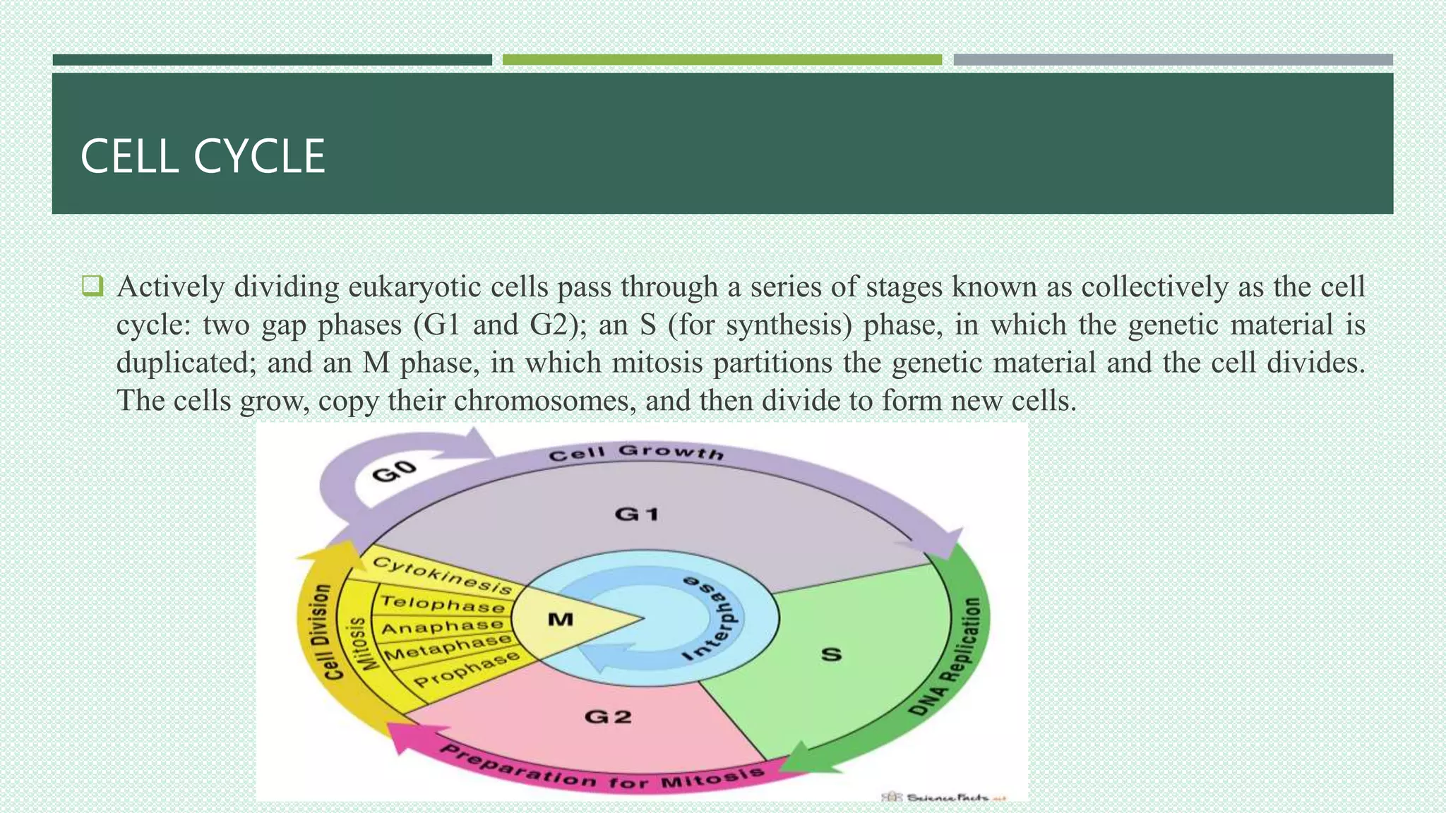
PRESENTATION ON CELL DIVISION
CONTENT
1. WHAT IS CELL?
2. CELL CYCLE
3. TYPES OF CELL DIVISION
4. MITOSIS
5. IMPORTANCE OF MITOSIS
6. STAGES OF MITOSIS
7. CYTOKINESIS
8. MEIOSIS
9. STAGES OF MEIOSIS
10. DIFFERENCE BETWEEN MITOSIS AND MEIOSIS
11. CONCLUSION
CELL DIVISION
WHAT IS CELL?
 Cell is the smallest structural and functional unit of an
organism, which is typically microscopic and consists of
cytoplasm and nucleus enclosed in a membrane.
CELL CYCLE
 Actively dividing eukaryotic cells pass through a series of stages known as collectively as the cell
cycle: two gap phases (G1 and G2); an S (for synthesis) phase, in which the genetic material is
duplicated; and an M phase, in which mitosis partitions the genetic material and the cell divides.
The cells grow, copy their chromosomes, and then divide to form new cells.
CELL CYCLE
 G1 phase: Metabolic changes prepare the cell for division.
At a certain point- the restriction point- the cell is committed to division
and moves into the S phase.
 S phase: DNA synthesis replicates the genetic material. Each chromosomes
now consists of two sister chromatids.
 G2 phase: Metabolic changes assemble the cytoplasmic materials
necessary for mitosis and cytokinesis.
 M phase: A nuclear division (mitosis) followed by a cell division
(cytokinesis).
 Interphase: The period between mitotic division that is, G1, S and G2 is known as interphase.
TYPES OF CELL DIVISION
 Cell divide into two different ways to make new cells.
 Mitosis: Mitosis is used to produce daughter cells that are genetically identical to the parent cells. The
cell copies or replicates its chromosomes, and then splits the copied chromosomes equally to make sure
that each daughter cell has a full set.
 Meiosis: Meiosis is used to make special cells –sperm cells and egg cells-that have the normal number
of chromosomes. It reduce number from 23 pairs of chromosomes to 23 single chromosomes. The cell
copies its chromosomes, but then separates the 23 pairs to ensure that each daughter cell has only one
copy of each chromosome. A second division that divides each daughter cell again to produce four
daughters cells.
MITOSIS
 In Mitosis two daughter cells with the same genetic component as the parent cell are produced.
Chromosomes replicated during the S phase are divided in such a way as to ensure that each daughter
cell receives a copy of every chromosomes.
 The replicated chromosomes are attached to a ‘mitotic apparatus’ that aligns them and then separates
the sister chromatids to produce an even portioning of the genetic material. This separation of the
genetic material in a mitotic nuclear division (or Karyokinesis) is followed by a separation of the cell
cytoplasm in a cellular division (or cytokinesis) to produce two daughter cells.
IMPORTANCE OF MITOSIS
I. In some single-celled organisms mitosis forms the basis of asexual reproduction.
II. In diploid multicellular organisms sexual reproduction involves the fusion of two haploid gametes to
produce a diploid zygote. Mitotic divisions of the zygote and daughter cells are then responsible for the
subsequent growth and development of the organism.
III. In the adult organism, mitosis plays a role in cell replacement, wound healing and tumor formation.
STAGES OF MITOSIS
 Although a continuous process, Mitosis is divided into five stages:
prophase, prometaphase, metaphase, anaphase and telophase.
Prophase
 Prophase occupies over half of mitosis. The nuclear membrane breaks
down to form a number of small vesicles and the nucleolus
disintegrates.
 A structure known as centrosome duplicates itself to form two
daughter centrosomes that migrates to opposite ends of the cell.
 The centrosomes organise the production of microtubules that form the
spindle fibres that constitute the mitotic spindle.
 The chromosomes condense into compact structures. Each replicated
chromosome can now be seen to consist of two identical chromatids
or (sister chromatids) held together by a structure known as the
centromere.
STAGES OF MITOSIS
Prometaphase
 The chromosomes, led by their centromeres, migrate to the equatorial plane in the mid-line of the cell
at right angles to the axis formed by the centrosomes. This regions of the mitotic spindle is known as
the metaphase plate.
 The spindle fibers bind to a structure associated with the centromere of the each chromosomes called a
kinetochore.
 Individuals spindle fibres bind to a kinetochore structure on each side of the centromere. The
chromosomes continue to condense.
Metaphase
 The chromosomes align themselves along the metaphase plate of the spindle apparatus.
STAGES OF MITOSIS
Anaphase
 It is the shortest stage of mitosis. The centromeres divide, and the sister chromatids of each
chromosomes are pulled apart or ‘disjoin’ and move to the opposite ends of the cell, pulled by spindle
fibres attached to the kinetochore regions.
 The separated sister chromatids are now referred to as daughter chromosomes.
Telophase
 The final stages of mitosis, and a reversal of many of the processes observed during prophase.
 The nuclear membrane reforms around the chromosomes grouped at either pole of the cell, the
chromosomes uncoil and become diffuse, and the spindle fibres disappear.
CYTOKINESIS
 It is the final cellular division to form two new cells.
In plants a cell plate forms along the line of the metaphase plate;
in animals there is a constriction of the cytoplasm.
The cell then enters interphase-the interval between mitotic division.
MEIOSIS
 It is the form of eukaryotic cell division that produces haploid sex cells or gametes (which contain a
single copy of each chromosome) from diploid cells (which contain two copies of each chromosomes).
 The process takes the form of one DNA replication followed by two successive nuclear and cellular
divisions (Meiosis I and Meiosis II). As in mitosis, meiosis is preceded by a process of DNA replication
that converts each chromosome into two sister chromatids.
STAGES OF MEIOSIS
 Meiosis I: Meiosis I separates the pairs of homologous chromosomes.
In Meiosis I, a special cell division reduces the cell from diploid to
haploid.
I. Prophase I: The homologous chromosomes pair and exchange DNA to
form recombinant chromosomes. Prophase I is divided into five
phases:
 Leptotene: Chromosomes start to condense.
 Zygotene: Homologous chromosomes become closely associated to
pairs of chromosomes consisting of four chromatids.
 Pachytene: Crossing over between pairs of homologous chromosomes
to form chiasmata.
 Diplotene: Homologous chromosomes start to separate but remain
attached by chiasmata.
 Diakinesis: Homologous chromosomes continue to separate, and
chiasmata move to the ends of the chromosomes.
STAGES OF MEIOSIS
II. Prometaphase I: Spindle apparatus formed, and chromosomes attached to spindle fibres by
kinetochores.
III. Metaphase I: Homologous pairs of chromosomes arranged as a double row along the metaphase
plate.
IV. Anaphase I: The homologous chromosomes in each bivalents are separated and move to the opposite
poles of the cell.
V. Telophase I: The chromosomes become diffuse and the nuclear membrane reforms.
 Cytokinesis: The final cellular division to form two new cells, followed by Meiosis II. Meiosis I is a
reduction division: the original diploid cell had two copies of each chromosomes; the newly formed
haploid cell have one copy of each chromosomes.
STAGES OF MEIOSIS
 Meiosis II
 Meiosis II separates each chromosomes into two chromatids. The events of Meiosis II are analogous to
those of mitotic division, although the number of chromosomes involved has been halved.
 Meiosis generates genetic diversity through:
(i). The exchange of genetic materials between homologous chromosomes during Meiosis I.
(ii). The random alignment of maternal and paternal chromosomes in Meiosis I.
(iii). The random alignment of the sister chromatids at Meiosis II.
DIFFERENCES BETWEEN MITOSIS AND MEIOSIS
Mitosis
 Mitosis takes place in the somatic cells.
 It is a single division which produces two cells.
 Interphase occurs prior to each division
 Mitosis is comparatively simple
 The number of chromosomes remains the same
after mitosis.
 A cell can undergo repeated mitosis.
Meiosis
 It occurs either in the reproductive cells or at the time
of germination of zygote.
 Meiosis is a double division. It gives rise to four
cells.
 Interphase precedes only meiosis 1. It does not occur
prior to meiosis 11.
 Meiosis occurs only once.
 The number of chromosomes is reduced to one half
after meiosis.
 Meiosis occurs only once.
CONCLUSION
 The cells in our body are highly intelligent. They know when to duplicate themselves and when to rest or
stop dividing altogether. This duplication occurs for many reasons but mainly it is to replace old cells. Cell
division is very important process to continue organisms’ life cycle.
 Cell division is a pre-requisite for the continuity of life and forms the basis of evolution to various life
forms.
 In unicellular organisms, cell division is the means of asexual reproduction, which produces two or more
new individuals from the mother cell. The group of such identical individuals is known as clone.
 In multicellular organisms, life starts from a single cell called zygote(fertilized egg). The zygote transforms
into an adult that is composed of millions of cells formed by successive divisions.
 Cell division is the basis of repair and regeneration of old and worn out tissues.
Bibliography
 Hartwell, Leland H., et al. 2000.‘Genetics: From Genes to Genomes’. New York:
McGraw-Hill.
 Hartwell, Leland H., and T.A. Weinert. 1989. ‘Checkpoints: Controls that ensure the
order of Cell Cycle Events’. vol..246: pp 629-634.
 Murry, Andrew, and Tim Hunt. 1993. ‘The cell cycle: An Introduction’. New York:
W. H. Freeman and Company.
 Snustad D. Peter, Simmons J. Michael and Robert H. Tamarin. 1981. ‘Principles of
Genetics’. UK: John Wiley and Sons, Inc.
THANK YOU

Cell division

  • 1.
  • 2.
    CONTENT 1. WHAT ISCELL? 2. CELL CYCLE 3. TYPES OF CELL DIVISION 4. MITOSIS 5. IMPORTANCE OF MITOSIS 6. STAGES OF MITOSIS 7. CYTOKINESIS 8. MEIOSIS 9. STAGES OF MEIOSIS 10. DIFFERENCE BETWEEN MITOSIS AND MEIOSIS 11. CONCLUSION
  • 3.
    CELL DIVISION WHAT ISCELL?  Cell is the smallest structural and functional unit of an organism, which is typically microscopic and consists of cytoplasm and nucleus enclosed in a membrane.
  • 4.
    CELL CYCLE  Activelydividing eukaryotic cells pass through a series of stages known as collectively as the cell cycle: two gap phases (G1 and G2); an S (for synthesis) phase, in which the genetic material is duplicated; and an M phase, in which mitosis partitions the genetic material and the cell divides. The cells grow, copy their chromosomes, and then divide to form new cells.
  • 5.
    CELL CYCLE  G1phase: Metabolic changes prepare the cell for division. At a certain point- the restriction point- the cell is committed to division and moves into the S phase.  S phase: DNA synthesis replicates the genetic material. Each chromosomes now consists of two sister chromatids.  G2 phase: Metabolic changes assemble the cytoplasmic materials necessary for mitosis and cytokinesis.  M phase: A nuclear division (mitosis) followed by a cell division (cytokinesis).  Interphase: The period between mitotic division that is, G1, S and G2 is known as interphase.
  • 6.
    TYPES OF CELLDIVISION  Cell divide into two different ways to make new cells.  Mitosis: Mitosis is used to produce daughter cells that are genetically identical to the parent cells. The cell copies or replicates its chromosomes, and then splits the copied chromosomes equally to make sure that each daughter cell has a full set.  Meiosis: Meiosis is used to make special cells –sperm cells and egg cells-that have the normal number of chromosomes. It reduce number from 23 pairs of chromosomes to 23 single chromosomes. The cell copies its chromosomes, but then separates the 23 pairs to ensure that each daughter cell has only one copy of each chromosome. A second division that divides each daughter cell again to produce four daughters cells.
  • 7.
    MITOSIS  In Mitosistwo daughter cells with the same genetic component as the parent cell are produced. Chromosomes replicated during the S phase are divided in such a way as to ensure that each daughter cell receives a copy of every chromosomes.  The replicated chromosomes are attached to a ‘mitotic apparatus’ that aligns them and then separates the sister chromatids to produce an even portioning of the genetic material. This separation of the genetic material in a mitotic nuclear division (or Karyokinesis) is followed by a separation of the cell cytoplasm in a cellular division (or cytokinesis) to produce two daughter cells.
  • 8.
    IMPORTANCE OF MITOSIS I.In some single-celled organisms mitosis forms the basis of asexual reproduction. II. In diploid multicellular organisms sexual reproduction involves the fusion of two haploid gametes to produce a diploid zygote. Mitotic divisions of the zygote and daughter cells are then responsible for the subsequent growth and development of the organism. III. In the adult organism, mitosis plays a role in cell replacement, wound healing and tumor formation.
  • 9.
    STAGES OF MITOSIS Although a continuous process, Mitosis is divided into five stages: prophase, prometaphase, metaphase, anaphase and telophase. Prophase  Prophase occupies over half of mitosis. The nuclear membrane breaks down to form a number of small vesicles and the nucleolus disintegrates.  A structure known as centrosome duplicates itself to form two daughter centrosomes that migrates to opposite ends of the cell.  The centrosomes organise the production of microtubules that form the spindle fibres that constitute the mitotic spindle.  The chromosomes condense into compact structures. Each replicated chromosome can now be seen to consist of two identical chromatids or (sister chromatids) held together by a structure known as the centromere.
  • 10.
    STAGES OF MITOSIS Prometaphase The chromosomes, led by their centromeres, migrate to the equatorial plane in the mid-line of the cell at right angles to the axis formed by the centrosomes. This regions of the mitotic spindle is known as the metaphase plate.  The spindle fibers bind to a structure associated with the centromere of the each chromosomes called a kinetochore.  Individuals spindle fibres bind to a kinetochore structure on each side of the centromere. The chromosomes continue to condense. Metaphase  The chromosomes align themselves along the metaphase plate of the spindle apparatus.
  • 11.
    STAGES OF MITOSIS Anaphase It is the shortest stage of mitosis. The centromeres divide, and the sister chromatids of each chromosomes are pulled apart or ‘disjoin’ and move to the opposite ends of the cell, pulled by spindle fibres attached to the kinetochore regions.  The separated sister chromatids are now referred to as daughter chromosomes. Telophase  The final stages of mitosis, and a reversal of many of the processes observed during prophase.  The nuclear membrane reforms around the chromosomes grouped at either pole of the cell, the chromosomes uncoil and become diffuse, and the spindle fibres disappear.
  • 12.
    CYTOKINESIS  It isthe final cellular division to form two new cells. In plants a cell plate forms along the line of the metaphase plate; in animals there is a constriction of the cytoplasm. The cell then enters interphase-the interval between mitotic division.
  • 13.
    MEIOSIS  It isthe form of eukaryotic cell division that produces haploid sex cells or gametes (which contain a single copy of each chromosome) from diploid cells (which contain two copies of each chromosomes).  The process takes the form of one DNA replication followed by two successive nuclear and cellular divisions (Meiosis I and Meiosis II). As in mitosis, meiosis is preceded by a process of DNA replication that converts each chromosome into two sister chromatids.
  • 14.
    STAGES OF MEIOSIS Meiosis I: Meiosis I separates the pairs of homologous chromosomes. In Meiosis I, a special cell division reduces the cell from diploid to haploid. I. Prophase I: The homologous chromosomes pair and exchange DNA to form recombinant chromosomes. Prophase I is divided into five phases:  Leptotene: Chromosomes start to condense.  Zygotene: Homologous chromosomes become closely associated to pairs of chromosomes consisting of four chromatids.  Pachytene: Crossing over between pairs of homologous chromosomes to form chiasmata.  Diplotene: Homologous chromosomes start to separate but remain attached by chiasmata.  Diakinesis: Homologous chromosomes continue to separate, and chiasmata move to the ends of the chromosomes.
  • 15.
    STAGES OF MEIOSIS II.Prometaphase I: Spindle apparatus formed, and chromosomes attached to spindle fibres by kinetochores. III. Metaphase I: Homologous pairs of chromosomes arranged as a double row along the metaphase plate. IV. Anaphase I: The homologous chromosomes in each bivalents are separated and move to the opposite poles of the cell. V. Telophase I: The chromosomes become diffuse and the nuclear membrane reforms.  Cytokinesis: The final cellular division to form two new cells, followed by Meiosis II. Meiosis I is a reduction division: the original diploid cell had two copies of each chromosomes; the newly formed haploid cell have one copy of each chromosomes.
  • 16.
    STAGES OF MEIOSIS Meiosis II  Meiosis II separates each chromosomes into two chromatids. The events of Meiosis II are analogous to those of mitotic division, although the number of chromosomes involved has been halved.  Meiosis generates genetic diversity through: (i). The exchange of genetic materials between homologous chromosomes during Meiosis I. (ii). The random alignment of maternal and paternal chromosomes in Meiosis I. (iii). The random alignment of the sister chromatids at Meiosis II.
  • 17.
    DIFFERENCES BETWEEN MITOSISAND MEIOSIS Mitosis  Mitosis takes place in the somatic cells.  It is a single division which produces two cells.  Interphase occurs prior to each division  Mitosis is comparatively simple  The number of chromosomes remains the same after mitosis.  A cell can undergo repeated mitosis. Meiosis  It occurs either in the reproductive cells or at the time of germination of zygote.  Meiosis is a double division. It gives rise to four cells.  Interphase precedes only meiosis 1. It does not occur prior to meiosis 11.  Meiosis occurs only once.  The number of chromosomes is reduced to one half after meiosis.  Meiosis occurs only once.
  • 18.
    CONCLUSION  The cellsin our body are highly intelligent. They know when to duplicate themselves and when to rest or stop dividing altogether. This duplication occurs for many reasons but mainly it is to replace old cells. Cell division is very important process to continue organisms’ life cycle.  Cell division is a pre-requisite for the continuity of life and forms the basis of evolution to various life forms.  In unicellular organisms, cell division is the means of asexual reproduction, which produces two or more new individuals from the mother cell. The group of such identical individuals is known as clone.  In multicellular organisms, life starts from a single cell called zygote(fertilized egg). The zygote transforms into an adult that is composed of millions of cells formed by successive divisions.  Cell division is the basis of repair and regeneration of old and worn out tissues.
  • 19.
    Bibliography  Hartwell, LelandH., et al. 2000.‘Genetics: From Genes to Genomes’. New York: McGraw-Hill.  Hartwell, Leland H., and T.A. Weinert. 1989. ‘Checkpoints: Controls that ensure the order of Cell Cycle Events’. vol..246: pp 629-634.  Murry, Andrew, and Tim Hunt. 1993. ‘The cell cycle: An Introduction’. New York: W. H. Freeman and Company.  Snustad D. Peter, Simmons J. Michael and Robert H. Tamarin. 1981. ‘Principles of Genetics’. UK: John Wiley and Sons, Inc.
  • 20.