
Recommended
More Related Content
What's hot
What's hot (20)
Similar to Mitosis and Meiosis Apoorva B. Vaghela
Similar to Mitosis and Meiosis Apoorva B. Vaghela (20)
Recently uploaded
Recently uploaded (20)
Mitosis and Meiosis Apoorva B. Vaghela
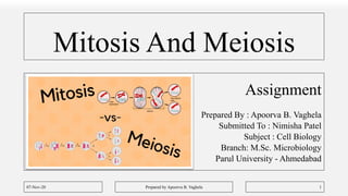
- 1. Mitosis And Meiosis Assignment Prepared By : Apoorva B. Vaghela Submitted To : Nimisha Patel Subject : Cell Biology Branch: M.Sc. Microbiology Parul University - Ahmedabad 07-Nov-20 Prepared by Apoorva B. Vaghela 1
- 2. What is Cell Cycle? • To understand what is mitosis and meiosis, one should first know what is cell cycle. • Cell cycle can be defined as the entire sequence of events happening from the end of one nuclear division to the beginning of the next. • The most basic function of the cell cycle is to duplicate accurately the vast amount of DNA in the chromosomes and then segregate the copies precisely into two genetically identical daughter cells. 07-Nov-20 Prepared by Apoorva B. Vaghela 2
- 3. Stages Of Cell Cycle • DNA duplication occurs during S phase, which requires 10–12 hours and occupies about half of the cell-cycle time in a typical mammalian cell. • After S phase, chromosome segregation and cell division occur in M phase (M for mitosis), which requires much less time. 07-Nov-20 Prepared by Apoorva B. Vaghela 3
- 4. Distribution of Cell Cycle • Howard and Pelc (1953) have divided cell cycle into four phases or stages : G1, S, G2 and M phase. 1. Interphase: Containing G1, S, G2 phases 2. Mitosis : Containing Mitotic phase 07-Nov-20 Prepared by Apoorva B. Vaghela 4
- 5. G1 Phase • After the M phase of previous cell cycle, the daughter cells begin G1 of interphase of new cell cycle. • G1 is a resting phase. It is called first gap phase, since no DNA synthesis takes place during this stage; currently, G1 is also called first growth phase. • Since it involves synthesis of RNA, proteins and membranes which leads to the growth of nucleus and cytoplasm of each daughter cell towards their mature size (see Maclean and Hall, 1987). 07-Nov-20 Prepared by Apoorva B. Vaghela 5
- 6. G1 Phase • During G1 phase, chromatin is fully extended and not distinguishable as discrete chromosomes with the light microscope. • G1 involves transcription of three types of RNAs,namely rRNA, tRNA and mRNA ; rRNA synthesis is indicated by the appearance of nucleolus in the interphase (G1 phase) nucleus. • Proteins synthesized during G1 phase (1) regulatory proteins which control various events of mitosis ; (2) enzymes ( e.g. , DNA polymerase) necessary for DNA synthesis of the next stage ; and (3) tubulin and other mitotic apparatus proteins. 07-Nov-20 Prepared by Apoorva B. Vaghela 6
- 7. S Phase • During the S phase or synthetic phase of interphase, replication of DNA and synthesis of histone proteins occur. • New histones are required in massive amounts immediately at the beginning of the S period of DNA synthesis to provide the new DNA with nucleosomes. • Thus, at the end of S phase, each chromosome has two DNA molecules and a duplicate set of genes. S phase occupies roughly 35 to 45 per cent of cell cycle. 07-Nov-20 Prepared by Apoorva B. Vaghela 7
- 8. G2 Phase • This is a second gap or growth phase or resting phase of interphase. • During G2 phase, synthesis of RNA and proteins continues which is required for cell growth. It may occupy 10 to 20 per cent time of cell cycle. • As the G2 phase draws to a close, the cell enters the M phase. 07-Nov-20 Prepared by Apoorva B. Vaghela 8
- 9. Mitosis • It takes place in somatic cells. • Mitosis begins with chromosome condensation: the duplicated DNA strands, packaged into elongated chromosomes, condense into the much more compact chromosomes required for their segregation. • For better understanding, it is divided in several stages based on it’s morphological as well as it’s compositional changes. 07-Nov-20 Prepared by Apoorva B. Vaghela 9
- 10. • The nuclear envelope then breaks down, and the replicated chromosomes, each consisting of a pair of sister chromatids, become attached to the microtubules of the mitotic spindle. • As mitosis proceeds, the cell pauses briefly in a state called metaphase, when the chromosomes are aligned at the equator of the mitotic spindle, poised for segregation. • The sudden separation of sister chromatids marks the beginning of anaphase, during which the chromosomes move to opposite poles of the spindle, where they decondense and reform intact nuclei. • The cell is then pinched in two by cytoplasmic division, or cytokinesis, and cell division is complete 07-Nov-20 Prepared by Apoorva B. Vaghela 10
- 11. 1. Prophase • The appearance of thin-thread like condensing chromosomes marks the first phase of mitosis, called prophase. • Each prophase chromosome is composed of two coiled filaments, the chromatids. • Chromatids become shorter and thicker and two sister chromatids of each chromosome are held together by a special DNA-containing region, called the centromere or primary constriction. 07-Nov-20 Prepared by Apoorva B. Vaghela 11
- 12. • During early prophase, the chromosomes are evenly distributed in the nuclear cavity. • As prophase progresses, the chromosomes approach the nuclear envelope, causing the central space of the nucleus to become empty. • Most conspicuous change is the formation of the spindle or mitotic apparatus. • In the early prophase, there are two pairs of centrioles. • The two pairs of centrioles migrate to opposite poles of the cell and become situated in antipodal positions. 07-Nov-20 Prepared by Apoorva B. Vaghela 12
- 13. • Between the separating centrioles forms a spindle. • Mitotic spindle contains three main types of fibers; 1. Polar Fibers : which extend from the two poles of the spindle toward the equator 2. Kinetochore Fibers: which attach to the kinetochores of centromeres of each mitotic chromosomes and extend toward the pole 3. Astral Fibers: which radiate outward from the poles toward the periphery or cortex of cell. 07-Nov-20 Prepared by Apoorva B. Vaghela 13
- 14. • Lastly, during prophase, the nucleolus gradually disintegrates. • Degeneration and disappearance of the nuclear envelope marks the end of prophase. 07-Nov-20 Prepared by Apoorva B. Vaghela 14
- 15. 2. Metaphase • During Metaphase; the chromosomes are shortest and thickest. • Their centromeres occupy the plane of the equator of the mitotic apparatus. • At this stage the sister chromatids are still held together by centromere and the kinetochores of the two sister chromatids face opposite poles. • As if the cell pause until all their chromosomes are lined up appropriately on the metaphase plate. 07-Nov-20 Prepared by Apoorva B. Vaghela 15
- 16. • At metaphase, subunits (tubulin dimers) are added to the plus end of a microtubule at the kinetochore and are removed from the minus end at the spindle pole. 07-Nov-20 Prepared by Apoorva B. Vaghela 16
- 17. 3. Anaphase • Anaphase begins abruptly with the synchronous splitting of each chromosome into its sister chromatids, called daughter chromosomes, each with one kinetochore. • Synchronous splitting of each centromere during prophase is evidently caused by an increase in cytosolic Ca2+. In fact, Ca2+ containing membrane vesicles accumulate at spindle poles and release calcium ions to initiate anaphase. 07-Nov-20 Prepared by Apoorva B. Vaghela 17
- 18. • Anaphase involves the following two steps; • Anaphase A: During it, there is poleward movement of chromatids due to shortening of the kinetochore microtubules. • During their poleward migration, the centromeres (and kinetochores) remain foremost so that the chromosomes characteristically appear U,V or J- shaped. • Anaphase B: It involves separation of poles themselves accompanied by the elongation of the polar microtubules. • The astral microtubules also help in anaphase B by their attractive interaction with cell cortex. 07-Nov-20 Prepared by Apoorva B. Vaghela 18
- 19. Anaphase 07-Nov-20 Prepared by Apoorva B. Vaghela 19
- 20. 4. Telophase • The end of the polar migration of the daughter chromosomes marks the beginning of the telophase; which in turn is terminated by the reorganization of two new nuclei and their entry into the G1 phase of interphase. • Events of prophase occur in reverse sequence during this phase. • A nuclear envelope reassembles around each group of chromosomes to form two daughter nuclei. 07-Nov-20 Prepared by Apoorva B. Vaghela 20
- 21. • The mitotic apparatus except the centrioles disappears ; high viscosity of the cytoplasm decreases; the chromosomes resume their long, slender, extended form as their coils relax; and RNA- synthesis restarts causing the nucleolus to reappear. 07-Nov-20 Prepared by Apoorva B. Vaghela 21
- 22. Cytokinesis • Cleavage is accomplished by the contraction of a ring composed mainly of actin filaments. This bundle of filaments, called contractile ring, is bound to the cytoplasmic face of the plasma membrane by unidentified attachment proteins. • Force is generated due to muscle-like sliding of actin and myosin filaments in the contractile ring. • During a normal cytokinesis, the contractile ring does not get thicker as the furrow invaginates, suggesting that it continuously reduces its volume by losing filaments. 07-Nov-20 Prepared by Apoorva B. Vaghela 22
- 23. • When cleavage ends, the contractile ring is finally dispensed with altogether and the plasma mem-brane of the cleavage furrow narrows to form the midbody, which remains as a tether. 07-Nov-20 Prepared by Apoorva B. Vaghela 23
- 24. Meiosis • It is a process of reduction division in which the number of chromosomes per cell is cut in half through the separation of homologous chromosomes in a diploid cell. • Meiosis produces a total of four haploid cells from each original diploid cell. • Meiosis superficially resembles two mitotic divisions without an intervening period of DNA replication. 07-Nov-20 Prepared by Apoorva B. Vaghela 24
- 25. Heterotypic Division or First Meiotic Division • The first meiotic division includes a long prophase in which the homologous chromosomes become closely associated to each other and interchange of hereditary material takes place between them. • Further, in the first meiotic division the reduction of chromosome number takes place and, thus, two haploid cells are resulted by this division. • The first meiotic division is also known as the Heterotypic Division. 07-Nov-20 Prepared by Apoorva B. Vaghela 25
- 26. Homotypic Division or Second Meiotic Division • In the second meiotic division the haploid cell divides mitotically and results into four haploid cells. • The second meiotic division is also known as the Homotypic Division. In the homotypic division pairing of chromosomes, exchange of the genetic material and reduction of the chromosome number do not occur. • Both the meiotic divisions occur continuously and each includes the usual stages of the meiosis, viz., prophase, metaphase, anaphase and telophase. 07-Nov-20 Prepared by Apoorva B. Vaghela 26
- 27. 07-Nov-20 Prepared by Apoorva B. Vaghela 27
- 28. Heterotypic Division or First Meiotic Division • Meiosis starts after an interphase, which is not very different from that of an intermitotic interphase. • During the premeiotic interphase DNA duplication has occurred at the S phase. • In the G2 phase of interphase apparently there is a decisive change that directs the cell toward meiosis, instead of toward mitosis 07-Nov-20 Prepared by Apoorva B. Vaghela 28
- 29. Heterotypic Division or First Meiotic Division • Further, in the beginning of the first meiotic division the nucleus of the meiocyte starts to swell up by absorbing the water from the cytoplasm and the nuclear volume increases about three folds. • After these changes the cell passes to the first stage of first meiotic division which is known as prophase. 07-Nov-20 Prepared by Apoorva B. Vaghela 29
- 30. Prophase I • The first prophase is the longest stage of the meiotic division. It includes following substages; 1. Leptotene or Leptonema: • In the leptotene stage the chromosomes become more uncoiled and assume a long thread-like shape. • The chromosomes at this stage take up a specific orientation inside the nucleus; the ends of the chromosomes converge toward one side of the nucleus, that side where the centrosome lies (the bouquet stage ). 07-Nov-20 Prepared by Apoorva B. Vaghela 30
- 31. Prophase I • The centriole duplicates and each daughter centriole migrates towards the opposite poles of the cell. • On reaching at the poles, each centriole duplicates and, thus, each pole of cell possesses two centrioles of a single diplosome. 2. Zygotene or Zygonema: • In the zygotene stage, the pairing of homologous chromosomes takes place. The homologous chromosomes which come from the mother and father are attracted towards each other and their pairing takes place. 07-Nov-20 Prepared by Apoorva B. Vaghela 31
- 32. Prophase I • The pairing of the homologous chromosomes is known as synapsis. • The synapsis begins at one or more points along the length of the homologous chromosomes. 3. Pachytene or Pachynema: • In the pachynema stage the pair of chromosomes become twisted spirally around each other and cannot be distinguished separately. 07-Nov-20 Prepared by Apoorva B. Vaghela 32
- 33. Prophase I • In the middle of the pachynema stage each homologous chromosome splits lengthwise to form two chromatids. • Through the earlier part of the meiotic prophase, however, the DNA molecule in each chromosome behaves as a single body. 07-Nov-20 Prepared by Apoorva B. Vaghela 33
- 34. Prophase I • In the pachynema stage, this is now changed, the two chromatids of each chromosome containing half of the DNA present in the chromosome at start, become partially independent of one another, although they still continue to be linked together by their common centromere. • Each synaptonemal pair at this point is commonly referred to as bivalent or dyads because it consists of two visible chromosomes ,or as a quadrivalent or tetrad because of the four visible chromatids. 07-Nov-20 Prepared by Apoorva B. Vaghela 34
- 35. Prophase I • During pachynema stage an important genetic phenomenon called “crossing over” takes place. • The crossing over involves reshuffling, redistribution and mutual exchange of hereditary material of two parents between two homologous chromosomes. • The process of interchange of chromatin material between one non- sister chromatid of each homologous chromosome is known as the crossing over which is accompanied by the chiasmata formation. 07-Nov-20 Prepared by Apoorva B. Vaghela 35
- 36. Prophase I 4. Diplotene or Diplonema: • In this phase, Unpairing or desynapsis of homologous chromosomes is started and chiasmata are first seen. • At this phase the chromatids of each tetrad are usually clearly visible, but the synaptonemal complex appears to be dissolved, leaving participating chromatids of the paired homologous chromosome physically joined at one or more discrete points called chiasmata. 07-Nov-20 Prepared by Apoorva B. Vaghela 36
- 37. Prophase I • These points are where crossing over took place. • Often there is some unfolding of the chromatids at this stage, allowing for RNA synthesis and cellular growth. 5. Diakinesis: • In the diakinesis stage the bivalent chromosomes become more condensed and evenly distributed in the nucleus. 07-Nov-20 Prepared by Apoorva B. Vaghela 37
- 38. Prophase I • The nucleolus detaches from the nucleolar organizer and ultimately disappears. • The nuclear envelope breaks down. During diakinesis the chiasma moves from the centromere towards the end of the chromosomes and the intermediate chiasmata diminish. • This type of movement of the chiasmata is known as terminalization . • The chromatids still remain connected by the terminal chiasmata and these exist up to the metaphase. 07-Nov-20 Prepared by Apoorva B. Vaghela 38
- 39. Metaphase I • Metaphase I consists of spindle fiber attachment to chromosomes and chromosomal alignment at the equator. • During metaphase I, the microtubules of the spindle are attached with the centromeres of the homologous chromosomes of each tetrad. • The centromere of each chromosome is directed towards the opposite poles. The repulsive forces between the homologous chromosomes increase greatly and the chromosomes become ready to separate. 07-Nov-20 Prepared by Apoorva B. Vaghela 39
- 40. Anaphase I • At anaphase I homologues are freed from each other and due to the shortening of chromosomal fibers or microtubules each homologous chromosome with its two chromatids and undivided centromere move towards the opposite poles of the cell. • The chromosomes with single or few terminal chiasma usually separate more frequently than the longer chromosomes containing many chiasmata. 07-Nov-20 Prepared by Apoorva B. Vaghela 40
- 41. Anaphase I • The actual reduction and disjunction occurs at this stage. • Moreover, because during the chiasma formation out of two chromatids of a chromosome, one has changed its counterpart, therefore, the two chromatids of a chromosome do not resemble with each other in the genetical terms. 07-Nov-20 Prepared by Apoorva B. Vaghela 41
- 42. Telophase I • The arrival of a haploid set of chromosomes at each pole defines the onset of telophase I, during which nuclei are reassembled. • The endoplasmic reticulum forms the nuclear envelope around the chromosomes and the chromosomes become uncoil. • The nucleolus reappears and, thus, two daughter chromosomes are formed. After the karyokinesis, cytokinesis occurs and two haploid cells are formed. 07-Nov-20 Prepared by Apoorva B. Vaghela 42
- 43. Telophase I • Both cells pass through a short resting phase of interphase. • During interphase, no DNA replication occurs, so that chromosomes at the second prophase are the same double stranded structures that disappeared at the first telophase. 07-Nov-20 Prepared by Apoorva B. Vaghela 43
- 44. Prophase I – Graphical Representation 07-Nov-20 Prepared by Apoorva B. Vaghela 44
- 45. Meiosis I 07-Nov-20 Prepared by Apoorva B. Vaghela 45
- 46. Homotypic or Second Meiotic Division • The homotypic or second meiotic division is actually the mitotic division which divides each haploid meiotic cell into two haploid cells. • The second meiotic division includes following four stages; 1. Prophase 2 2. Metaphase 2 3. Anaphase 2 4. Telophase 2 07-Nov-20 Prepared by Apoorva B. Vaghela 46
- 47. Prophase II • In the prophase second, each centriole divides into two and, thus, two pairs of centrioles are formed. • Each pair of centrioles migrates to the opposite pole. The microtubules get arranged in the form of spindle at the right angle of the spindle of first meiosis. • The nuclear membrane and the nucleolus disappear. The chromosomes with two chromatids become short and thick. 07-Nov-20 Prepared by Apoorva B. Vaghela 47
- 48. Metaphase II • During Metaphase II, the chromosomes get arranged on the equator of the spindle. • The centromere divides into two and, thus, each chromosome produces two monads or daughter chromosomes. • The microtubules of the spindle are attached with the centromere of the chromosomes. 07-Nov-20 Prepared by Apoorva B. Vaghela 48
- 49. Anaphase II • The daughter chromosomes move towards the opposite poles due to the shortening of chromosomal microtubules and stretching of interzonal microtubules of the spindle. 07-Nov-20 Prepared by Apoorva B. Vaghela 49
- 50. Telophase II • The chromatids migrate to the opposite poles and now known as chromosomes. • The endoplasmic reticulum forms the nuclear envelope around the chromosomes and the nucleolus reappears due to synthesis of ribosomal RNA (rRNA) by rDNA and also due to accumulation of ribosomal proteins. 07-Nov-20 Prepared by Apoorva B. Vaghela 50
- 51. Telophase II • After the karyokinesis, in each haploid meiotic cell, the cytokinesis occurs and, thus, four haploid cells are resulted. • These cells have different types of chromosomes due to the crossing over in the prophase I. 07-Nov-20 Prepared by Apoorva B. Vaghela 51
- 52. Meiosis 2 - Graphical Representation 07-Nov-20 Prepared by Apoorva B. Vaghela 52
- 53. | Thank You | 07-Nov-20 Prepared by Apoorva B. Vaghela 53