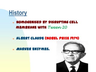
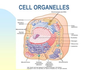
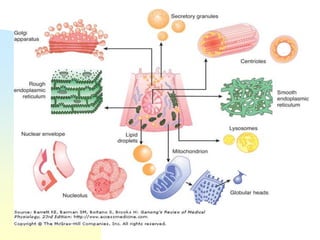
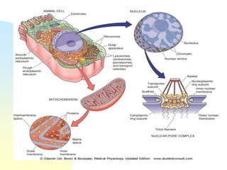
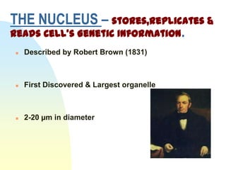
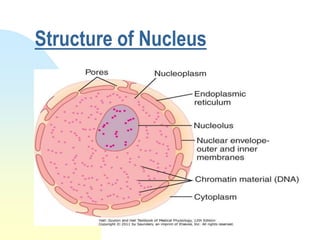
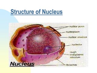
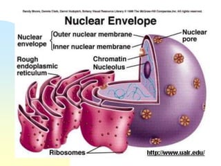
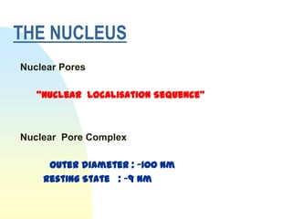
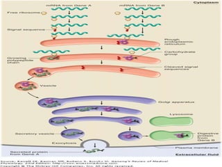
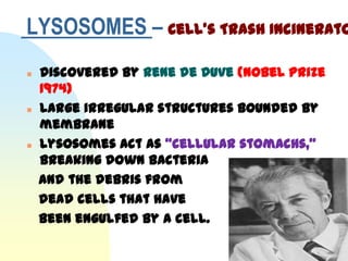
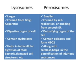
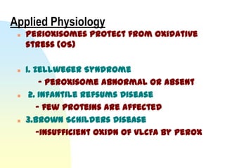
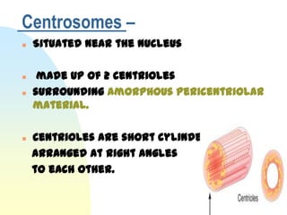
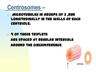
Seminar on
CELL ORGANELLES


Dr.Simi M
Dept of Physiology
Sree Gokulam Medical College & Research Foundation.
26th August 2011.
Introduction
   Discrete organelles that subserve
    distinct functions.

   Bounded by limiting membrane
    “Membrane-enclosed organelles”

   Contain enzymes.
History
    Homogenised by disrupting cell
     membrane with Tween 20

    Albert Claude (Nobel Prize 1974)

    Marker enzymes.
CELL ORGANELLES
      Nucleus
      Endoplasmic reticulum
      Golgi Apparatus
      Mitochondria
      Lysosomes
      Peroxisome
      Ribosome
      Centrosomes
CELL ORGANELLES
THE NUCLEUS – stores,replicates &
reads Cell’s Genetic Information.
   Described by Robert Brown (1831)


   First Discovered & Largest organelle


   2-20 µm in diameter
Structure of Nucleus
    Sorrounded by double membrane
                 - outer membrane
                 - inner membrane

    Space b/w 2 membranes

            "Perinuclear Cistern"
Structure of Nucleus
THE NUCLEUS
Nuclear Pores

   “Nuclear Localisation Sequence”


Nuclear Pore Complex

     Outer diameter : ~100 nm
    Resting State : ~9 nm
Nucleus : Storehouse of DNA
    Chromosomal DNA present inside
     nucleus

    Chromatin
         - Heterochromatin
         - Euchromatin
    Transport pathways
            -Importins
          -Exportins
Nucleolus

    described by Gabriel Valentin 1836

     prominent in cells actively
     synthesizing proteins

    Function :
     - RNA Processing
     - Ribosome synthesis
ENDOPLASMIC RETICULUM
     Web of Tubules or Saccules
      sorrounding the nucleus.

     “ Railway Track Appearance ” - EM

  George Palade
  (Nobel Prize 1974)

     2 forms : RER & SER
RER                  SER
Granular            Agranular

Ribosome studded   Not Ribosome
                    studded

PROTEIN SYN        LIPID &STERIOD SYN
                    Egs:
Egs:                Leydig cells
 Nissl Granules      Cells of Adrenal
 Russel bodies      Cortex
 Acinar cells
Structure of Endoplasmic Reticulum
ENDOPLASMIC RETICULUM
     Functions:

  1.Synthesis of proteins,Glycoprotein &
     Lipoproteins

  2.Detoxification of various drugs

  3.Major Reservoir of Ca2+ ions
     Modified ER (Sarcoplasmic Reticulum)
Microsomes :

Formed by automatic reassembly of
 disrupted complex ER on cell
 fractionation.

Func:
1.Valuable tool for understanding
   metabolism of compounds.
2.Study drug – drug interactions
MITOCHONDRIA -             Site of
Oxidative Energy Production.

     “Power House of the Cell”

     Richard Altman (1886)
     Carl Benda coined the term
     0.5 – 1 µm in diameter &
     7 µm in length.
Structure of Mitochondria

   Described as a “Balloon with in a
    balloon”

   Cigar shaped organelles/
    Sausage shaped organelles
   2 membrane
         - outer membrane
         - inner membrane (Cristae)
       Lollipop shaped globular structures
   Define 2 distinct internal compartments
                  - inter membrane space
                  - matrix space

   Capable of “Self replication”

   Short Life span
Structure of Mitochondria
Mitochondrial Enzymes
   A. Membrane enzymes
     1.Enzymes of outer membrane
     a) Cytochrome b5 & b5 reductase
     b) Fatty acid coA synthase
     c) Phospholipase A
     d) Nucleoside diphosphokinase
Mitochondrial Enzymes
   2. Enzymes of inner membrane
     a) Cytochrome b,C1,C,a& a3
     b) NADPH dehydrogenase
     c) Succinate dehydrogenase
     d) Electron transferring flavoproteins
     e) β-OH-butyrate dehydrogenase
     f) Carnitine – palmitoyl transferase
     g)All translocases
Mitochondrial Enzymes

   B. Enzymes of inter membrane space
     a) Adenylate kinase
     b) Nucleoside diphoshokinase
     c)Sulfite oxidase
Mitochondrial Enzymes
   C. Enzymes in the matrix
     a) Pyruvate dehydrogenase complex
     b) Citrate synthase
     c) Isocitrate dehydrogenase
     d)Malate dehydrogenase
     e) Fatty acid oxidation system
     f) Ornithine transcarbamoylase
     g) α-oxoglutarate dehydrogenase
     h) Aconitase
MITOCHONDRIA
    Functions:

    Critical manufacturer of ATP.

    Programmed Cell death (Apoptosis)
       intracellular Ca 2+ stores.
   ETC (innermemb )
   TCA Cycle (matrix )
   β-Oxidation of FA (matrix )
   Ketone body production
   Urea,Heme,Pyramidine syn
   Gluconeogenesis
Mitochondrial diseases dt mtdna
mutations
   Leber hereditary optic neuropathy (LHON)
   NARP, Leigh's disease
   MELAS
   MERRF
   Progressive sensorineural deafness
   Chronic progressive external
    ophthalmoplegia (PEO)
   Pearson syndrome
   Kearn-Sayre syndrome (KSS)
GOLGI APPARATUS – stack of
pancakes/stack of dinner plates

   Also called “Dyctyosome”
 Collection of membrane enclosed sac
(abt 6 sacs )
 Camillo Golgi (Nobel Prize 1906)

 Continuous with ER

 >200 enzymes

 Processing station
Structure of Golgi Apparatus
   Polarised structure with cis & trans
    sides
   Membrane vesicle containing proteins
    bud off from ER
               ↓
   fuse with the cistern on the cis side of
    the apparatus.
               ↓
   passed via other vesicles to the middle
    cisterns
              ↓
finally to the cistern on the trans side,
  from which vesicles branch off into the
Functions :
   Site for Packaging of proteins
    synthesized in ER into vesicles

   Formation of Lysosomal enzymes

   Transport to other organelles

   Glycosylation of proteins
LYSOSOMES – Cell’s Trash Incinerato
   Discovered by Rene de Duve (Nobel Prize
    1974)
   Large irregular structures bounded by
    membrane
   Lysosomes act as “cellular stomachs,”
    breaking down bacteria
    and the debris from
    dead cells that have
    been engulfed by a cell.
LYSOSOMES
   Proton Pump or H+-ATP ase
       acidic interior (ph 5.0)
   40 types of enz
        “Acid Hydrolases”
   Specially adapted lys.membrane
     Clinical app: Gout(loss of memb
    integrity)

   Endocytic Vesicle

   Autophagy : “Multivesicular body”
LYSOSOMES
   Particularly abundant in cells involved
    in phagocytic activity. (Eg: Neutrophils &
    Macrophages)

   3 Forms of lysosomes

    Primary
    Secondary
    Residual Bodies
Important Lysosomal Enzymes
Proteolytic    1.Cathepsins 2.Collagenas    3.Elastases
Enzymes                     es

Lipolytic      1.Lipases     2.Phospholip 3.Fatty acyl
Enzymes                      ase          Esterases

Carbohydrat    1.α-          2. β-          3.Hyaluronid   4.Aryl
e splitting    glycosidase   galactosidas   ase            Sulphatase
enzymes                      e
Nucleic acid   1.Ribonuclea 2.Deoxy
Hydrolysing    e            Ribonucleas
enzymes                     e
Other          1.Acid        2.Catalase
enzymes        Phosphatse
Lysosomal storage diseases
 - Congenital absence of Lysosomal
enzymes
Disease            Enzyme               Unique Features
                   deficiency
Fabry disease      α-galactosidase A    Cutaneous
                                        Angiokeratomas&
                                        hypohydrosis
Gaucher disease    Acid β glucosidase   HSM & Skeletal Dysplasia


Niemann - Pick     Sphingomyelinase     MR,Seizures & Lung
                                        Failure
disease

Tay- Sachs disease β-hexaminidase A     MR,Macrocephaly &
                                        Hypercuisis in infants


Hurler disease     α-L-iduronidase      MR,Coarse facies,CVS inv


Pompe disease      Acid α-glucosidase   Myocardiopathy
Functions of Lysosomes:

      1.Contain enzymes essential for
      intracellular digestion

      2.Kill & remove foreign bodies

      3.Acrosome :- specialised lysosome

      4.Autolysis

      5.remove IC pdts of metabolism
Ribosomes – Sites of Protein
Synthesis.
    Granular structures present on surface
     of ER & also as free
    1st observed by George Palade (1953)

    Non membrane bound organelle

    Measure ~ 22 x 32 nm.
    Contain 85% RNA of cell
Ribosomes
   Each is made up of a large and a small
    subunit
    on the basis of their rates of
    sedimentation in the ultracentrifuge, the
    60S and 40S subunits
    Polyribosomes(3-5)
   Func
    Main Site of Protein Synthesis
PEROXISOMES
   also known as microbodies

   “Subcellular respiratory organelles”

   0.5 µm in diameter

   Predominantly present in Hepatocytes &
    Tubular Epithelial cells.

   surrounded by a membrane.
   This membrane contains a no of
    peroxisome-specific proteins


    PEROXINS-
    the protein Chaperones,various proteins
     with specifc signal sequence are directed
     to peroxisome.
Function:
    Essentially contain two types of Enzymes
     :

     Oxidases : which are active in oxidation
     of lipid

     Catalase : which act on Hydrogen
     Peroxide to liberate Oxygen.
   Several years ago, a number of synthetic
    compounds were found to cause
    proliferation of peroxisomes by acting
    on receptors in the nuclei of cells.

   These receptors Peroxisome
    Proliferation Activated Receptors
    (PPARs)
Peroxisome Proliferation
  Activated Receptors (PPARs)
• Three PPAR receptors α ,β and γ have been
  characterized.

• When activated, they bind to DNA,
  producing changes in the production of
  mRNAs.

• Mutations of the peroxisome
  proliferators' activator receptor γ
  (PPARγ) cause insulin resistance
Lysosomes               Peroxisomes
Larger                     Smaller
Formed from Golgi         Formed by self-
Apparatus                  replication or budding
                           from smooth ER
Digestive organ of cell   Detoxifying organ of the
                           cell
Contain Hydrolases        Contain oxidases and
                           form H2O2
Helps in intracellular    Along with
digestion of food,         catalase,helps in the
bacteria,damaged cell      detoxification of injurious
structures etc             substances
Applied Physiology
    Perioxisomes protect from oxidative
     stress (OS)

    1. Zellweger Syndrome
          - peroxisome abnormal or absent
     2. Infantile Refsums Disease
         - few proteins are affected
    3.Brown Schilders Disease
         -insufficient oxidn of VLCFA by perox
Centrosomes –
   Situated near the nucleus

    made up of 2 centrioles
   surrounding amorphous pericentriolar
    material.

   centrioles are short cylinders,
    arranged at right angles
    to each other.
Centrosomes –
    Microtubules in groups of 3 ,run
    longitudinally in the walls of each
    centriole.

   9 of these triplets
    are spaced at regular intervals
    around the circumference.
Centrosomes –
   “microtubule-organizing
    centers” (MTOCs) that contain γ-
    tubulin.

    Func:
    monitor steps in cell division.
    regulate chromosome movement
References
 1.Walter.F.Boron & Emile .L.Boulpaep
 Medical Physiology 2nd edition
 2.Ganong’s Review of Physiology 23
                                      rd

  edition
 3.GK Pal Textbook of Medical Physiology

  2nd edition
 4.Guyton & Hall Text book of Physiology

  12th edition
 5.Vanders Human Physiology 8
                                 th edition
References
   6.Sreekumari & Vasudevan Textbook of
    Biochemistry 6th edition
   7.Harrison’s Principles of Internal
    Medicine 17th edition
   8.Indu Khurana Textbook of Medical
    physiology
   9.N Geetha Textbook of Medical Physiology
    2nd edition.
   10.A.K Jain Textbook of Physiology
    4th edition.
RECENT ADVANCES
   Organellar dysfunction in the pathogenesis of
    pancreatitis.
   Acute pancreatitis is an inflammatory
    disease of exocrine pancreas that carries
    considerable morbidity and mortality; its
    pathophysiology remains poorly
    understood.
   Recent findings obtained on experimental
    models, which reveal disordering of key cellular
    organelles, namely, mitochondria,
    autophagosomes, and lysosomes, in pancreatitis.
   Ref
RECENT ADVANCES
   (PPAR) represented by 3 types: PPAR alpha, PPAR
    beta, PPAR gamma.
   PPAR alpha is a key regulator of fatty acid beta-
    oxidation, participates in development of
    inflammatory reaction and atherosclerosis
    formation.
    PPAR gamma plays important role in lipid
    metabolism, processes of cell differentiation
    and growth, participates in glucose utilization
    and mechanisms of insulin resistance. Specific
    activators of PPAR gamma are glytazones
   Ref :http://www.ncbi.nlm.nih.gov/pubmed/14671562
RECENT ADVANCES
   The nuclear (PPAR gamma) in DM,HTN,atherosclerosis

   is a transcription factor that is activated by
    polyunsaturated fatty acids and their
    metabolites and is essential for fat cell
    formation.
   PPAR gamma activators such as the glitazone
    drugs lower glucose and lipid levels in patients
    with type 2 diabetes and also have
    antiatherosclerotic and antihypertensive effects.

   Ref :http://www.ncbi.nlm.nih.gov/pubmed/11395411.
Thank You !!!

Cell organelles

  • 1.
    Seminar on CELL ORGANELLES Dr.SimiM Dept of Physiology Sree Gokulam Medical College & Research Foundation. 26th August 2011.
  • 2.
    Introduction  Discrete organelles that subserve distinct functions.  Bounded by limiting membrane “Membrane-enclosed organelles”  Contain enzymes.
  • 3.
    History  Homogenised by disrupting cell membrane with Tween 20  Albert Claude (Nobel Prize 1974)  Marker enzymes.
  • 4.
    CELL ORGANELLES  Nucleus  Endoplasmic reticulum  Golgi Apparatus  Mitochondria  Lysosomes  Peroxisome  Ribosome  Centrosomes
  • 5.
  • 8.
    THE NUCLEUS –stores,replicates & reads Cell’s Genetic Information.  Described by Robert Brown (1831)  First Discovered & Largest organelle  2-20 µm in diameter
  • 9.
    Structure of Nucleus  Sorrounded by double membrane - outer membrane - inner membrane  Space b/w 2 membranes "Perinuclear Cistern"
  • 10.
  • 11.
  • 12.
  • 13.
    THE NUCLEUS Nuclear Pores “Nuclear Localisation Sequence” Nuclear Pore Complex Outer diameter : ~100 nm Resting State : ~9 nm
  • 15.
    Nucleus : Storehouseof DNA  Chromosomal DNA present inside nucleus  Chromatin - Heterochromatin - Euchromatin  Transport pathways -Importins -Exportins
  • 17.
    Nucleolus  described by Gabriel Valentin 1836  prominent in cells actively synthesizing proteins  Function : - RNA Processing - Ribosome synthesis
  • 18.
    ENDOPLASMIC RETICULUM  Web of Tubules or Saccules sorrounding the nucleus.  “ Railway Track Appearance ” - EM George Palade (Nobel Prize 1974)  2 forms : RER & SER
  • 19.
    RER SER Granular  Agranular Ribosome studded Not Ribosome studded PROTEIN SYN LIPID &STERIOD SYN Egs: Egs: Leydig cells Nissl Granules Cells of Adrenal Russel bodies Cortex Acinar cells
  • 20.
  • 21.
    ENDOPLASMIC RETICULUM  Functions: 1.Synthesis of proteins,Glycoprotein & Lipoproteins 2.Detoxification of various drugs 3.Major Reservoir of Ca2+ ions Modified ER (Sarcoplasmic Reticulum)
  • 22.
    Microsomes : Formed byautomatic reassembly of disrupted complex ER on cell fractionation. Func: 1.Valuable tool for understanding metabolism of compounds. 2.Study drug – drug interactions
  • 23.
    MITOCHONDRIA - Site of Oxidative Energy Production.  “Power House of the Cell”  Richard Altman (1886)  Carl Benda coined the term  0.5 – 1 µm in diameter &  7 µm in length.
  • 24.
    Structure of Mitochondria  Described as a “Balloon with in a balloon”  Cigar shaped organelles/ Sausage shaped organelles  2 membrane - outer membrane - inner membrane (Cristae) Lollipop shaped globular structures
  • 25.
    Define 2 distinct internal compartments - inter membrane space - matrix space  Capable of “Self replication”  Short Life span
  • 26.
  • 30.
    Mitochondrial Enzymes  A. Membrane enzymes 1.Enzymes of outer membrane a) Cytochrome b5 & b5 reductase b) Fatty acid coA synthase c) Phospholipase A d) Nucleoside diphosphokinase
  • 31.
    Mitochondrial Enzymes  2. Enzymes of inner membrane a) Cytochrome b,C1,C,a& a3 b) NADPH dehydrogenase c) Succinate dehydrogenase d) Electron transferring flavoproteins e) β-OH-butyrate dehydrogenase f) Carnitine – palmitoyl transferase g)All translocases
  • 32.
    Mitochondrial Enzymes  B. Enzymes of inter membrane space a) Adenylate kinase b) Nucleoside diphoshokinase c)Sulfite oxidase
  • 33.
    Mitochondrial Enzymes  C. Enzymes in the matrix a) Pyruvate dehydrogenase complex b) Citrate synthase c) Isocitrate dehydrogenase d)Malate dehydrogenase e) Fatty acid oxidation system f) Ornithine transcarbamoylase g) α-oxoglutarate dehydrogenase h) Aconitase
  • 35.
    MITOCHONDRIA  Functions:  Critical manufacturer of ATP.  Programmed Cell death (Apoptosis) intracellular Ca 2+ stores.
  • 36.
    ETC (innermemb )  TCA Cycle (matrix )  β-Oxidation of FA (matrix )  Ketone body production  Urea,Heme,Pyramidine syn  Gluconeogenesis
  • 37.
    Mitochondrial diseases dtmtdna mutations  Leber hereditary optic neuropathy (LHON)  NARP, Leigh's disease  MELAS  MERRF  Progressive sensorineural deafness  Chronic progressive external ophthalmoplegia (PEO)  Pearson syndrome  Kearn-Sayre syndrome (KSS)
  • 39.
    GOLGI APPARATUS –stack of pancakes/stack of dinner plates  Also called “Dyctyosome”  Collection of membrane enclosed sac (abt 6 sacs )  Camillo Golgi (Nobel Prize 1906)  Continuous with ER  >200 enzymes  Processing station
  • 40.
  • 41.
    Polarised structure with cis & trans sides  Membrane vesicle containing proteins bud off from ER ↓  fuse with the cistern on the cis side of the apparatus. ↓  passed via other vesicles to the middle cisterns ↓ finally to the cistern on the trans side, from which vesicles branch off into the
  • 44.
    Functions :  Site for Packaging of proteins synthesized in ER into vesicles  Formation of Lysosomal enzymes  Transport to other organelles  Glycosylation of proteins
  • 45.
    LYSOSOMES – Cell’sTrash Incinerato  Discovered by Rene de Duve (Nobel Prize 1974)  Large irregular structures bounded by membrane  Lysosomes act as “cellular stomachs,” breaking down bacteria and the debris from dead cells that have been engulfed by a cell.
  • 46.
    LYSOSOMES  Proton Pump or H+-ATP ase acidic interior (ph 5.0)  40 types of enz “Acid Hydrolases”  Specially adapted lys.membrane Clinical app: Gout(loss of memb integrity)  Endocytic Vesicle  Autophagy : “Multivesicular body”
  • 47.
    LYSOSOMES  Particularly abundant in cells involved in phagocytic activity. (Eg: Neutrophils & Macrophages)  3 Forms of lysosomes Primary Secondary Residual Bodies
  • 48.
    Important Lysosomal Enzymes Proteolytic 1.Cathepsins 2.Collagenas 3.Elastases Enzymes es Lipolytic 1.Lipases 2.Phospholip 3.Fatty acyl Enzymes ase Esterases Carbohydrat 1.α- 2. β- 3.Hyaluronid 4.Aryl e splitting glycosidase galactosidas ase Sulphatase enzymes e Nucleic acid 1.Ribonuclea 2.Deoxy Hydrolysing e Ribonucleas enzymes e Other 1.Acid 2.Catalase enzymes Phosphatse
  • 49.
    Lysosomal storage diseases - Congenital absence of Lysosomal enzymes
  • 50.
    Disease Enzyme Unique Features deficiency Fabry disease α-galactosidase A Cutaneous Angiokeratomas& hypohydrosis Gaucher disease Acid β glucosidase HSM & Skeletal Dysplasia Niemann - Pick Sphingomyelinase MR,Seizures & Lung Failure disease Tay- Sachs disease β-hexaminidase A MR,Macrocephaly & Hypercuisis in infants Hurler disease α-L-iduronidase MR,Coarse facies,CVS inv Pompe disease Acid α-glucosidase Myocardiopathy
  • 51.
    Functions of Lysosomes:  1.Contain enzymes essential for intracellular digestion 2.Kill & remove foreign bodies 3.Acrosome :- specialised lysosome 4.Autolysis 5.remove IC pdts of metabolism
  • 52.
    Ribosomes – Sitesof Protein Synthesis.  Granular structures present on surface of ER & also as free  1st observed by George Palade (1953)  Non membrane bound organelle  Measure ~ 22 x 32 nm.  Contain 85% RNA of cell
  • 53.
    Ribosomes  Each is made up of a large and a small subunit  on the basis of their rates of sedimentation in the ultracentrifuge, the 60S and 40S subunits  Polyribosomes(3-5)  Func Main Site of Protein Synthesis
  • 54.
    PEROXISOMES  also known as microbodies  “Subcellular respiratory organelles”  0.5 µm in diameter  Predominantly present in Hepatocytes & Tubular Epithelial cells.  surrounded by a membrane.
  • 55.
    This membrane contains a no of peroxisome-specific proteins  PEROXINS- the protein Chaperones,various proteins with specifc signal sequence are directed to peroxisome.
  • 56.
    Function:  Essentially contain two types of Enzymes :  Oxidases : which are active in oxidation of lipid  Catalase : which act on Hydrogen Peroxide to liberate Oxygen.
  • 57.
    Several years ago, a number of synthetic compounds were found to cause proliferation of peroxisomes by acting on receptors in the nuclei of cells.  These receptors Peroxisome Proliferation Activated Receptors (PPARs)
  • 58.
    Peroxisome Proliferation Activated Receptors (PPARs) • Three PPAR receptors α ,β and γ have been characterized. • When activated, they bind to DNA, producing changes in the production of mRNAs. • Mutations of the peroxisome proliferators' activator receptor γ (PPARγ) cause insulin resistance
  • 59.
    Lysosomes Peroxisomes Larger  Smaller Formed from Golgi Formed by self- Apparatus replication or budding from smooth ER Digestive organ of cell Detoxifying organ of the cell Contain Hydrolases Contain oxidases and form H2O2 Helps in intracellular Along with digestion of food, catalase,helps in the bacteria,damaged cell detoxification of injurious structures etc substances
  • 60.
    Applied Physiology  Perioxisomes protect from oxidative stress (OS)  1. Zellweger Syndrome - peroxisome abnormal or absent  2. Infantile Refsums Disease - few proteins are affected  3.Brown Schilders Disease -insufficient oxidn of VLCFA by perox
  • 61.
    Centrosomes –  Situated near the nucleus  made up of 2 centrioles  surrounding amorphous pericentriolar material.  centrioles are short cylinders, arranged at right angles to each other.
  • 62.
    Centrosomes –  Microtubules in groups of 3 ,run longitudinally in the walls of each centriole.  9 of these triplets are spaced at regular intervals around the circumference.
  • 64.
    Centrosomes –  “microtubule-organizing centers” (MTOCs) that contain γ- tubulin.  Func: monitor steps in cell division. regulate chromosome movement
  • 65.
    References  1.Walter.F.Boron &Emile .L.Boulpaep Medical Physiology 2nd edition  2.Ganong’s Review of Physiology 23 rd edition  3.GK Pal Textbook of Medical Physiology 2nd edition  4.Guyton & Hall Text book of Physiology 12th edition  5.Vanders Human Physiology 8 th edition
  • 66.
    References  6.Sreekumari & Vasudevan Textbook of Biochemistry 6th edition  7.Harrison’s Principles of Internal Medicine 17th edition  8.Indu Khurana Textbook of Medical physiology  9.N Geetha Textbook of Medical Physiology 2nd edition.  10.A.K Jain Textbook of Physiology 4th edition.
  • 67.
    RECENT ADVANCES  Organellar dysfunction in the pathogenesis of pancreatitis.  Acute pancreatitis is an inflammatory disease of exocrine pancreas that carries considerable morbidity and mortality; its pathophysiology remains poorly understood.  Recent findings obtained on experimental models, which reveal disordering of key cellular organelles, namely, mitochondria, autophagosomes, and lysosomes, in pancreatitis.  Ref
  • 68.
    RECENT ADVANCES  (PPAR) represented by 3 types: PPAR alpha, PPAR beta, PPAR gamma.  PPAR alpha is a key regulator of fatty acid beta- oxidation, participates in development of inflammatory reaction and atherosclerosis formation.  PPAR gamma plays important role in lipid metabolism, processes of cell differentiation and growth, participates in glucose utilization and mechanisms of insulin resistance. Specific activators of PPAR gamma are glytazones  Ref :http://www.ncbi.nlm.nih.gov/pubmed/14671562
  • 69.
    RECENT ADVANCES  The nuclear (PPAR gamma) in DM,HTN,atherosclerosis  is a transcription factor that is activated by polyunsaturated fatty acids and their metabolites and is essential for fat cell formation.  PPAR gamma activators such as the glitazone drugs lower glucose and lipid levels in patients with type 2 diabetes and also have antiatherosclerotic and antihypertensive effects.  Ref :http://www.ncbi.nlm.nih.gov/pubmed/11395411.
  • 70.