Download as PDF, PPTX


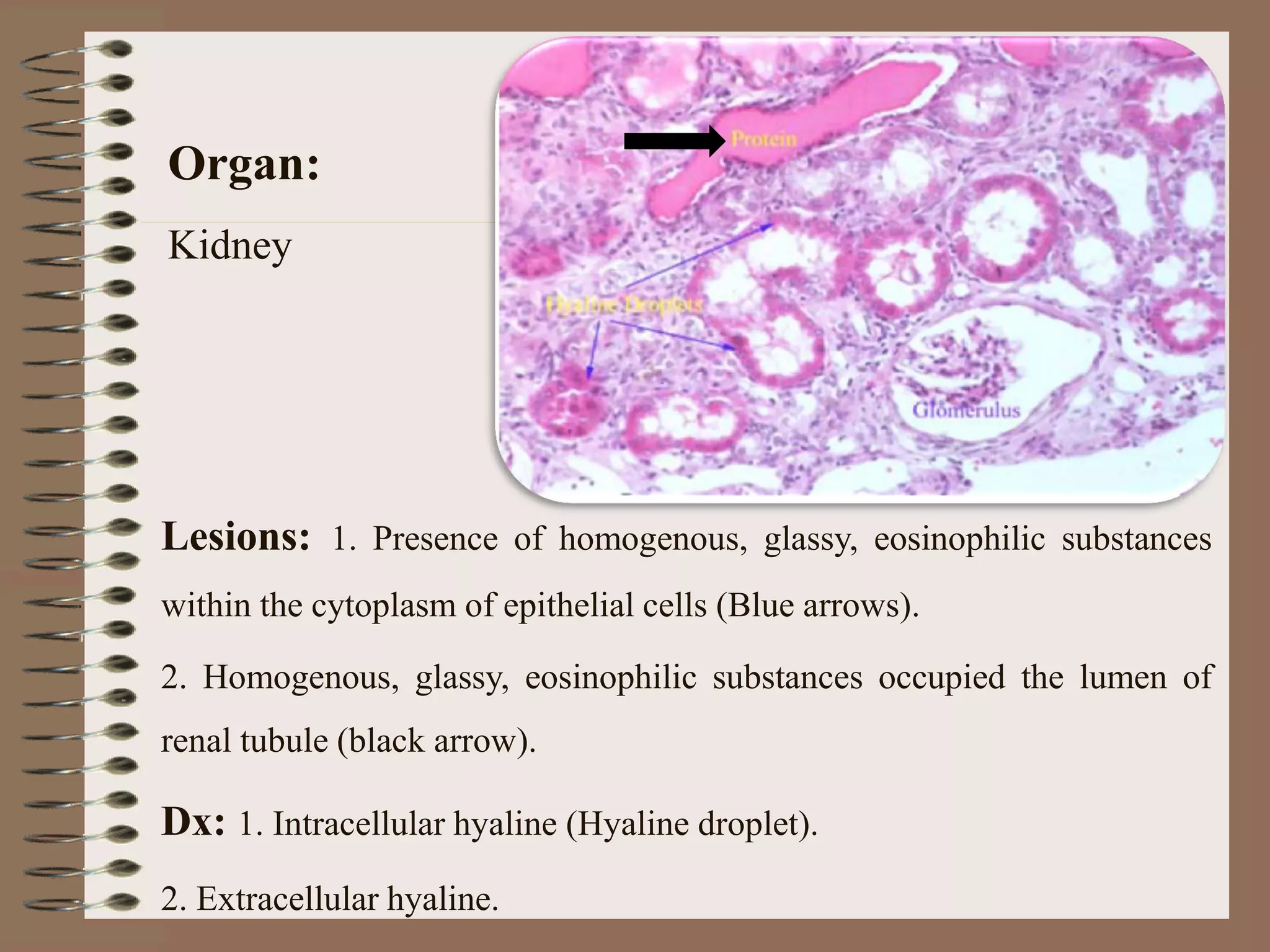


The document discusses intracellular and extracellular accumulation in various organs. It describes cases of glycogen accumulation seen as vacuolation in hepatocyte cytoplasm on H&E staining of the liver. Kidney samples show intracellular hyaline droplets and extracellular hyaline occupying renal tubules. Amyloidosis is seen in the pancreas, kidney, lymph node, duodenum and heart, appearing as extracellular deposition of amorphous eosinophilic material that stains pink with Congo Red.


