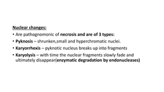
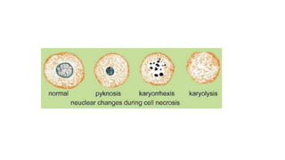
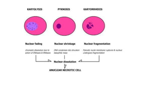
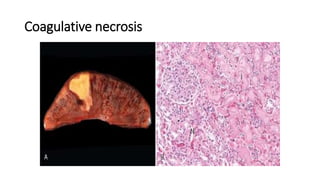
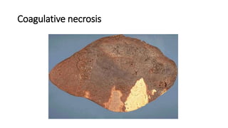
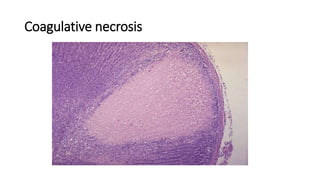
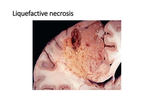
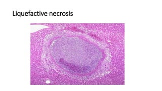
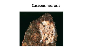
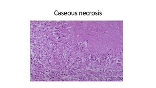
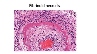
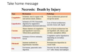
Necrosis
Dr. S Poude, MD
Department of Pathology
Irreversible changes
•Necrosis and
•Apoptosis
Irreversible injury
• Severe injury or persistent injury causes irreversible damage.
• Extensive damage to cell membrane
• Vacuolisation of mitochondria
• Reduced capacity to generate ATP
• Necrosis
Morphogical changes are :
• Nuclear changes and cytoplasmic changes
Nuclear changes:
• Are pathognomonic of necrosis and are of 3 types:
• Pyknosis – shrunken,small and hyperchromatic nuclei.
• Karyorrhexis – pyknotic nucleus breaks up into fragments
• Karyolysis – with time the nuclear fragments slowly fade and
ultimately disappear(enzymatic degradation by endonucleases)
Cytoplasmic changes:
• Dense pink cytoplasm ( eosinophilic cytoplasm)
• Glassy and homogeneous
• Becomes vacuolated and motheaten in appearance
• Dead cells may be replaced by large, whorled phospholipid masses
called myelin figures
Necrosis
• Is the result of denaturation of intracellular proteins and enzymatic
digestion of the lethally injured cell
• unable to maintain membrane integrity
• their contents often leak out, a process that may elicit inflammation
in the surrounding tissue.
Patterns of Tissue Necrosis
• Coagulative necrosis
• Liquefactive necrosis
• Gangrenous necrosis
• Caseous necrosis
• Fat necrosis
• Fibrinoid necrosis
Coagulative necrosis
• the architecture of dead tissues is preserved
for a span of at least some days
• the injury denatures enzymes also and so
blocks the proteolysis of the dead cells
• as a result, eosinophilic, anucleate cells may
persist for days or weeks
• E.g Renal infarction, Myocardial infarction
• Caused by obstruction of blood supply
• A localized area of coagulative necrosis is
called an infarct
Coagulative necrosis
Liquefactive necrosis
• is characterized by digestion of the dead cells,
resulting in transformation of the tissue into a
liquid viscous mass.
• No structural detail can be made out
• The necrotic material is frequently creamy
yellow because of the presence of dead
leukocytes and is called pus
• CNS often manifests as liquefactive necrosis
• E.g., Bacterial, fungal and ischemic injury of
CNS
Liquefactive necrosis
Gangrenous necrosis
• not a specific pattern of cell death
• usually applied to a limb, generally the lower leg, that has lost its
blood supply and has undergone necrosis (typically coagulative
necrosis) involving multiple tissue planes.
Caseous necrosis
• Most often in foci of tuberculous infection
• The term “caseous” (cheeselike) is derived from the friable white
appearance of the area of necrosis
• microscopic examination, the necrotic area appears as a collection
of fragmented or lysed cells and amorphous granular debris
Caseous necrosis
Caseous necrosis
Fat necrosis
• The focal areas of fat destruction, typically resulting from release of
activated pancreatic lipases into the substance of the pancreas and
the peritoneal cavity
• The fatty acids, so derived, combine with calcium to produce grossly
visible chalky-white areas (fat saponification)
Fat necrosis
Fat necrosis
Fibrinoid necrosis
• Special form of necrosis usually seen in immune reactions involving
blood vessels
• Deposits of these “immune complexes,” together with fibrin that
has leaked out of vessels, result in a bright pink and amorphous
appearance in H&E stains, called “fibrinoid” (fibrin-like) by
pathologists
Fibrinoid necrosis
Take home message
• Thank You

Necrosis.pptx definition, types, examples

  • 1.
    Necrosis Dr. S Poude,MD Department of Pathology
  • 2.
  • 3.
    Irreversible injury • Severeinjury or persistent injury causes irreversible damage. • Extensive damage to cell membrane • Vacuolisation of mitochondria • Reduced capacity to generate ATP • Necrosis Morphogical changes are : • Nuclear changes and cytoplasmic changes
  • 4.
    Nuclear changes: • Arepathognomonic of necrosis and are of 3 types: • Pyknosis – shrunken,small and hyperchromatic nuclei. • Karyorrhexis – pyknotic nucleus breaks up into fragments • Karyolysis – with time the nuclear fragments slowly fade and ultimately disappear(enzymatic degradation by endonucleases)
  • 7.
    Cytoplasmic changes: • Densepink cytoplasm ( eosinophilic cytoplasm) • Glassy and homogeneous • Becomes vacuolated and motheaten in appearance • Dead cells may be replaced by large, whorled phospholipid masses called myelin figures
  • 8.
    Necrosis • Is theresult of denaturation of intracellular proteins and enzymatic digestion of the lethally injured cell • unable to maintain membrane integrity • their contents often leak out, a process that may elicit inflammation in the surrounding tissue.
  • 9.
    Patterns of TissueNecrosis • Coagulative necrosis • Liquefactive necrosis • Gangrenous necrosis • Caseous necrosis • Fat necrosis • Fibrinoid necrosis
  • 10.
    Coagulative necrosis • thearchitecture of dead tissues is preserved for a span of at least some days • the injury denatures enzymes also and so blocks the proteolysis of the dead cells • as a result, eosinophilic, anucleate cells may persist for days or weeks • E.g Renal infarction, Myocardial infarction • Caused by obstruction of blood supply • A localized area of coagulative necrosis is called an infarct
  • 11.
  • 12.
  • 13.
  • 14.
    Liquefactive necrosis • ischaracterized by digestion of the dead cells, resulting in transformation of the tissue into a liquid viscous mass. • No structural detail can be made out • The necrotic material is frequently creamy yellow because of the presence of dead leukocytes and is called pus • CNS often manifests as liquefactive necrosis • E.g., Bacterial, fungal and ischemic injury of CNS
  • 15.
  • 16.
  • 17.
    Gangrenous necrosis • nota specific pattern of cell death • usually applied to a limb, generally the lower leg, that has lost its blood supply and has undergone necrosis (typically coagulative necrosis) involving multiple tissue planes.
  • 18.
    Caseous necrosis • Mostoften in foci of tuberculous infection • The term “caseous” (cheeselike) is derived from the friable white appearance of the area of necrosis • microscopic examination, the necrotic area appears as a collection of fragmented or lysed cells and amorphous granular debris
  • 19.
  • 20.
  • 21.
    Fat necrosis • Thefocal areas of fat destruction, typically resulting from release of activated pancreatic lipases into the substance of the pancreas and the peritoneal cavity • The fatty acids, so derived, combine with calcium to produce grossly visible chalky-white areas (fat saponification)
  • 22.
  • 23.
  • 24.
    Fibrinoid necrosis • Specialform of necrosis usually seen in immune reactions involving blood vessels • Deposits of these “immune complexes,” together with fibrin that has leaked out of vessels, result in a bright pink and amorphous appearance in H&E stains, called “fibrinoid” (fibrin-like) by pathologists
  • 25.
  • 26.
  • 28.