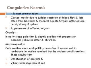
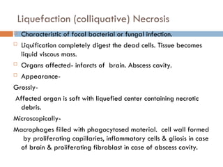
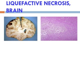
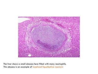
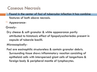
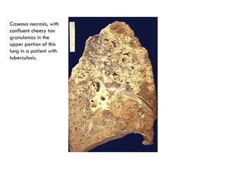
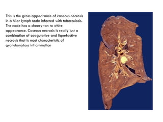
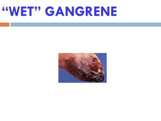
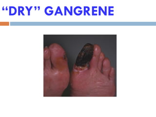
CELL DEATH & NECROSIS
DR. NITIN PATEL
CELL DEATH
 Autolysis
 Disintegration of cell by its own hydrolytic enzyme
liberated from lysosomes. It can occur in living body
surrounded by inflammatory reaction or postmortem
change where complete absence of inflammatory
response. It is rapid in some tissues e.g. Pancreas,
gastric mucosa. Intermediate in tissues e.g. Heart, liver
& kidney. It is slow in fibrous tissue. Morphologically it
is identified by homogenous & eosinophilic cytoplasm
with loss of cellular details & remains of cell debris.
APOPTOSIS
 Programmed death of cell. In this process normal abnormal or unwanted
cells are eliminated. It is different from necrosis.
 -Developmental,Homeostatic mechanism to maintain cell population,As a
defence mechanism in immune reactions, Aging.
 Physiological, Pathological or adaptive.
 Apoptosis in health-
1)metamorphosis of tadpole to frog.
2)loss of auto reactive response of T cells in thymus preventing autoimmune
attack.
3)atrophy & involution often in withdrawal of hormones.
- Endom.cell breakdown in menstrual cycle, ovarian follicular cell atresia in
menopause, prostatic atrophy after castration.
 Apoptosis in disease-
1)Irradiation.
2)virus infection – viral hepatitis – councilman bodies.
3)Action of cytotoxic T cells e.g. in rejection of transplant organ.
4)in tumour apoptosis & proliferation rate control the rata of tumour growth.
 Apoptosis cont.
- Inflammation : Death of neutrophils.
- Pathological atrophy after duct obstruction in panc.,
parotid, kidney.
MORPHOLOGY
 Cell shrinkage : small, cytoplasm dense, organelles
tighhtely packed.
 - Chromatin condensation.
 Formation of cytoplasmic blebs and apoptotiic bodies
 Phagocytosis of apoptotic cells or bodies.
- Cell memb. Remain intact during apoptosis.
- Apoptotic cells : Round or oval mass of intenseley
eosinophilic cytoplasm with dense nuclear chromatin
fragment.
- No inflammation.
Biochemical features
 Protein cleavage : By caspases (Cysteine proteases)
lead to nuclear and cytoplasmic alteration.
Protein cross linking : Transglutaminase activation lead
to shrinkage of cells that break into apoptotic
bodies.
DNA Breakdown
Phagocytic recognition :
MECHANISMS
 Signaling pathways that initiate apoptosis.
 Control and intgration stage : By specific proteins
 Execution phase : actual death programe by
caspase family of proteases.
 Removal of dead cell by phogocytosis.
NECROSIS
It is a spectrum of morphologic changes that follow cell
death in living tissue, resulting from the progressive
degradative action of enzymes on the lethally injured cell.
Autolysis : enzymes derived from the lysozomes of the dead
cells.
Heterolysis : enzymes from the lysozomes of the immigrent
leucocytes.
 There are 5 types of Necrosis-
1)Coagulative necrosis
2)liquification (colliquative) necrosis.
3)Caseous necrosis.
4)Fat necrosis
5)Fibrinoid necrosis.
 MORPHOLOGY :
 Increased eosinophilia. Glassy homogenous
appearance.
 Cytoplasm – vacuolated – Moth eaten appearance.
 Nuclear changes :
- Karyolysis : basophilia fade.
- Pyknosis : nuclear shrinkage and increased
basophilia.
- Karyorrhexis : Nuclear fragmentation.
- Total disappearance of nucleus.
Coagulative Necrosis
 It is most common type.
 Causes- mostly due to sudden cessation of blood flow & less
often from bacterial & chemical agents. Organs affected are
heart, kidney & spleen
 Appearance of affected organ-
Grossly-:
In early stage pale firm & slightly swollen with progression
becomes yellowish softer & shrunken.
Microscopically-
Cells swollen, more eosinphillic, conversion of normal cell to
tombstone i.e. outline retained but the nuclear details are lost.
Those results from
1) Denaturation of proteins &
2) 2)Enzymatic digestion of cell
Liquefaction (colliquative) Necrosis
 Characteristic of focal bacterial or fungal infection.
 Liquification completely digest the dead cells. Tissue becomes
liquid viscous mass.
 Organs affected- infarcts of brain. Abscess cavity.
 Appearance-
Grossly-
Affected organ is soft with liquefied center containing necrotic
debris.
Microscopically-
Macrophages filled with phagocytosed material. cell wall formed
by proliferating capillaries, inflammatory cells & gliosis in case
of brain & proliferating fibroblast in case of abscess cavity.
LIQUEFACTIVE NECROSIS,
BRAIN
MORE LIQUID  MORE WATER 
MORE PROTONS
The liver shows a small abscess here filled with many neutrophils.
This abscess is an example of localized liquefactive necrosis
Caseous Necrosis
 Found in the center of foci of tubercular infection it has combine
features of both above necrosis.
 Appearance-
Grossly-
Dry cheese & soft granular & white appearance partly
attributed to histotoxic effect of lipopolyscchariedes present in
capsule of tubercle bacilli.
Microscopically-
Foci are eosinophilic structureless & contain granular debris.
Surrounding tissue shows inflammatory reaction consisting of
epitheloid cells with interspersed giant cells of langerhans &
foreign body & peripheral mantle of lymphocytes.
Caseous necrosis, with
confluent cheesy tan
granulomas in the
upper portion of this
lung in a patient with
tuberculosis.
This is the gross appearance of caseous necrosis
in a hilar lymph node infected with tuberculosis.
The node has a cheesy tan to white
appearance. Caseous necrosis is really just a
combination of coagulative and liquefactive
necrosis that is most characteristic of
granulomatous inflammation
Fat Necrosis
 Occurs in two types-
1)Accute pancreatic necrosis-
there is liberation of pancreatic lipase from injured or inflamed
tissue results in necrosis.
2)Tramatic fat necrosis-
Damage adipose cells assume cloudy appearance when only free
fatty acids complex with Ca+ to form calcium soaps.
 Appearance –
Grossly- Yellowish white & firm deposits. Chalky white
appearance.
Microscopically- Cloudy appearance & surrounded by
inflammatory tissue. Formation of calcium soap as amorphous
granular & basophilic material.
Fibrinoid Necrosis
 Characterised by deposition of fibrinlike material
which has staining properties of fibrin. It occurs in
immunologic tissue injury.
E.g. Immune complex vasculitis.
Autoimmune diseases.
Appearance –
Microscopically –
Identified by brightly eosinophilic hyaline like
deposition in vessel wall or on luminal surface of
peptic ulcer. Local hemorrhage occur due to rupture of
these blood vessels.
FIBRINOID NECROSIS
GANGRENE
 It is form of necrosis of tissue with superadded putrefaction.
There are 3 types of gangrene:
 1)Dry gangrene
 2)Wet gangrene
 3)Gas gangrene
1)Dry gangrene-Begins in distal part of limb due to ischemia
occurs in toes & feet of old patient.
Causes -Atherosclerosis, thromboangitis obliterance, rayaund’s
disease, trauma, ergot poisoning. Line of separation present
between gangrenous & viable part.
Pathologic changes-Dry, shrunken & dark black due to liberation
of Hb from hemolysed RBCs acted by H2S produced by
bacteria result in iron sulphied. Histologically- Line of
separation consist of inflammatory tissue.
Wet Gangrene
Occurs in moist tissues & organs such as mouth, bowel, lungs,
cervix, vulva etc. diabetic foot is an another example of wet
gangrene. It develops rapidly due to the blockage of venous &
less commonly due to arterial flow from thrombosis & embolism.
There is no line of demarcation between the gangrenous &
viable part.
Pathologic changes –
organ becomes soft, swollen, putrid, rotten, dark.
Histologically-
Coagulative necrosis with shifting of affected part is with
blood. There is ulceration of mucosa & intence inflammatory
infiltration.
“WET” GANGRENE
“DRY” GANGRENE
Gas Gangrene
It is the special form of gangrene caused by gas
forming clostridia which gain entry in tissues through
open contaminated wound. Clostridia produces
various toxins which produce necrosis & edema locally
& also absorb producing profound systemic
manifestation.
 Pathologic changes-
Area is swollen edematous painful & crepitant due to
accumulation of gas bubbles within the tissues.
Affected tisse become dark, black, & foul smelling.

cell death & necrosis.pptddrghfddetygfdeee

  • 1.
    CELL DEATH &NECROSIS DR. NITIN PATEL
  • 2.
    CELL DEATH  Autolysis Disintegration of cell by its own hydrolytic enzyme liberated from lysosomes. It can occur in living body surrounded by inflammatory reaction or postmortem change where complete absence of inflammatory response. It is rapid in some tissues e.g. Pancreas, gastric mucosa. Intermediate in tissues e.g. Heart, liver & kidney. It is slow in fibrous tissue. Morphologically it is identified by homogenous & eosinophilic cytoplasm with loss of cellular details & remains of cell debris.
  • 3.
    APOPTOSIS  Programmed deathof cell. In this process normal abnormal or unwanted cells are eliminated. It is different from necrosis.  -Developmental,Homeostatic mechanism to maintain cell population,As a defence mechanism in immune reactions, Aging.  Physiological, Pathological or adaptive.  Apoptosis in health- 1)metamorphosis of tadpole to frog. 2)loss of auto reactive response of T cells in thymus preventing autoimmune attack. 3)atrophy & involution often in withdrawal of hormones. - Endom.cell breakdown in menstrual cycle, ovarian follicular cell atresia in menopause, prostatic atrophy after castration.  Apoptosis in disease- 1)Irradiation. 2)virus infection – viral hepatitis – councilman bodies. 3)Action of cytotoxic T cells e.g. in rejection of transplant organ. 4)in tumour apoptosis & proliferation rate control the rata of tumour growth.
  • 4.
     Apoptosis cont. -Inflammation : Death of neutrophils. - Pathological atrophy after duct obstruction in panc., parotid, kidney.
  • 5.
    MORPHOLOGY  Cell shrinkage: small, cytoplasm dense, organelles tighhtely packed.  - Chromatin condensation.  Formation of cytoplasmic blebs and apoptotiic bodies  Phagocytosis of apoptotic cells or bodies. - Cell memb. Remain intact during apoptosis. - Apoptotic cells : Round or oval mass of intenseley eosinophilic cytoplasm with dense nuclear chromatin fragment. - No inflammation.
  • 6.
    Biochemical features  Proteincleavage : By caspases (Cysteine proteases) lead to nuclear and cytoplasmic alteration. Protein cross linking : Transglutaminase activation lead to shrinkage of cells that break into apoptotic bodies. DNA Breakdown Phagocytic recognition :
  • 7.
    MECHANISMS  Signaling pathwaysthat initiate apoptosis.  Control and intgration stage : By specific proteins  Execution phase : actual death programe by caspase family of proteases.  Removal of dead cell by phogocytosis.
  • 8.
    NECROSIS It is aspectrum of morphologic changes that follow cell death in living tissue, resulting from the progressive degradative action of enzymes on the lethally injured cell. Autolysis : enzymes derived from the lysozomes of the dead cells. Heterolysis : enzymes from the lysozomes of the immigrent leucocytes.  There are 5 types of Necrosis- 1)Coagulative necrosis 2)liquification (colliquative) necrosis. 3)Caseous necrosis. 4)Fat necrosis 5)Fibrinoid necrosis.
  • 9.
     MORPHOLOGY : Increased eosinophilia. Glassy homogenous appearance.  Cytoplasm – vacuolated – Moth eaten appearance.  Nuclear changes : - Karyolysis : basophilia fade. - Pyknosis : nuclear shrinkage and increased basophilia. - Karyorrhexis : Nuclear fragmentation. - Total disappearance of nucleus.
  • 10.
    Coagulative Necrosis  Itis most common type.  Causes- mostly due to sudden cessation of blood flow & less often from bacterial & chemical agents. Organs affected are heart, kidney & spleen  Appearance of affected organ- Grossly-: In early stage pale firm & slightly swollen with progression becomes yellowish softer & shrunken. Microscopically- Cells swollen, more eosinphillic, conversion of normal cell to tombstone i.e. outline retained but the nuclear details are lost. Those results from 1) Denaturation of proteins & 2) 2)Enzymatic digestion of cell
  • 11.
    Liquefaction (colliquative) Necrosis Characteristic of focal bacterial or fungal infection.  Liquification completely digest the dead cells. Tissue becomes liquid viscous mass.  Organs affected- infarcts of brain. Abscess cavity.  Appearance- Grossly- Affected organ is soft with liquefied center containing necrotic debris. Microscopically- Macrophages filled with phagocytosed material. cell wall formed by proliferating capillaries, inflammatory cells & gliosis in case of brain & proliferating fibroblast in case of abscess cavity.
  • 12.
  • 13.
    MORE LIQUID MORE WATER  MORE PROTONS
  • 14.
    The liver showsa small abscess here filled with many neutrophils. This abscess is an example of localized liquefactive necrosis
  • 15.
    Caseous Necrosis  Foundin the center of foci of tubercular infection it has combine features of both above necrosis.  Appearance- Grossly- Dry cheese & soft granular & white appearance partly attributed to histotoxic effect of lipopolyscchariedes present in capsule of tubercle bacilli. Microscopically- Foci are eosinophilic structureless & contain granular debris. Surrounding tissue shows inflammatory reaction consisting of epitheloid cells with interspersed giant cells of langerhans & foreign body & peripheral mantle of lymphocytes.
  • 16.
    Caseous necrosis, with confluentcheesy tan granulomas in the upper portion of this lung in a patient with tuberculosis.
  • 17.
    This is thegross appearance of caseous necrosis in a hilar lymph node infected with tuberculosis. The node has a cheesy tan to white appearance. Caseous necrosis is really just a combination of coagulative and liquefactive necrosis that is most characteristic of granulomatous inflammation
  • 18.
    Fat Necrosis  Occursin two types- 1)Accute pancreatic necrosis- there is liberation of pancreatic lipase from injured or inflamed tissue results in necrosis. 2)Tramatic fat necrosis- Damage adipose cells assume cloudy appearance when only free fatty acids complex with Ca+ to form calcium soaps.  Appearance – Grossly- Yellowish white & firm deposits. Chalky white appearance. Microscopically- Cloudy appearance & surrounded by inflammatory tissue. Formation of calcium soap as amorphous granular & basophilic material.
  • 19.
    Fibrinoid Necrosis  Characterisedby deposition of fibrinlike material which has staining properties of fibrin. It occurs in immunologic tissue injury. E.g. Immune complex vasculitis. Autoimmune diseases. Appearance – Microscopically – Identified by brightly eosinophilic hyaline like deposition in vessel wall or on luminal surface of peptic ulcer. Local hemorrhage occur due to rupture of these blood vessels.
  • 20.
  • 21.
    GANGRENE  It isform of necrosis of tissue with superadded putrefaction. There are 3 types of gangrene:  1)Dry gangrene  2)Wet gangrene  3)Gas gangrene 1)Dry gangrene-Begins in distal part of limb due to ischemia occurs in toes & feet of old patient. Causes -Atherosclerosis, thromboangitis obliterance, rayaund’s disease, trauma, ergot poisoning. Line of separation present between gangrenous & viable part. Pathologic changes-Dry, shrunken & dark black due to liberation of Hb from hemolysed RBCs acted by H2S produced by bacteria result in iron sulphied. Histologically- Line of separation consist of inflammatory tissue.
  • 22.
    Wet Gangrene Occurs inmoist tissues & organs such as mouth, bowel, lungs, cervix, vulva etc. diabetic foot is an another example of wet gangrene. It develops rapidly due to the blockage of venous & less commonly due to arterial flow from thrombosis & embolism. There is no line of demarcation between the gangrenous & viable part. Pathologic changes – organ becomes soft, swollen, putrid, rotten, dark. Histologically- Coagulative necrosis with shifting of affected part is with blood. There is ulceration of mucosa & intence inflammatory infiltration.
  • 23.
  • 24.
  • 25.
    Gas Gangrene It isthe special form of gangrene caused by gas forming clostridia which gain entry in tissues through open contaminated wound. Clostridia produces various toxins which produce necrosis & edema locally & also absorb producing profound systemic manifestation.  Pathologic changes- Area is swollen edematous painful & crepitant due to accumulation of gas bubbles within the tissues. Affected tisse become dark, black, & foul smelling.

Editor's Notes

  • #12 When the brain tissue dies it tends to soften, then liquefy, so there can be more water (i.e., protons) to cause increased MRI signals.
  • #13 T2 weighted MRI images emphasize water density but some anatomic resolution is lost.
  • #20 Fibrinoid necrosis looks like fibrin microscopically. In fact, often, it IS fibrin.
  • #23 Wet gangrene comes BEFORE dry gangrene. Why? Gangrene is also a type of necrosis, and the term “gangrenous necrosis” is common.
  • #24 Dry gangrene is deader, i.e., longer standing, than the death of wet gangrene.