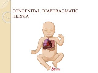
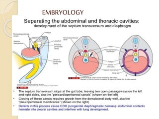
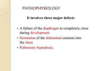
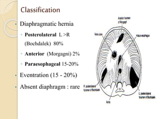
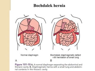
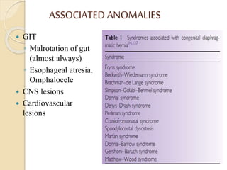
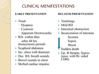
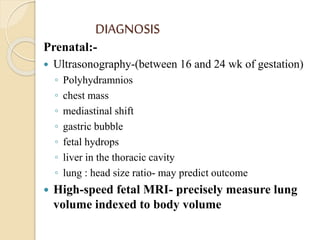
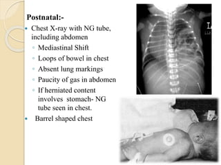
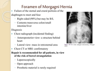
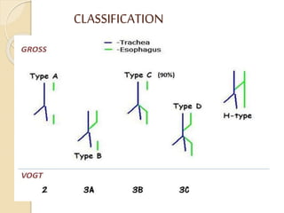
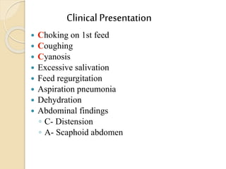
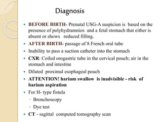
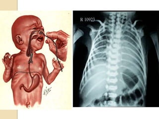
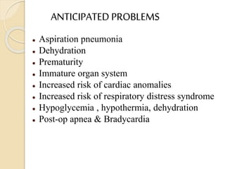
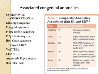
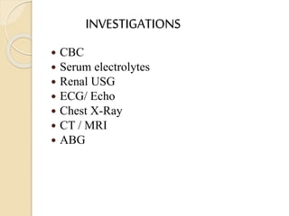
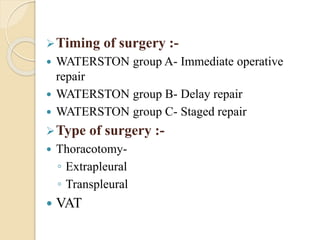
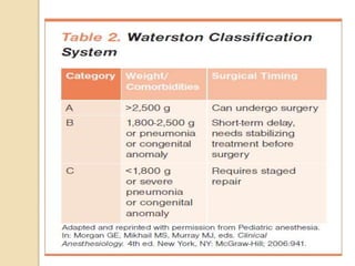
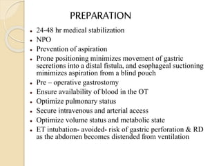
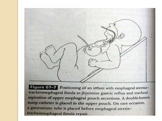
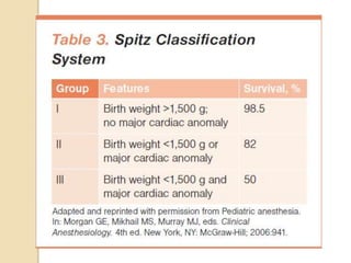
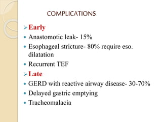
CONGENITAL DIAPHRAGMATIC
HERNIA
AND
TRACHEO ESOPHAGEAL
FISTULA
Presentor- Dr. Bikramjit Singh Jafr
Moderator- Dr. Anand Khushwaha
CONGENITAL DIAPHRAGMATIC
HERNIA
 A diaphragmatic hernia is defined as a
communication between the abdominal and thoracic
cavities with or without abdominal contents in the thorax.
◦ Congenital
◦ Traumatic
 Location of the defect
◦ Esophageal hiatus (hiatal)
◦ Paraesophageal (adjacent to the hiatus)
◦ Retrosternal (Morgagni)
◦ At the posterolateral (Bochdalek) portion of the
diaphragm
HISTORY
 1679-LAZARUS first described in postmorterm
examination of 24 yr old man.
 1888-The first attempt at surgical repair by
Nauman of Sweden in 19 yrs old.
 1940- first successful repair of neonatal hernia
within 24 hrs of birth byWilliam Ladd and
Robert Gross.
EMBRYOLOGY
PATHOPHYSIOLOGY
It involves three major defects
 A failure of the diaphragm to completely close
during development.
 Herniation of the abdominal contents into
the chest.
 Pulmonary hypoplasia.
Classification
• Diaphragmatic hernia
◦ Posterolateral L >R
(Bochdalek) 80%
◦ Anterior (Morgagni) 2%
◦ Paraesophageal 15-20%
• Eventration (15 - 20%)
• Absent diaphragm : rare
Bochdalek hernia
ASSOCIATED ANOMALIES
 GIT
◦ Malrotation of gut
(almost always)
◦ Esophageal atresia,
Omphalocele
 CNS lesions
 Cardiovascular
lesions
CLINICAL MENIFESTATIONS
EARLY PRESENTATION
 Triad-
◦ Dyspnea
◦ Cyanosis
◦ Apparent Dextrocardia
 RD- within 6hrs
-after 48 hrs
(honeymoon period)
 Scaphoid abdomen
 Inc. chest wall diameter
 Dec. B/L breath sounds
 Bowel sounds in chest
 Shifted cardiac impulse
DELAYED PRESENTATION
 Vomitings
 Mild RD
 Intestinal obstruction
 Incarceration of intestine
Iscemia
Sepsis
Shock
 Sudden death
 Gp. B Strepto. Sepsis
(asso. with Rt. sided
CDH)
DIAGNOSIS
Prenatal:-
 Ultrasonography-(between 16 and 24 wk of gestation)
◦ Polyhydramnios
◦ chest mass
◦ mediastinal shift
◦ gastric bubble
◦ fetal hydrops
◦ liver in the thoracic cavity
◦ lung : head size ratio- may predict outcome
 High-speed fetal MRI- precisely measure lung
volume indexed to body volume
Postnatal:-
 Chest X-ray with NG tube,
including abdomen
◦ Mediastinal Shift
◦ Loops of bowel in chest
◦ Absent lung markings
◦ Paucity of gas in abdomen
◦ If herniated content
involves stomach- NG
tube seen in chest.
 Barrel shaped chest
LAB. STUDIES
 Arterial blood gas (ABG) measurements: To assess
for pH, PaCO 2 & PaO2
 Serum lactate: for assessing circulatory
insufficiency or severe hypoxemia associated with
tissue hypoxia
 Serum electrolytes, ionized calcium & glucose
 Continuous pulse oximetry- for diagnosis and
management of PPHN
 ECHO
 Chromosome studies & microarray analysis
DIFFERENTIAL DIAGNOSIS
 Eventration of diaphragm
 Cystic lung lesions
◦ Pulmonary sequestration
◦ Cystic adenomatoid malformation
TREATMENT
 PRENATAL CARE
 Councelling of parents
 Support fetus & mother
 Refer to a tertiary centre
 NVD preferred (CDH is not an indication for CS)
 Fetal intervention with in-utero correction
(experimental)
IMMEDIATE
 Intubation + Stomach Decompression
Pre- op care:-
Resuscitation
 ET & NG tube insertion
 Bag & Mask C/I
 Umbilical arterial &
venous access
 Maintain proper temp.,
glucose & volume
homeostasis
 Sedation with Midaz/
Fentanyl/ Morphine
 Muscle paralysis C/I
 Correct acid-base
disturbances
 Ventilation
◦ Conventional mechanical ventilation
◦ HFOV
◦ ECMO
(Using a combination of high respiratory rate,
modest peak airway pressure & no PEEP)
 NO
 Inotropes
 Surfactant
NOVEL STRATEGIES
◦ Tracheal occlusion in-utero
◦ Partial liquid ventilation
◦ Extracorporeal Membrane Oxygenation
(ECMO)
OPERATIVE REPAIR
Timing of Surgery
 CDH= physiologic emergency NOT surgical emergency
 The ideal time to repair the diaphragmatic defect is under
debate.
 MC = Wait at least 48 hr after stabilization and resolution
of the pulmonary hypertension
 Indicators of stability:-
◦ the requirement for conventional ventilation only
◦ a low peak inspiratory pressure
◦ Fio2 <50
 Newborn on ECMO- consider weaning off before Sx
repair
APPROACH
 Subcostal approach = Most Common
 If the abdominal cavity cannot accommodate the
herniated contents, a polymeric silicone (Silastic)
patch can be placed.
 In case of stable infants
◦ Laparoscopic repair
◦ Thoracoscopic repair
 Whenever possible, a primary repair using native
tissue is performed.
 If the defect is too large, a porous
polytetrafluoroethylene (Gore-Tex) patch is used.
COMPLICATIONS
Short Term
 Post-op adhesions
 Recurrence
◦ more in PTFE
◦ loose fitted patch
 Pulm. HT
 Abdominal Compartment
Syndrome
 Bleeding
 Chylothorax
 Bowel obstruction
Long Term
 Pulmonary HT
 Pulmonary Hypoplasia
 Volutrauma
 Need for O2 / ECMO
 BPD
 GERD
 Delayed growth
 Neurocognitive defects
◦ transient and permanent
developmental delay
◦ abnormal hearing or vision
◦ seizures
 Pectus excavatum & Scoliosis
POOR PROGNOSTIC INDICATORS
 Asso. Major anomaly
 Symptomatic before 24 hrs of age
 Pulmonary Hypoplasia
 Herniation to C/L lung
 Need for ECMO
 Stomach in the contents
 Liver in thoracic cavity
 Fetal- Lung: Head size ratio <1
FOLLOW-UP
 Failure to thrive is common
 Need for supplemental oxygen at the time of discharge
is a significant predictor for subsequent growth failure.
 Possible causes include
◦ increased caloric requirements due to chronic lung
disease
◦ oral aversion after prolonged intubation
◦ poor oral feeding due to neurologic delays
◦ gastroesophageal reflux
 Risk for CNS insult and sensorineural hearing loss,
infants should be closely monitored for the first 3 years
of life
Foramen of Morgagni Hernia
 Failure of the sternal and crural portions of the
diaphragm to meet and fuse
◦ Right-sided (90%) but may be B/L
◦ Contents-transverse colon/small
intestine/liver
Diagnosis
 Chest radiograph (incidental finding)
◦ Anteroposterior view- a structure behind
heart
◦ Lateral view- mass in retrosternal area
 Chest CT or MRI- confirmatory
Repair is recommended for all patients, in view
of the risk of bowel strangulation
◦ Laparoscopically
◦ Open approach
◦ Prosthetic material is rarely required
TRACHEO-ESOPHAGEAL FISTULA
 mc congenital anomaly of esophagus
 Incidence: 1:3000 live births
 M > F (25:3)
 10-40% are preterm
 Antenatal history: polyhydramnios (60%)
EMBRYOLOGY
PATHOPHYSIOLOGY
 Two main pathologic entities
◦ Dehydration
◦ Aspiration pneumonitis
 EA- Aspiration-RD,Atelectasis and pneumonia
 TEF-
◦ Proximal-aspiration
◦ Distal-stomach/bowel distension and elevation of
diaphragm
 Abnormal Esophageal peristalsis
 Maldevelopment of cartilage rings
(tracheomalacia)
CLASSIFICATION
Clinical Presentation
 Choking on 1st feed
 Coughing
 Cyanosis
 Excessive salivation
 Feed regurgitation
 Aspiration pneumonia
 Dehydration
 Abdominal findings
◦ C- Distension
◦ A- Scaphoid abdomen
Diagnosis
 BEFORE BIRTH- Prenatal USG-A suspicion is based on the
presence of polyhydramnios and a fetal stomach that either is
absent or shows reduced filling.
 AFTER BIRTH- passage of 8 French oral tube
 Inability to pass a suction catheter into the stomach
 CXR: Coiled orogastric tube in the cervical pouch; air in the
stomach and intestine
 Dilated proximal esophageal pouch
 ATTENTION! barium swallow is inadvisible - risk of
barium aspiration
 For H- type fistula
◦ Bronchoscopy
◦ Dye test
 CT - sagittal computed tomography scan
ANTICIPATED PROBLEMS
 Aspiration pneumonia
 Dehydration
 Prematurity
 Immature organ system
 Increased risk of cardiac anomalies
 Increased risk of respiratory distress syndrome
 Hypoglycemia , hypothermia, dehydration
 Post-op apnea & Bradycardia
Associated congenital anomalies
SYNDROMIC
ASSOCIATION :-
DiGeorge sequence
Feingold syndrome
Pierre-robbin sequence
Polysplenia sequence
Holt Oram sequence
Trisomy 13,18,21
VACTERL
CHARGE
Anorectal- Triple atresia
(EA+DA+AA)
INVESTIGATIONS
 CBC
 Serum electrolytes
 Renal USG
 ECG/ Echo
 Chest X-Ray
 CT / MRI
 ABG
Timing of surgery :-
 WATERSTON group A- Immediate operative
repair
 WATERSTON group B- Delay repair
 WATERSTON group C- Staged repair
Type of surgery :-
 Thoracotomy-
◦ Extrapleural
◦ Transpleural
 VAT
PREPARATION
 24-48 hr medical stabilization
 NPO
 Prevention of aspiration
 Prone positioning minimizes movement of gastric
secretions into a distal fistula, and esophageal suctioning
minimizes aspiration from a blind pouch
 Pre – operative gastrostomy
 Ensure availability of blood in the OT
 Optimize pulmonary status
 Secure intravenous and arterial access
 Optimize volume status and metabolic state
 ET intubation- avoided- risk of gastric perforation & RD
as the abdomen becomes distended from ventilation
OPERATIVE REPAIR
 Depends on specific type
 Surgical ligation & division of TEF and primary end-to-end
anastomosis of the esophagus via right-sided thoracotomy
constitute the current standard surgical approach.
 In the premature or otherwise complicated infant, a primary
closure may be delayed by temporizing with fistula ligation and
gastrostomy tube placement.
 If the gap between the atretic ends of the esophagus is >3-4 cm,
primary repair cannot be done; options include using gastric,
jejunal, or colonic segments interposed as a neoesophagus.
 Thoracoscopic surgical repair is now considered feasible and
associated with favorable long-term outcomes.
 Careful search must be undertaken for the common associated
cardiac and other anomalies.
COMPLICATIONS
Early
 Anastomotic leak- 15%
 Esophageal stricture- 80% require eso.
dilatation
 Recurrent TEF
Late
 GERD with reactive airway disease- 30-70%
 Delayed gastric emptying
 Tracheomalacia
THANK YOU

Cdh and tef

  • 1.
    CONGENITAL DIAPHRAGMATIC HERNIA AND TRACHEO ESOPHAGEAL FISTULA Presentor-Dr. Bikramjit Singh Jafr Moderator- Dr. Anand Khushwaha
  • 2.
  • 3.
     A diaphragmatichernia is defined as a communication between the abdominal and thoracic cavities with or without abdominal contents in the thorax. ◦ Congenital ◦ Traumatic  Location of the defect ◦ Esophageal hiatus (hiatal) ◦ Paraesophageal (adjacent to the hiatus) ◦ Retrosternal (Morgagni) ◦ At the posterolateral (Bochdalek) portion of the diaphragm
  • 4.
    HISTORY  1679-LAZARUS firstdescribed in postmorterm examination of 24 yr old man.  1888-The first attempt at surgical repair by Nauman of Sweden in 19 yrs old.  1940- first successful repair of neonatal hernia within 24 hrs of birth byWilliam Ladd and Robert Gross.
  • 5.
  • 6.
    PATHOPHYSIOLOGY It involves threemajor defects  A failure of the diaphragm to completely close during development.  Herniation of the abdominal contents into the chest.  Pulmonary hypoplasia.
  • 7.
    Classification • Diaphragmatic hernia ◦Posterolateral L >R (Bochdalek) 80% ◦ Anterior (Morgagni) 2% ◦ Paraesophageal 15-20% • Eventration (15 - 20%) • Absent diaphragm : rare
  • 8.
  • 9.
    ASSOCIATED ANOMALIES  GIT ◦Malrotation of gut (almost always) ◦ Esophageal atresia, Omphalocele  CNS lesions  Cardiovascular lesions
  • 10.
    CLINICAL MENIFESTATIONS EARLY PRESENTATION Triad- ◦ Dyspnea ◦ Cyanosis ◦ Apparent Dextrocardia  RD- within 6hrs -after 48 hrs (honeymoon period)  Scaphoid abdomen  Inc. chest wall diameter  Dec. B/L breath sounds  Bowel sounds in chest  Shifted cardiac impulse DELAYED PRESENTATION  Vomitings  Mild RD  Intestinal obstruction  Incarceration of intestine Iscemia Sepsis Shock  Sudden death  Gp. B Strepto. Sepsis (asso. with Rt. sided CDH)
  • 11.
    DIAGNOSIS Prenatal:-  Ultrasonography-(between 16and 24 wk of gestation) ◦ Polyhydramnios ◦ chest mass ◦ mediastinal shift ◦ gastric bubble ◦ fetal hydrops ◦ liver in the thoracic cavity ◦ lung : head size ratio- may predict outcome  High-speed fetal MRI- precisely measure lung volume indexed to body volume
  • 12.
    Postnatal:-  Chest X-raywith NG tube, including abdomen ◦ Mediastinal Shift ◦ Loops of bowel in chest ◦ Absent lung markings ◦ Paucity of gas in abdomen ◦ If herniated content involves stomach- NG tube seen in chest.  Barrel shaped chest
  • 13.
    LAB. STUDIES  Arterialblood gas (ABG) measurements: To assess for pH, PaCO 2 & PaO2  Serum lactate: for assessing circulatory insufficiency or severe hypoxemia associated with tissue hypoxia  Serum electrolytes, ionized calcium & glucose  Continuous pulse oximetry- for diagnosis and management of PPHN  ECHO  Chromosome studies & microarray analysis
  • 14.
    DIFFERENTIAL DIAGNOSIS  Eventrationof diaphragm  Cystic lung lesions ◦ Pulmonary sequestration ◦ Cystic adenomatoid malformation
  • 15.
    TREATMENT  PRENATAL CARE Councelling of parents  Support fetus & mother  Refer to a tertiary centre  NVD preferred (CDH is not an indication for CS)  Fetal intervention with in-utero correction (experimental) IMMEDIATE  Intubation + Stomach Decompression
  • 16.
    Pre- op care:- Resuscitation ET & NG tube insertion  Bag & Mask C/I  Umbilical arterial & venous access  Maintain proper temp., glucose & volume homeostasis  Sedation with Midaz/ Fentanyl/ Morphine  Muscle paralysis C/I  Correct acid-base disturbances
  • 17.
     Ventilation ◦ Conventionalmechanical ventilation ◦ HFOV ◦ ECMO (Using a combination of high respiratory rate, modest peak airway pressure & no PEEP)  NO  Inotropes  Surfactant
  • 18.
    NOVEL STRATEGIES ◦ Trachealocclusion in-utero ◦ Partial liquid ventilation ◦ Extracorporeal Membrane Oxygenation (ECMO)
  • 19.
    OPERATIVE REPAIR Timing ofSurgery  CDH= physiologic emergency NOT surgical emergency  The ideal time to repair the diaphragmatic defect is under debate.  MC = Wait at least 48 hr after stabilization and resolution of the pulmonary hypertension  Indicators of stability:- ◦ the requirement for conventional ventilation only ◦ a low peak inspiratory pressure ◦ Fio2 <50  Newborn on ECMO- consider weaning off before Sx repair
  • 20.
    APPROACH  Subcostal approach= Most Common  If the abdominal cavity cannot accommodate the herniated contents, a polymeric silicone (Silastic) patch can be placed.  In case of stable infants ◦ Laparoscopic repair ◦ Thoracoscopic repair  Whenever possible, a primary repair using native tissue is performed.  If the defect is too large, a porous polytetrafluoroethylene (Gore-Tex) patch is used.
  • 21.
    COMPLICATIONS Short Term  Post-opadhesions  Recurrence ◦ more in PTFE ◦ loose fitted patch  Pulm. HT  Abdominal Compartment Syndrome  Bleeding  Chylothorax  Bowel obstruction Long Term  Pulmonary HT  Pulmonary Hypoplasia  Volutrauma  Need for O2 / ECMO  BPD  GERD  Delayed growth  Neurocognitive defects ◦ transient and permanent developmental delay ◦ abnormal hearing or vision ◦ seizures  Pectus excavatum & Scoliosis
  • 22.
    POOR PROGNOSTIC INDICATORS Asso. Major anomaly  Symptomatic before 24 hrs of age  Pulmonary Hypoplasia  Herniation to C/L lung  Need for ECMO  Stomach in the contents  Liver in thoracic cavity  Fetal- Lung: Head size ratio <1
  • 23.
    FOLLOW-UP  Failure tothrive is common  Need for supplemental oxygen at the time of discharge is a significant predictor for subsequent growth failure.  Possible causes include ◦ increased caloric requirements due to chronic lung disease ◦ oral aversion after prolonged intubation ◦ poor oral feeding due to neurologic delays ◦ gastroesophageal reflux  Risk for CNS insult and sensorineural hearing loss, infants should be closely monitored for the first 3 years of life
  • 24.
    Foramen of MorgagniHernia  Failure of the sternal and crural portions of the diaphragm to meet and fuse ◦ Right-sided (90%) but may be B/L ◦ Contents-transverse colon/small intestine/liver Diagnosis  Chest radiograph (incidental finding) ◦ Anteroposterior view- a structure behind heart ◦ Lateral view- mass in retrosternal area  Chest CT or MRI- confirmatory Repair is recommended for all patients, in view of the risk of bowel strangulation ◦ Laparoscopically ◦ Open approach ◦ Prosthetic material is rarely required
  • 25.
  • 26.
    TRACHEO-ESOPHAGEAL FISTULA  mccongenital anomaly of esophagus  Incidence: 1:3000 live births  M > F (25:3)  10-40% are preterm  Antenatal history: polyhydramnios (60%)
  • 27.
  • 28.
    PATHOPHYSIOLOGY  Two mainpathologic entities ◦ Dehydration ◦ Aspiration pneumonitis  EA- Aspiration-RD,Atelectasis and pneumonia  TEF- ◦ Proximal-aspiration ◦ Distal-stomach/bowel distension and elevation of diaphragm  Abnormal Esophageal peristalsis  Maldevelopment of cartilage rings (tracheomalacia)
  • 29.
  • 30.
    Clinical Presentation  Chokingon 1st feed  Coughing  Cyanosis  Excessive salivation  Feed regurgitation  Aspiration pneumonia  Dehydration  Abdominal findings ◦ C- Distension ◦ A- Scaphoid abdomen
  • 31.
    Diagnosis  BEFORE BIRTH-Prenatal USG-A suspicion is based on the presence of polyhydramnios and a fetal stomach that either is absent or shows reduced filling.  AFTER BIRTH- passage of 8 French oral tube  Inability to pass a suction catheter into the stomach  CXR: Coiled orogastric tube in the cervical pouch; air in the stomach and intestine  Dilated proximal esophageal pouch  ATTENTION! barium swallow is inadvisible - risk of barium aspiration  For H- type fistula ◦ Bronchoscopy ◦ Dye test  CT - sagittal computed tomography scan
  • 33.
    ANTICIPATED PROBLEMS  Aspirationpneumonia  Dehydration  Prematurity  Immature organ system  Increased risk of cardiac anomalies  Increased risk of respiratory distress syndrome  Hypoglycemia , hypothermia, dehydration  Post-op apnea & Bradycardia
  • 34.
    Associated congenital anomalies SYNDROMIC ASSOCIATION:- DiGeorge sequence Feingold syndrome Pierre-robbin sequence Polysplenia sequence Holt Oram sequence Trisomy 13,18,21 VACTERL CHARGE Anorectal- Triple atresia (EA+DA+AA)
  • 35.
    INVESTIGATIONS  CBC  Serumelectrolytes  Renal USG  ECG/ Echo  Chest X-Ray  CT / MRI  ABG
  • 36.
    Timing of surgery:-  WATERSTON group A- Immediate operative repair  WATERSTON group B- Delay repair  WATERSTON group C- Staged repair Type of surgery :-  Thoracotomy- ◦ Extrapleural ◦ Transpleural  VAT
  • 38.
    PREPARATION  24-48 hrmedical stabilization  NPO  Prevention of aspiration  Prone positioning minimizes movement of gastric secretions into a distal fistula, and esophageal suctioning minimizes aspiration from a blind pouch  Pre – operative gastrostomy  Ensure availability of blood in the OT  Optimize pulmonary status  Secure intravenous and arterial access  Optimize volume status and metabolic state  ET intubation- avoided- risk of gastric perforation & RD as the abdomen becomes distended from ventilation
  • 40.
    OPERATIVE REPAIR  Dependson specific type  Surgical ligation & division of TEF and primary end-to-end anastomosis of the esophagus via right-sided thoracotomy constitute the current standard surgical approach.  In the premature or otherwise complicated infant, a primary closure may be delayed by temporizing with fistula ligation and gastrostomy tube placement.  If the gap between the atretic ends of the esophagus is >3-4 cm, primary repair cannot be done; options include using gastric, jejunal, or colonic segments interposed as a neoesophagus.  Thoracoscopic surgical repair is now considered feasible and associated with favorable long-term outcomes.  Careful search must be undertaken for the common associated cardiac and other anomalies.
  • 42.
    COMPLICATIONS Early  Anastomotic leak-15%  Esophageal stricture- 80% require eso. dilatation  Recurrent TEF Late  GERD with reactive airway disease- 30-70%  Delayed gastric emptying  Tracheomalacia
  • 43.