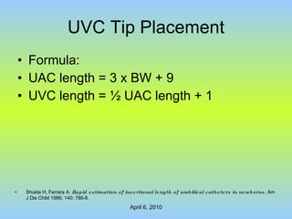
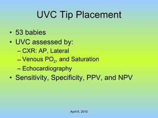
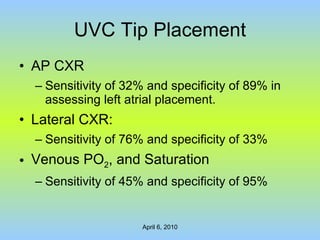
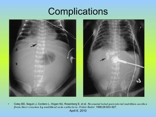
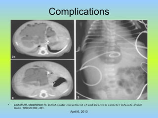
Umbilical Venous Catheter Ayman Abou Mehrem, MD, CABP Neonatology Fellow University of Manitoba
Case 1 Mom: 36 yr-old, G 4  P 3 , uneventful pregnancy Presented in labour Good CTG    Sudden Fetal Bradycardia Emergency LSCS
Case 1 Baby: Apnoeic, No Heart Beats Resus: PPV, Chest Compression, ET Epi Apgars: 0/1’, 4/5’, 4/10’, 4/20’ Cord pH: 6.9 Birth Weight: 4.260 kg
Case 1 Impression Perinatal Asphyxia
Case 1 Management: Mechanical Ventilation Body Cooling Judicious Fluid Restriction BC + Abx Double lumen 4 Fr. UVC was inserted
Case 1 X-rays
Case 1 UVC was pulled 1 cm. X-ray has not been repeated.
Case 1 On day 4 of life : HIE: Sarnat stage 3 Minimal ventilatory support Normal blood gas Never needed inotrops
Case 1 Rapid deterioration within 3 hours: Metabolic acidosis Poor perfusion    Fluid bolus Desaturation Bradycardia Cardiac arrest
Case 1 Management: Standard resuscitation Epinephrine, NS bolus, NaHCO 3 After 15 min,  empirical pericardiocentesis  resulted in 30 ml of clear fluids. Heart started to beat, perfusion improved. Few hrs later, No metabolic acidosis.
Case 1
Case 1 Biochemichal analysis Glucose 80 mmol/L Sodium 142 mmol/L, Calcium 2.3 mmol/L Protein undetectable No organism Composition similar to the fluid infused through the UVC
Case 2 Preterm baby boy 35 weeks Uneventful Pregnancy Referred from a secondary affiliating hospital
Case 2 Multiple congenital anomalies: Dandy Walker cyst Brain atrophy Large midline cleft palate Micrognathia Low set ears, low posterior hair line, webbed neck.
Case 2 On day 3 of life UVC + UAC were inserted. The position was confirmed by x-ray.
Case 2 On day 13 of life cystoperitonial shunt was inserted Nine hours after operation: Marked abdominal distension Signs of dehydration   several NS boluses Hyponatremia and modest hyperglycemia
Case 2 Abdominal US: Massive ascites Hypoechoic lesion in the right hepatic lobe
Case 2 Paracentesis: 100 ml of clear yellow fluids. WBC 56/mm3 Glucose 77.6 mmol/L Protein < 8 g/L Gram stain showed no organisms
Case 2 Water soluble contrast material (Lobitridol) was injected via UVC and x-ray showed 14 mm cavity in the right hepatic lobe with spillage to the peritoneum cavity    TPN ascites.
Case 2 UVC was removed. Abdominal CT scan on day 23 of life: cystic lesion in the liver was getting smaller
Case 3 Mom: 32 yr old, Primigravida Primary infertility, paternal reason IVF pregnancy   Triplet APH and PT labour    LSCS @ 24 wks
Case 3 All babies were intubated Prophylactic Surfactant UVC + UAC BC + Abx
Case 3
Case 3
Case 3 Echocardiography: UVC in Rt pulmonary vein!
Anatomy of The Umbilical Vein
UVC Tip Placement In the IVC just below the junction of IVC and RA T-8 to T-9: 90% of UVCs @ the IVC-RA junction T-7: 80% of UVCs are in the RA Bradshaw WT, Furdon SA.  A nurse's guide to early detection of umbilical venous catheter complications in infants.  Adv Neonatal Care. 2006 Jun;6(3):127-38. Meerstadt PWD, Gyll C.  Manual of Neonatal Emergency X-Ray Interpretation .  London, UK: WB Saunders Co. Ltd; 2000:252.
UVC Tip Placement Dunn P:  Localisation of the umbilical catheter by post-mortem measurement.  Arch Dis Child 1966; 41:69–75
UVC Tip Placement Lopriore E et al. Neonatology . 2008; 94(1):35-7. 101 pediatric professionals in the Netherlands The method used by the participants to measure the S-U length was  highly inconsistent.
UVC Tip Placement Formula: UAC length = 3 x BW + 9 UVC length =  ½ UAC length + 1 Shukla H, Ferrara A.  Rapid estimation of insertional length of umbilical catheters in newborns.  Am J Dis Child 1986; 140: 786-8.
UVC Tip Placement Radiography: AP Lateral or Cross-table Echocardiography: Ades A, Sable C, Cummings S, Cross R, Markle B, et al. Echocardiographic evaluation of umbilical venous catheter placement.   J Perinatol . 2003;23:24 –28.
UVC Tip Placement 53 babies UVC assessed by: CXR: AP, Lateral Venous PO 2 , and Saturation Echocardiography Sensitivity, Specificity, PPV, and NPV
UVC Tip Placement Catheters properly placed at the RA/IVC junction or in the inferior vena cava, as documented by echocardiography, were located at a wide range of vertebral bodies by CXR (T6–T11)
UVC Tip Placement AP CXR Sensitivity of 32% and specificity of 89% in assessing left atrial placement. Lateral CXR: Sensitivity of 76% and specificity of 33% Venous PO 2 , and Saturation Sensitivity of 45% and specificity of 95%
UVC Tip Placement
Complications Blood loss Perforation of UV: Hemoperitoneum TPN/ IVF ascites 7 Retroperitoneal fluid extravasation: abdominal, genital, buttock, and thigh edema 8 Mohan MS, Patole SK.  Neonatal ascites and hyponatremia following umbilical venous catheterization.   J Paediatr Child Health . 2002;38:612– 614. Liao CH, Sy LB, Tsou KI.  Umbilical vein catheter malposition: report of one case .  Acta Paediatr Taiwan . 2003;44:38–40.
Complications Cardiac: Dysrrhythmia: Atrial flutter reported in 2 cases  9, 10 Tamponade: several case reports Sinha A, Fernandes CJ, Kim JJ, Fenrich AL Jr, Enciso J.  Atrial flutter following placement of an umbilical venous catheter.   Am J Perinatol . 2005;22:275–277. Leroy V, Belin V, Farnoux C, Magnier S, Auburtin B, Gondon E, Saizou C, Dauger S.  Une observation de flutter auriculaire après pose de cathéter veineux ombilical .  Arch Pediatr.  2002 Feb;9(2):147-50
Complications Pulmonary:  due to malposition  11 Pulmonary edema, localized Pulmonary hemorrhage Pulmonary infarction  ± hydrothorax Possible systemic embolism Björklund LJ, Malmgren N, Lindroth M.  Pulmonary complications of umbilical venous catheters.  Pediatr Radiol.  1995;25(2):149-52.
Complications Hepatic: Necrosis Calcification Infusate encystment Infusate ascites Laceration Biliary venous fistula formation Abscess formation
Complications Coley BD, Seguin J, Cordero L, Hogan MJ, Rosenberg E, et al.  Neonatal total parenteral nutrition ascites from liver erosion by umbilical vein catheters.   Pediatr Radiol.  1998;28:923–927.
Complications Yiğiter M, Arda IS, Hiçsönmez A.  Hepatic laceration because of malpositioning of the umbilical vein catheter: case report and literature review.  J Pediatr Surg.  2008 May;43(5):E39-41
Complications Levkoff AH, Macpherson RI.  Intrahepatic encystment of umbilical vein catheter infusate.   Pediatr Radiol.  1990;20:360 –361.
Complications Air embolism: iatrogenic Thrombosis: Intimal damage Bacterial colonization, slime-forming organisms The low-flow nature of the venous system
Complications Thrombosis may lead to: Occlusion Portal vein thrombosis Intracardiac thrombi Renal vein thrombosis 15 Pulmonary and systemic embolism Marks SD, Massicotte MP, Steele BT, Matsell DG, Filler G, et al.  Neonatal renal venous thrombosis: clinical outcomes and prevalence of prothrombotic disorders .  J Pediatr.  2005;146:811– 816.
Complications Infection: Aseptic technique during insertion and care Increased after 14 days: CDC A study showed safety up to 28 days 16 Butler-O'Hara M, Buzzard CJ, Reubens L, McDermott MP, DiGrazio W, D'Angio CT.  A randomized trial comparing long-term and short-term use of umbilical venous catheters in premature infants with birth weights of less than 1251 grams . Pediatrics . 2006 Jul;118(1):e25-35.
Complications Recent study introduced a multimodal approach to reduce CR-BSI 17 : 15/1000 to 10/1000 catheter-days Sannoh S, Clones B, Munoz J, Montecalvo M, Parvez B.  A multimodal approach to central venous catheter hub care can decrease catheter-related bloodstream infection . Am J Infect Control. 2010 Feb 3. [Epub ahead of print].
Summary UV is good, easily accessible route for parenteral therapy and nutrition during neonatal period. Predicting the required length for insertion is not easy. Radiography is relatively unreliable in confirming the catheter tip position. Complications of malposition are devastating.
Recommendation Further studies are required to determine the required length of insertion. Repeated radiographs to ensure the tip in correct position. How frequent? Bedside echocardiography may be a useful tool to confirm the tip position. Strict infection control policies to reduce CR-BSI.
THANK YOU

Umbilical Venous Catheter

  • 1.
    Umbilical Venous CatheterAyman Abou Mehrem, MD, CABP Neonatology Fellow University of Manitoba
  • 2.
    Case 1 Mom:36 yr-old, G 4 P 3 , uneventful pregnancy Presented in labour Good CTG  Sudden Fetal Bradycardia Emergency LSCS
  • 3.
    Case 1 Baby:Apnoeic, No Heart Beats Resus: PPV, Chest Compression, ET Epi Apgars: 0/1’, 4/5’, 4/10’, 4/20’ Cord pH: 6.9 Birth Weight: 4.260 kg
  • 4.
    Case 1 ImpressionPerinatal Asphyxia
  • 5.
    Case 1 Management:Mechanical Ventilation Body Cooling Judicious Fluid Restriction BC + Abx Double lumen 4 Fr. UVC was inserted
  • 6.
  • 7.
    Case 1 UVCwas pulled 1 cm. X-ray has not been repeated.
  • 8.
    Case 1 Onday 4 of life : HIE: Sarnat stage 3 Minimal ventilatory support Normal blood gas Never needed inotrops
  • 9.
    Case 1 Rapiddeterioration within 3 hours: Metabolic acidosis Poor perfusion  Fluid bolus Desaturation Bradycardia Cardiac arrest
  • 10.
    Case 1 Management:Standard resuscitation Epinephrine, NS bolus, NaHCO 3 After 15 min, empirical pericardiocentesis resulted in 30 ml of clear fluids. Heart started to beat, perfusion improved. Few hrs later, No metabolic acidosis.
  • 11.
  • 12.
    Case 1 Biochemichalanalysis Glucose 80 mmol/L Sodium 142 mmol/L, Calcium 2.3 mmol/L Protein undetectable No organism Composition similar to the fluid infused through the UVC
  • 13.
    Case 2 Pretermbaby boy 35 weeks Uneventful Pregnancy Referred from a secondary affiliating hospital
  • 14.
    Case 2 Multiplecongenital anomalies: Dandy Walker cyst Brain atrophy Large midline cleft palate Micrognathia Low set ears, low posterior hair line, webbed neck.
  • 15.
    Case 2 Onday 3 of life UVC + UAC were inserted. The position was confirmed by x-ray.
  • 16.
    Case 2 Onday 13 of life cystoperitonial shunt was inserted Nine hours after operation: Marked abdominal distension Signs of dehydration  several NS boluses Hyponatremia and modest hyperglycemia
  • 17.
    Case 2 AbdominalUS: Massive ascites Hypoechoic lesion in the right hepatic lobe
  • 18.
    Case 2 Paracentesis:100 ml of clear yellow fluids. WBC 56/mm3 Glucose 77.6 mmol/L Protein < 8 g/L Gram stain showed no organisms
  • 19.
    Case 2 Watersoluble contrast material (Lobitridol) was injected via UVC and x-ray showed 14 mm cavity in the right hepatic lobe with spillage to the peritoneum cavity  TPN ascites.
  • 20.
    Case 2 UVCwas removed. Abdominal CT scan on day 23 of life: cystic lesion in the liver was getting smaller
  • 21.
    Case 3 Mom:32 yr old, Primigravida Primary infertility, paternal reason IVF pregnancy  Triplet APH and PT labour  LSCS @ 24 wks
  • 22.
    Case 3 Allbabies were intubated Prophylactic Surfactant UVC + UAC BC + Abx
  • 23.
  • 24.
  • 25.
    Case 3 Echocardiography:UVC in Rt pulmonary vein!
  • 26.
    Anatomy of TheUmbilical Vein
  • 27.
    UVC Tip PlacementIn the IVC just below the junction of IVC and RA T-8 to T-9: 90% of UVCs @ the IVC-RA junction T-7: 80% of UVCs are in the RA Bradshaw WT, Furdon SA. A nurse's guide to early detection of umbilical venous catheter complications in infants. Adv Neonatal Care. 2006 Jun;6(3):127-38. Meerstadt PWD, Gyll C. Manual of Neonatal Emergency X-Ray Interpretation . London, UK: WB Saunders Co. Ltd; 2000:252.
  • 28.
    UVC Tip PlacementDunn P: Localisation of the umbilical catheter by post-mortem measurement. Arch Dis Child 1966; 41:69–75
  • 29.
    UVC Tip PlacementLopriore E et al. Neonatology . 2008; 94(1):35-7. 101 pediatric professionals in the Netherlands The method used by the participants to measure the S-U length was highly inconsistent.
  • 30.
    UVC Tip PlacementFormula: UAC length = 3 x BW + 9 UVC length = ½ UAC length + 1 Shukla H, Ferrara A. Rapid estimation of insertional length of umbilical catheters in newborns. Am J Dis Child 1986; 140: 786-8.
  • 31.
    UVC Tip PlacementRadiography: AP Lateral or Cross-table Echocardiography: Ades A, Sable C, Cummings S, Cross R, Markle B, et al. Echocardiographic evaluation of umbilical venous catheter placement. J Perinatol . 2003;23:24 –28.
  • 32.
    UVC Tip Placement53 babies UVC assessed by: CXR: AP, Lateral Venous PO 2 , and Saturation Echocardiography Sensitivity, Specificity, PPV, and NPV
  • 33.
    UVC Tip PlacementCatheters properly placed at the RA/IVC junction or in the inferior vena cava, as documented by echocardiography, were located at a wide range of vertebral bodies by CXR (T6–T11)
  • 34.
    UVC Tip PlacementAP CXR Sensitivity of 32% and specificity of 89% in assessing left atrial placement. Lateral CXR: Sensitivity of 76% and specificity of 33% Venous PO 2 , and Saturation Sensitivity of 45% and specificity of 95%
  • 35.
  • 36.
    Complications Blood lossPerforation of UV: Hemoperitoneum TPN/ IVF ascites 7 Retroperitoneal fluid extravasation: abdominal, genital, buttock, and thigh edema 8 Mohan MS, Patole SK. Neonatal ascites and hyponatremia following umbilical venous catheterization. J Paediatr Child Health . 2002;38:612– 614. Liao CH, Sy LB, Tsou KI. Umbilical vein catheter malposition: report of one case . Acta Paediatr Taiwan . 2003;44:38–40.
  • 37.
    Complications Cardiac: Dysrrhythmia:Atrial flutter reported in 2 cases 9, 10 Tamponade: several case reports Sinha A, Fernandes CJ, Kim JJ, Fenrich AL Jr, Enciso J. Atrial flutter following placement of an umbilical venous catheter. Am J Perinatol . 2005;22:275–277. Leroy V, Belin V, Farnoux C, Magnier S, Auburtin B, Gondon E, Saizou C, Dauger S. Une observation de flutter auriculaire après pose de cathéter veineux ombilical . Arch Pediatr. 2002 Feb;9(2):147-50
  • 38.
    Complications Pulmonary: due to malposition 11 Pulmonary edema, localized Pulmonary hemorrhage Pulmonary infarction ± hydrothorax Possible systemic embolism Björklund LJ, Malmgren N, Lindroth M. Pulmonary complications of umbilical venous catheters. Pediatr Radiol. 1995;25(2):149-52.
  • 39.
    Complications Hepatic: NecrosisCalcification Infusate encystment Infusate ascites Laceration Biliary venous fistula formation Abscess formation
  • 40.
    Complications Coley BD,Seguin J, Cordero L, Hogan MJ, Rosenberg E, et al. Neonatal total parenteral nutrition ascites from liver erosion by umbilical vein catheters. Pediatr Radiol. 1998;28:923–927.
  • 41.
    Complications Yiğiter M,Arda IS, Hiçsönmez A. Hepatic laceration because of malpositioning of the umbilical vein catheter: case report and literature review. J Pediatr Surg. 2008 May;43(5):E39-41
  • 42.
    Complications Levkoff AH,Macpherson RI. Intrahepatic encystment of umbilical vein catheter infusate. Pediatr Radiol. 1990;20:360 –361.
  • 43.
    Complications Air embolism:iatrogenic Thrombosis: Intimal damage Bacterial colonization, slime-forming organisms The low-flow nature of the venous system
  • 44.
    Complications Thrombosis maylead to: Occlusion Portal vein thrombosis Intracardiac thrombi Renal vein thrombosis 15 Pulmonary and systemic embolism Marks SD, Massicotte MP, Steele BT, Matsell DG, Filler G, et al. Neonatal renal venous thrombosis: clinical outcomes and prevalence of prothrombotic disorders . J Pediatr. 2005;146:811– 816.
  • 45.
    Complications Infection: Aseptictechnique during insertion and care Increased after 14 days: CDC A study showed safety up to 28 days 16 Butler-O'Hara M, Buzzard CJ, Reubens L, McDermott MP, DiGrazio W, D'Angio CT. A randomized trial comparing long-term and short-term use of umbilical venous catheters in premature infants with birth weights of less than 1251 grams . Pediatrics . 2006 Jul;118(1):e25-35.
  • 46.
    Complications Recent studyintroduced a multimodal approach to reduce CR-BSI 17 : 15/1000 to 10/1000 catheter-days Sannoh S, Clones B, Munoz J, Montecalvo M, Parvez B. A multimodal approach to central venous catheter hub care can decrease catheter-related bloodstream infection . Am J Infect Control. 2010 Feb 3. [Epub ahead of print].
  • 47.
    Summary UV isgood, easily accessible route for parenteral therapy and nutrition during neonatal period. Predicting the required length for insertion is not easy. Radiography is relatively unreliable in confirming the catheter tip position. Complications of malposition are devastating.
  • 48.
    Recommendation Further studiesare required to determine the required length of insertion. Repeated radiographs to ensure the tip in correct position. How frequent? Bedside echocardiography may be a useful tool to confirm the tip position. Strict infection control policies to reduce CR-BSI.
  • 49.