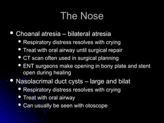
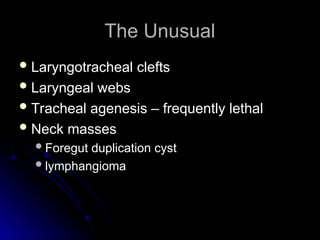
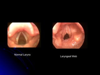
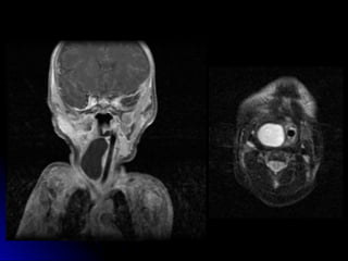
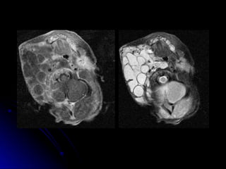
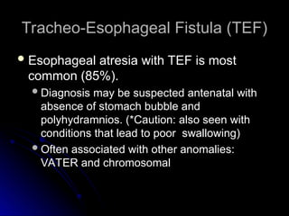
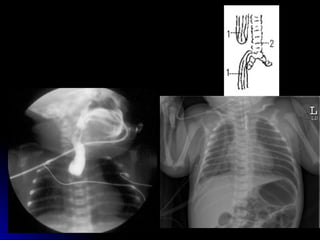
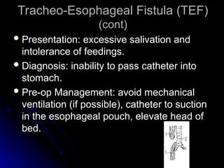
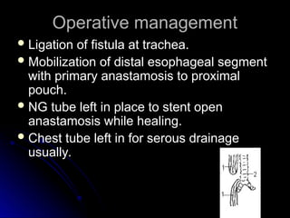
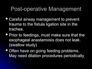
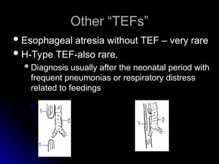
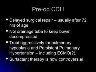
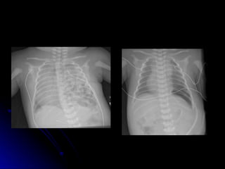
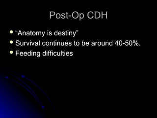
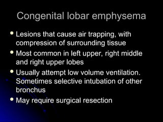
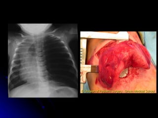
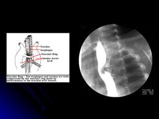
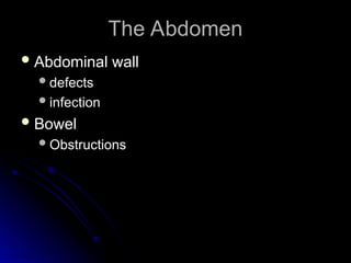
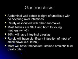
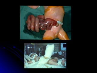
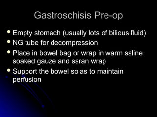
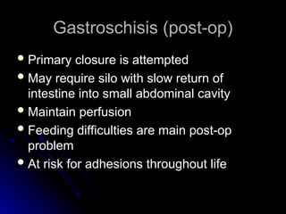
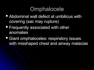
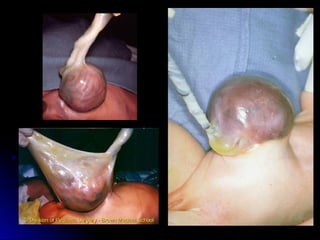
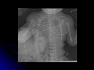
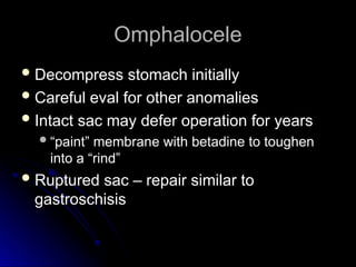
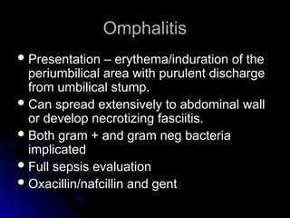
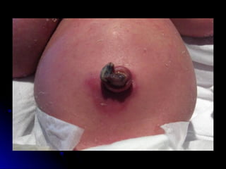
Neonatal Surgical Issues
(Part 1)
(Part 1)
Sue Ann Smith, MD
Sue Ann Smith, MD
Neonatologist
Neonatologist
An anatomic survey
An anatomic survey
 Head and Neck lesions
Head and Neck lesions
 Chest lesions
Chest lesions
 Abdomen
Abdomen
Abdominal wall defects and infection
Abdominal wall defects and infection
The Nose
The Nose
 Choanal atresia – bilateral atresia
Choanal atresia – bilateral atresia
 Respiratory distress resolves with crying
Respiratory distress resolves with crying
 Treat with oral airway until surgical repair
Treat with oral airway until surgical repair
 CT scan often used in surgical planning
CT scan often used in surgical planning
 ENT surgeons make opening in bony plate and stent
ENT surgeons make opening in bony plate and stent
open during healing
open during healing
 Nasolacrimal duct cysts – large and bilat
Nasolacrimal duct cysts – large and bilat
 Respiratory distress resolves with crying
Respiratory distress resolves with crying
 Treat with oral airway
Treat with oral airway
 Can usually be seen with otoscope
Can usually be seen with otoscope
Robin sequence
Robin sequence
 AKA Pierre Robin syndrome
AKA Pierre Robin syndrome
 Hypoplastic mandible with U-shaped
Hypoplastic mandible with U-shaped
midline cleft palate
Respiratory and feeding difficulties
Respiratory and feeding difficulties
Position prone, may require nasopharyngeal
Position prone, may require nasopharyngeal
tube, oral airway, LMA, or endotracheal tube
Mandibular distraction is now treatment of
Mandibular distraction is now treatment of
choice at OHSU
choice at OHSU
The Unusual
The Unusual
 Laryngotracheal clefts
Laryngotracheal clefts
 Laryngeal webs
Laryngeal webs
 Tracheal agenesis – frequently lethal
Tracheal agenesis – frequently lethal
 Neck masses
Neck masses
Foregut duplication cyst
Foregut duplication cyst
lymphangioma
lymphangioma
Normal Larynx
Laryngeal Web
Congenital Chest Lesions
 Tracheo-esophageal fistula
Tracheo-esophageal fistula
 Diaphragmatic Hernia (briefly)
Diaphragmatic Hernia (briefly)
 Congenital lobar emphysema
Congenital lobar emphysema
 Cystic adenomatoid malformation
Cystic adenomatoid malformation
 Vascular rings
Vascular rings
Tracheo-Esophageal Fistula (TEF)
 Esophageal atresia with TEF is most
Esophageal atresia with TEF is most
common (85%).
common (85%).
Diagnosis may be suspected antenatal with
Diagnosis may be suspected antenatal with
absence of stomach bubble and
polyhydramnios. (*Caution: also seen with
conditions that lead to poor swallowing)
Often associated with other anomalies:
Often associated with other anomalies:
VATER and chromosomal
Tracheo-Esophageal Fistula (TEF)
(cont)
(cont)
 Presentation: excessive salivation and
Presentation: excessive salivation and
intolerance of feedings.
 Diagnosis: inability to pass catheter into
Diagnosis: inability to pass catheter into
stomach.
stomach.
 Pre-op Management: avoid mechanical
Pre-op Management: avoid mechanical
ventilation (if possible), catheter to suction
in the esophageal pouch, elevate head of
bed.
bed.
Operative management
 Ligation of fistula at trachea.
Ligation of fistula at trachea.
 Mobilization of distal esophageal segment
Mobilization of distal esophageal segment
with primary anastamosis to proximal
pouch.
pouch.
 NG tube left in place to stent open
NG tube left in place to stent open
anastamosis while healing.
 Chest tube left in for serous drainage
Chest tube left in for serous drainage
usually.
usually.
Post-operative Management
 Careful airway management to prevent
Careful airway management to prevent
trauma to the fistula ligation site in the
trachea.
trachea.
 Prior to feedings, must make sure that the
Prior to feedings, must make sure that the
esophageal anastamosis does not leak.
(swallow study)
(swallow study)
 Often have on going feeding problems.
Often have on going feeding problems.
May need dilation procedures periodically
Other “TEFs”
Other “TEFs”
 Esophageal atresia without TEF – very rare
Esophageal atresia without TEF – very rare
 H-Type TEF-also rare.
H-Type TEF-also rare.
Diagnosis usually after the neonatal period with
Diagnosis usually after the neonatal period with
frequent pneumonias or respiratory distress
related to feedings
related to feedings
Congenital Diaphragmatic Hernia
(CDH)
(CDH)
 Most commonly on left side
Most commonly on left side
 Incidence 1:2000 to 1:5000
Incidence 1:2000 to 1:5000
 Often associated with other malformations
Often associated with other malformations
 Frequently diagnosed prenatally
Frequently diagnosed prenatally
 Avoid bag-mask PPV
Avoid bag-mask PPV
Pre-op CDH
Pre-op CDH
 Delayed surgical repair – usually after 72
Delayed surgical repair – usually after 72
hrs of age
hrs of age
 NG drainage tube to keep bowel
NG drainage tube to keep bowel
decompressed
decompressed
 Treat aggressively for pulmonary
Treat aggressively for pulmonary
hypoplasia and Persistent Pulmonary
Hypertension – including ECMO(?).
 Surfactant therapy is now controversial
Surfactant therapy is now controversial
Post-Op CDH
Post-Op CDH
 “
“Anatomy is destiny”
Anatomy is destiny”
 Survival continues to be around 40-50%.
Survival continues to be around 40-50%.
 Feeding difficulties
Feeding difficulties
Congenital lobar emphysema
 Lesions that cause air trapping, with
Lesions that cause air trapping, with
compression of surrounding tissue
 Most common in left upper, right middle
Most common in left upper, right middle
and right upper lobes
 Usually attempt low volume ventilation.
Usually attempt low volume ventilation.
Sometimes selective intubation of other
bronchus
bronchus
 May require surgical resection
May require surgical resection
Congenital Cystic Adenomatoid
Malformation (CCAM)
Malformation (CCAM)
 May be confused with CDH
May be confused with CDH
 Abnormal lung tissue that forms fluid filled
Abnormal lung tissue that forms fluid filled
cysts. May be large cysts, or many small
cysts and solid areas
 Space occupying lesion
Space occupying lesion
 May cause shifting of mediastium
May cause shifting of mediastium
 May spontaneously regress in fetus
May spontaneously regress in fetus
 May require surgical removal
May require surgical removal
Vascular Rings
Vascular Rings
 Uncommon
Uncommon
 Signs include stridor, vomiting and difficulty
Signs include stridor, vomiting and difficulty
swallowing.
swallowing.
 Barium swallow can be diagnostic, but may
Barium swallow can be diagnostic, but may
need chest MRI.
need chest MRI.
 Sometimes may need cardiac
Sometimes may need cardiac
catheterization
catheterization
The Abdomen
The Abdomen
 Abdominal wall
Abdominal wall
defects
defects
infection
infection
 Bowel
Bowel
Obstructions
Obstructions
Gastroschisis
Gastroschisis
 Abdominal wall defect to right of umbilicus with
Abdominal wall defect to right of umbilicus with
no covering over intestines
 Rarely associated with other anomalies
Rarely associated with other anomalies
 Most babies are SGA and born to young
Most babies are SGA and born to young
mothers (why?)
mothers (why?)
 10% will have intestinal atresias
10% will have intestinal atresias
 Rarely will have significant infarction of most of
Rarely will have significant infarction of most of
small bowel (i.e. lethal)
 Most will have “meconium” stained amniotic fluid
Most will have “meconium” stained amniotic fluid
(really bile)
(really bile)
Gastroschisis Pre-op
 Empty stomach (usually lots of bilious fluid)
Empty stomach (usually lots of bilious fluid)
 NG tube for decompression
NG tube for decompression
 Place in bowel bag or wrap in warm saline
Place in bowel bag or wrap in warm saline
soaked gauze and saran wrap
 Support the bowel so as to maintain
Support the bowel so as to maintain
perfusion
perfusion
Gastroschisis (post-op)
 Primary closure is attempted
Primary closure is attempted
 May require silo with slow return of
May require silo with slow return of
intestine into small abdominal cavity
 Maintain perfusion
Maintain perfusion
 Feeding difficulties are main post-op
Feeding difficulties are main post-op
problem
problem
 At risk for adhesions throughout life
At risk for adhesions throughout life
Omphalocele
Omphalocele
 Abdominal wall defect at umbilicus with
Abdominal wall defect at umbilicus with
covering (sac may rupture)
 Frequently associated with other
Frequently associated with other
anomalies
anomalies
 Giant omphaloceles: respiratory issues
Giant omphaloceles: respiratory issues
with misshaped chest and airway malacias
Omphalocele
Omphalocele
 Decompress stomach initially
Decompress stomach initially
 Careful eval for other anomalies
Careful eval for other anomalies
 Intact sac may defer operation for years
Intact sac may defer operation for years
“
“paint” membrane with betadine to toughen
paint” membrane with betadine to toughen
into a “rind”
into a “rind”
 Ruptured sac – repair similar to
Ruptured sac – repair similar to
gastroschisis
gastroschisis
Omphalitis
Omphalitis
 Presentation – erythema/induration of the
Presentation – erythema/induration of the
periumbilical area with purulent discharge
from umbilical stump.
 Can spread extensively to abdominal wall
Can spread extensively to abdominal wall
or develop necrotizing fasciitis.
 Both gram + and gram neg bacteria
Both gram + and gram neg bacteria
implicated
implicated
 Full sepsis evaluation
Full sepsis evaluation
 Oxacillin/nafcillin and gent
Oxacillin/nafcillin and gent
Normal Larynx (upside down) Laryngeal Web (also upside down)

f6072576_Neonatal_Surgical_Issues_not_including_cardiovascular.ppt

  • 1.
    Neonatal Surgical Issues NeonatalSurgical Issues (Part 1) (Part 1) Sue Ann Smith, MD Sue Ann Smith, MD Neonatologist Neonatologist
  • 2.
    An anatomic survey Ananatomic survey  Head and Neck lesions Head and Neck lesions  Chest lesions Chest lesions  Abdomen Abdomen Abdominal wall defects and infection Abdominal wall defects and infection
  • 3.
    The Nose The Nose Choanal atresia – bilateral atresia Choanal atresia – bilateral atresia  Respiratory distress resolves with crying Respiratory distress resolves with crying  Treat with oral airway until surgical repair Treat with oral airway until surgical repair  CT scan often used in surgical planning CT scan often used in surgical planning  ENT surgeons make opening in bony plate and stent ENT surgeons make opening in bony plate and stent open during healing open during healing  Nasolacrimal duct cysts – large and bilat Nasolacrimal duct cysts – large and bilat  Respiratory distress resolves with crying Respiratory distress resolves with crying  Treat with oral airway Treat with oral airway  Can usually be seen with otoscope Can usually be seen with otoscope
  • 4.
    Robin sequence Robin sequence AKA Pierre Robin syndrome AKA Pierre Robin syndrome  Hypoplastic mandible with U-shaped Hypoplastic mandible with U-shaped midline cleft palate midline cleft palate Respiratory and feeding difficulties Respiratory and feeding difficulties Position prone, may require nasopharyngeal Position prone, may require nasopharyngeal tube, oral airway, LMA, or endotracheal tube tube, oral airway, LMA, or endotracheal tube Mandibular distraction is now treatment of Mandibular distraction is now treatment of choice at OHSU choice at OHSU
  • 6.
    The Unusual The Unusual Laryngotracheal clefts Laryngotracheal clefts  Laryngeal webs Laryngeal webs  Tracheal agenesis – frequently lethal Tracheal agenesis – frequently lethal  Neck masses Neck masses Foregut duplication cyst Foregut duplication cyst lymphangioma lymphangioma
  • 7.
  • 10.
    Congenital Chest Lesions CongenitalChest Lesions  Tracheo-esophageal fistula Tracheo-esophageal fistula  Diaphragmatic Hernia (briefly) Diaphragmatic Hernia (briefly)  Congenital lobar emphysema Congenital lobar emphysema  Cystic adenomatoid malformation Cystic adenomatoid malformation  Vascular rings Vascular rings
  • 11.
    Tracheo-Esophageal Fistula (TEF) Tracheo-EsophagealFistula (TEF)  Esophageal atresia with TEF is most Esophageal atresia with TEF is most common (85%). common (85%). Diagnosis may be suspected antenatal with Diagnosis may be suspected antenatal with absence of stomach bubble and absence of stomach bubble and polyhydramnios. (*Caution: also seen with polyhydramnios. (*Caution: also seen with conditions that lead to poor swallowing) conditions that lead to poor swallowing) Often associated with other anomalies: Often associated with other anomalies: VATER and chromosomal VATER and chromosomal
  • 13.
    Tracheo-Esophageal Fistula (TEF) Tracheo-EsophagealFistula (TEF) (cont) (cont)  Presentation: excessive salivation and Presentation: excessive salivation and intolerance of feedings. intolerance of feedings.  Diagnosis: inability to pass catheter into Diagnosis: inability to pass catheter into stomach. stomach.  Pre-op Management: avoid mechanical Pre-op Management: avoid mechanical ventilation (if possible), catheter to suction ventilation (if possible), catheter to suction in the esophageal pouch, elevate head of in the esophageal pouch, elevate head of bed. bed.
  • 14.
    Operative management Operative management Ligation of fistula at trachea. Ligation of fistula at trachea.  Mobilization of distal esophageal segment Mobilization of distal esophageal segment with primary anastamosis to proximal with primary anastamosis to proximal pouch. pouch.  NG tube left in place to stent open NG tube left in place to stent open anastamosis while healing. anastamosis while healing.  Chest tube left in for serous drainage Chest tube left in for serous drainage usually. usually.
  • 15.
    Post-operative Management Post-operative Management Careful airway management to prevent Careful airway management to prevent trauma to the fistula ligation site in the trauma to the fistula ligation site in the trachea. trachea.  Prior to feedings, must make sure that the Prior to feedings, must make sure that the esophageal anastamosis does not leak. esophageal anastamosis does not leak. (swallow study) (swallow study)  Often have on going feeding problems. Often have on going feeding problems. May need dilation procedures periodically May need dilation procedures periodically
  • 16.
    Other “TEFs” Other “TEFs” Esophageal atresia without TEF – very rare Esophageal atresia without TEF – very rare  H-Type TEF-also rare. H-Type TEF-also rare. Diagnosis usually after the neonatal period with Diagnosis usually after the neonatal period with frequent pneumonias or respiratory distress frequent pneumonias or respiratory distress related to feedings related to feedings
  • 17.
    Congenital Diaphragmatic Hernia CongenitalDiaphragmatic Hernia (CDH) (CDH)  Most commonly on left side Most commonly on left side  Incidence 1:2000 to 1:5000 Incidence 1:2000 to 1:5000  Often associated with other malformations Often associated with other malformations  Frequently diagnosed prenatally Frequently diagnosed prenatally  Avoid bag-mask PPV Avoid bag-mask PPV
  • 18.
    Pre-op CDH Pre-op CDH Delayed surgical repair – usually after 72 Delayed surgical repair – usually after 72 hrs of age hrs of age  NG drainage tube to keep bowel NG drainage tube to keep bowel decompressed decompressed  Treat aggressively for pulmonary Treat aggressively for pulmonary hypoplasia and Persistent Pulmonary hypoplasia and Persistent Pulmonary Hypertension – including ECMO(?). Hypertension – including ECMO(?).  Surfactant therapy is now controversial Surfactant therapy is now controversial
  • 20.
    Post-Op CDH Post-Op CDH “ “Anatomy is destiny” Anatomy is destiny”  Survival continues to be around 40-50%. Survival continues to be around 40-50%.  Feeding difficulties Feeding difficulties
  • 21.
    Congenital lobar emphysema Congenitallobar emphysema  Lesions that cause air trapping, with Lesions that cause air trapping, with compression of surrounding tissue compression of surrounding tissue  Most common in left upper, right middle Most common in left upper, right middle and right upper lobes and right upper lobes  Usually attempt low volume ventilation. Usually attempt low volume ventilation. Sometimes selective intubation of other Sometimes selective intubation of other bronchus bronchus  May require surgical resection May require surgical resection
  • 23.
    Congenital Cystic Adenomatoid CongenitalCystic Adenomatoid Malformation (CCAM) Malformation (CCAM)  May be confused with CDH May be confused with CDH  Abnormal lung tissue that forms fluid filled Abnormal lung tissue that forms fluid filled cysts. May be large cysts, or many small cysts. May be large cysts, or many small cysts and solid areas cysts and solid areas  Space occupying lesion Space occupying lesion  May cause shifting of mediastium May cause shifting of mediastium  May spontaneously regress in fetus May spontaneously regress in fetus  May require surgical removal May require surgical removal
  • 25.
    Vascular Rings Vascular Rings Uncommon Uncommon  Signs include stridor, vomiting and difficulty Signs include stridor, vomiting and difficulty swallowing. swallowing.  Barium swallow can be diagnostic, but may Barium swallow can be diagnostic, but may need chest MRI. need chest MRI.  Sometimes may need cardiac Sometimes may need cardiac catheterization catheterization
  • 27.
    The Abdomen The Abdomen Abdominal wall Abdominal wall defects defects infection infection  Bowel Bowel Obstructions Obstructions
  • 28.
    Gastroschisis Gastroschisis  Abdominal walldefect to right of umbilicus with Abdominal wall defect to right of umbilicus with no covering over intestines no covering over intestines  Rarely associated with other anomalies Rarely associated with other anomalies  Most babies are SGA and born to young Most babies are SGA and born to young mothers (why?) mothers (why?)  10% will have intestinal atresias 10% will have intestinal atresias  Rarely will have significant infarction of most of Rarely will have significant infarction of most of small bowel (i.e. lethal) small bowel (i.e. lethal)  Most will have “meconium” stained amniotic fluid Most will have “meconium” stained amniotic fluid (really bile) (really bile)
  • 30.
    Gastroschisis Pre-op Gastroschisis Pre-op Empty stomach (usually lots of bilious fluid) Empty stomach (usually lots of bilious fluid)  NG tube for decompression NG tube for decompression  Place in bowel bag or wrap in warm saline Place in bowel bag or wrap in warm saline soaked gauze and saran wrap soaked gauze and saran wrap  Support the bowel so as to maintain Support the bowel so as to maintain perfusion perfusion
  • 31.
    Gastroschisis (post-op) Gastroschisis (post-op) Primary closure is attempted Primary closure is attempted  May require silo with slow return of May require silo with slow return of intestine into small abdominal cavity intestine into small abdominal cavity  Maintain perfusion Maintain perfusion  Feeding difficulties are main post-op Feeding difficulties are main post-op problem problem  At risk for adhesions throughout life At risk for adhesions throughout life
  • 32.
    Omphalocele Omphalocele  Abdominal walldefect at umbilicus with Abdominal wall defect at umbilicus with covering (sac may rupture) covering (sac may rupture)  Frequently associated with other Frequently associated with other anomalies anomalies  Giant omphaloceles: respiratory issues Giant omphaloceles: respiratory issues with misshaped chest and airway malacias with misshaped chest and airway malacias
  • 35.
    Omphalocele Omphalocele  Decompress stomachinitially Decompress stomach initially  Careful eval for other anomalies Careful eval for other anomalies  Intact sac may defer operation for years Intact sac may defer operation for years “ “paint” membrane with betadine to toughen paint” membrane with betadine to toughen into a “rind” into a “rind”  Ruptured sac – repair similar to Ruptured sac – repair similar to gastroschisis gastroschisis
  • 36.
    Omphalitis Omphalitis  Presentation –erythema/induration of the Presentation – erythema/induration of the periumbilical area with purulent discharge periumbilical area with purulent discharge from umbilical stump. from umbilical stump.  Can spread extensively to abdominal wall Can spread extensively to abdominal wall or develop necrotizing fasciitis. or develop necrotizing fasciitis.  Both gram + and gram neg bacteria Both gram + and gram neg bacteria implicated implicated  Full sepsis evaluation Full sepsis evaluation  Oxacillin/nafcillin and gent Oxacillin/nafcillin and gent
  • 40.
    Normal Larynx (upsidedown) Laryngeal Web (also upside down)