Downloaded 729 times


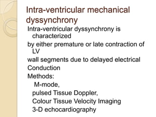


This document discusses various types and assessment of left ventricular dyssynchrony. It defines electrical and mechanical dyssynchrony. It describes different types of dyssynchrony including atrioventricular, interventricular, and intraventricular dyssynchrony. It discusses various echocardiography techniques to demonstrate and quantify each type of dyssynchrony, including M-mode, tissue Doppler, speckle tracking, and 3D echocardiography. It also mentions the use of MRI to assess dyssynchrony. The key application of assessing dyssynchrony is to predict response to cardiac resynchronization therapy in patients with heart failure.