This summary covers a set of lecture notes for internal medicine created for medical students at Weill Bugando School of Medicine. The notes were created to teach the basic concepts of internal medicine and cover major topic areas. They are focused on teaching clinical management algorithms and are intended to be used alongside textbooks. The notes include sections on history taking, physical examination, cardiology, renal medicine, gastroenterology, infectious diseases and other areas. The goal is to provide a useful guide for both students and faculty in teaching internal medicine essentials.
![1
Introduction
The following lecture notes are based on the topics described in the official curriculum of the Weill
Bugando School of Medicine. The target audience for these lecture notes is medical students in their
final 2 years of medical school. These lecture notes contain the basic concepts of internal medicine
that every medical student should know by the time of graduation. They cover most of the major
topic areas, but are far from comprehensive in scope. Students are still encouraged to consult one of
the recommended textbooks of internal medicine for in depth explanations.
These lecture notes were written for use by the faculty and students of Bugando, but we hope that
they are useful for faculty and students at other medical schools in East Africa. We have attempted
to adapt these lecture notes and clinical cases to the diseases and resources that are commonly
available at Bugando and in East Africa. In addition we hope that these notes provide a useful guide
to the most essential learning points for any faculty asked to give a lecture on short notice.
We believe that the best setting for learning medicine is at the bedside of a patient, and that this
material would be best taught at the bedside. These handouts can then serve as a useful
reinforcement of key learning points for students. When an illustrative patient is not available, the
teaching cases may be used to cover the most salient clinical details for each condition.
These lecture notes are focused on teaching algorithms for management of common problems with
specific focus on differential diagnosis, diagnostic workup, treatment and natural history. Sessions
on the physical examination of the major body systems are included with the expectation that these
can be taught and modeled at the bedside. This will allow further instruction in communication skills
and ethical standards of care.
These lecture notes are a labor of love that has been completed over the course of the past 5 years.
Many people have contributed and we do not have space to thank them all. In particular we would
like to thank our Vice Chancellor (Prof. Jacob Mtabaji), Hospital Director (Dr. Charles Majinge) , Dean
(Prof. J.B. Kataraihya) and Department Head (Prof. Samuel Kalluvya). We would also like to thank all
of the other members of our Department: Drs. Hyasinta Jaka, Dr. Rodrick Kabangila, Dr. Bahati
Wajanga, Dr. Andrew Luhanga and Dr. Mubarak Janmohamed. This is a first edition and we hope
these lecture notes will be improved with the contributions of additional editors in the 2nd
edition.
Those who have prepared these notes have used a variety of resources. In particular, we have relied
on:
Swash M., Glynn M. Hutchison’s Clinical Methods: An integrated approach to clinical practice.
22nd
ed. Edinburgh: Saunders Elsevier; 2007. [for physical examination topics]
Eddleston M, Davidson R, Brent A, Wilkinson R. Oxford Handbook of Tropical Medicine. 3rd ed.
New York, NY: Oxford University Press USA; 2008. [for clinical topics]
Sincerely,
Drs. Robert Peck, Luke Smart and Riaz Aziz (The Editors)](https://image.slidesharecdn.com/lecturenotesinmedicine-160317031130/85/Lecture-notes-in-medicine-2-320.jpg)


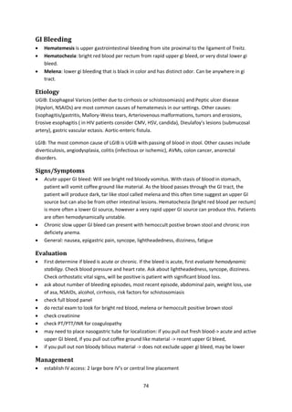
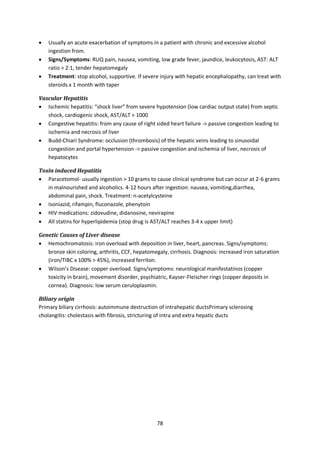

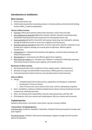
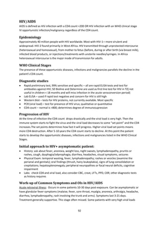
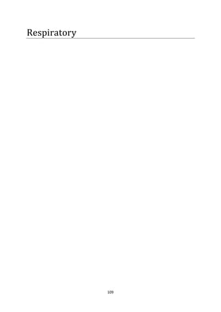
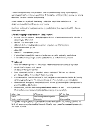
![91
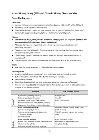
Fever
Temperature > 37.5C (axillary or oral) is definied as fever; Temperature > 38.3C usually indicates
and infectious etiology [T>38 (neutropenic pts)]
Fever of unknown origin (FUO): T>38.3 for >3 weeks and after 1 week of inpatient diagnostic
workup/3 outpatient visits
Etiologies
Infection (bacterial, viral, fungal, parasitic)
Connective Tissue Disease
Malignancy (leukemia, lymphoma, renal cell carcinoma, metastatic disease)
Miscellaneous: (DVT/PE, Medications, Drug withdrawal, hyperthyroidism)
History
Travel, sick contacts, tuberculosis history/contacts, IDS risk factors, pets, occupation,
medications, trauma, environmental contacts
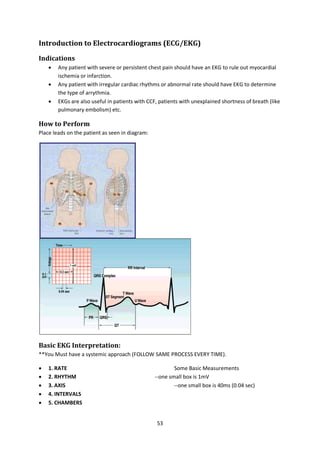
Localizing features: HA, vomiting, diarrhea, cough, hemoptysis, skin rash, joint/bone pains,
confusion, sore throat
Physical Exam
Rash, lymphadenopathy, murmurs, hepatosplenomegaly, joint exam, pelvic exam (women with
abdominal pain)
Laboratory
FBP with differential; peripheral smear
Blood Cultures (3sets sequentially for
endocarditis)
BUN/Cr
Urinalysis, urine culture
Rapid Test
MPS
Sputum cultures with AFB (pts with
cough)
Fungal Cultures
(IDS/immunocompromised pts)
Stool studies (diarrhea)
LFTs
ESR
ANA/RF
Fluid analysis (pleural, peritoneal, csf)
Bone marrow biopsy
Imaging
CXR
Echocardiogram (new murmurs)
Abdominal ultrasound (Abdominal pain)
Joint xrays
Arthrocentesis (joint effusions)
Treatment
Antipyretic
Cooling blankets for T>39
Empiric antibiotics for hemodynamically unstable patients in whom infection is the primary
concern and in neutropenic/asplenic patients.](https://image.slidesharecdn.com/lecturenotesinmedicine-160317031130/85/Lecture-notes-in-medicine-92-320.jpg)
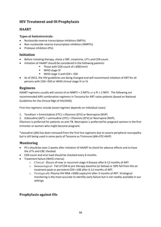


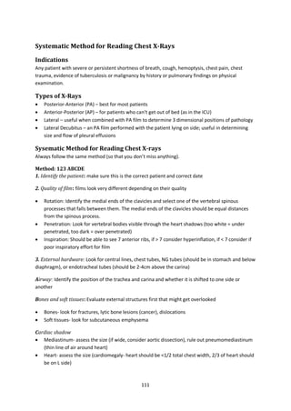


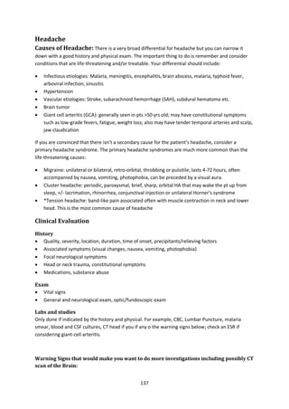

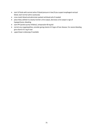
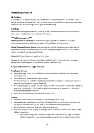
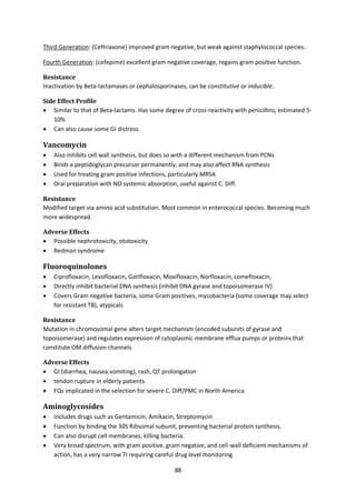
![143
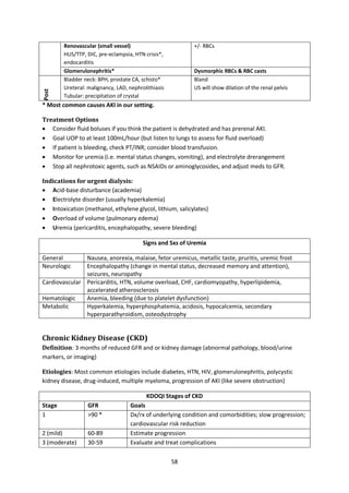
Acute Complications of Diabetes Mellitus in Adults
Diabetic Ketoacidosis (DKA) and Hyperosmolar Hyperglycemic State (HHS)
Definitions
HHS: extreme hyperglycemia w/o ketoacidosis, but with hyperosmolar state and altered mental
status in type 2 diabetics. Usually hyperglycemia osmotic diuresis dehydration more
hyperglycemia.
DKA: a triad of hyperglycemia, anion gap metabolic acidosis, and ketonemia. Occurs mostly in type 1
Diabetics. Hyperglycemia develops from decreased glucose uptake into the cells, increased
gluconeogenesis. Ketosis develops because of inability to use glucose mobilization and oxidation
of fatty acids and increased ketogenic state of the liver with decreased ketone clearance.
Precipitating events (VERY similar for both)
HHS: Inadequate insulin intake (under treated OR noncompliant), DEHYDRATION, infection
(pneumonia, UTI, sepsis), acute illness (myocardial infarction, stroke, acute pancreatitis, acute renal
failure, mesenteric ischemia, cholecystitis, etc), mediations (steroids)
DKA: Inadequate insulin intake (under treated OR noncompliant OR newly diagnosed diabetes [20-
25%]), infection (pneumonia, UTI, sepsis), acute illness (myocardial infarction, stroke, acute
pancreatitis, acute renal failure mesenteric ischemia, cholecystitis, etc), medications, steroids
Clinical Presentation
HHS: Usually insidious (subacute) presentation of polyuria, polydipsia, and weight loss a few days
before hospital admission. Present with dehydration and altered mental status. Usually sugar > 600
but even > 1000!
DKA: Usually acute presentation over 24 hours. Present with h/o polyuria, polydipsia, nausea,
vomiting, abdominal pain, hyperventilation (2/2 metabolic acidosis) called Kussmaul’s respirations
(with odor of acetone), eventually altered mental status, somnolence as academia progresses.
Usually sugar >500 but < 800
Physical Exam
HHS: Signs of volume depletion (low skin turgor, hypotensive, tachycardic, dry mucous membranes),
altered mental status
DKA: Signs of volume depletion (low skin turgor, hypotensive, tachycardic, dry mucous membranes),
+/- altered mental status, Kussmaul’s respirations (deep and fast) to compensate for metabolic
acidosis, fruity odor of breat from acetone
Laboratory investigations
Hyperglycemia and hyperosmolality are the two primary laboratory findings in patients with DKA or
HHS; patients with DKA also have a high anion gap metabolic acidosis (Na – [Cl +HCO3] )Most
patients: acute increase BUN and creatinine ( reduction in glomerular filtration rate induced by
hypovolemia).](https://image.slidesharecdn.com/lecturenotesinmedicine-160317031130/85/Lecture-notes-in-medicine-144-320.jpg)