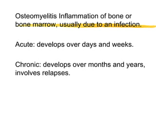
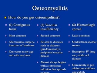
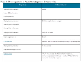
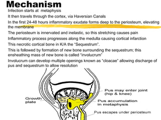
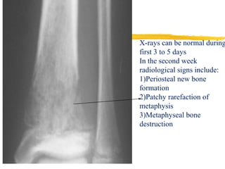
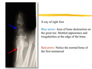
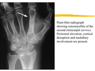
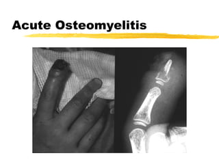
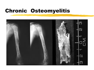
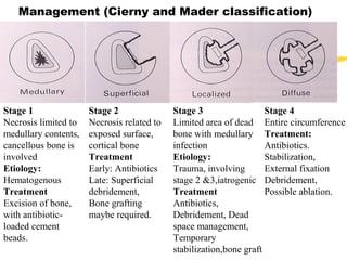
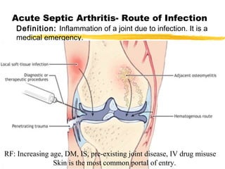
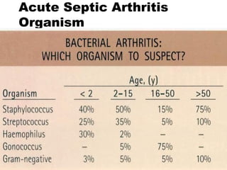
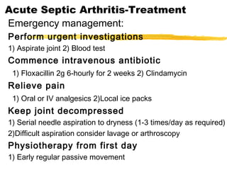
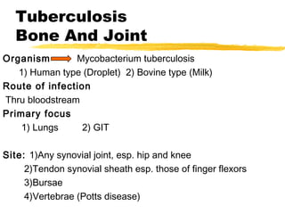
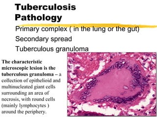
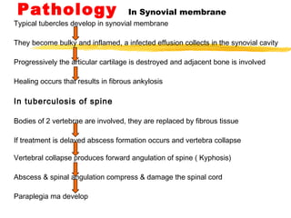
Infection of Bone and Joint
By
Darayus P.Gazder
Osteomyelitis Inflammation of bone or
bone marrow, usually due to an infection.
Acute: develops over days and weeks.
Chronic: develops over months and years,
involves relapses.
Mechanism
Infection starts at metaphysis
It then travels through the cortex, via Haversian Canals
In the first 24-48 hours inflammatory exudate forms deep to the periosteum, elevating
the membrane
The periosteum is innervated and inelastic, so this stretching causes pain
Inflammatory process progresses along the medulla causing cortical infarction
This necrotic cortical bone in K/A the “Sequestrum”.
This is followed by formation of new bone surrounding the sequestrum; this
ensheathing mass of new bone is called “involucrum”
Involucrum can develop multiple openings known as “cloacae” allowing discharge of
pus and sequestrum to allow resolution
Symptoms
1) Local pain---) gradual increase in intensity.
2) Fever, pulse rate will increase.
Signs:
1) Site: Bones commonly infected are tibia, femur, humerus, radius and
ulna.
2) Color: Skin over area is red or reddish-brown.
3) Temperature: Raised.
4) Tenderness: Infected area is tender and the whole bone is painful.
5) Swelling: Maybe soft and indistinct initially and later becomes
noticeable if there is pus at the surface, in which it fluctuates.
6) Adjacent joints: May contain an effusion.
Acute Osteomyelitis
Diagnosis
History and clinical examination
FBC, ESR, CRP.
X-ray (normal in the first week)
Ultrasound (Biopsy and aspiration)
Bone scan Tc-99m methylene diphosphonate
CT scan can define extent of bone
sequestration and cavitation
MRI is the investigation of choice, MRI of bone
shows edema and periosteal elevation. (Highly
sensitive and specific)
X-rays can be normal during first 3 to 5
days
In the second week radiological signs
include:
Periosteal new bone formation
Patchy rarefaction of metaphysis
Metaphyseal bone destruction
X-rays can be normal during
first 3 to 5 days
In the second week
radiological signs include:
1)Periosteal new bone
formation
2)Patchy rarefaction of
metaphysis
3)Metaphyseal bone
destruction
X-ray of right foot
Blue arrow: Area of bone destruction on
the great toe. Mottled appearance and
irregularities at the edge of the bone.
Red arrow: Notice the normal bone of
the first metatarsal
Plain-film radiograph
showing osteomyelitis of the
second metacarpal (arrow).
Periosteal elevation, cortical
disruption and medullary
involvement are present.
Acute Osteomyelitis
Bone scans, both anterior (A) and lateral (B), showing the
accumulation of radioactive tracer at the right ankle (arrow). This
focal accumulation is characteristic of osteomyelitis.
However, a bone scan will be positive despite the absence of bone or
joint abnormality for ex: gout, degenerative joint disease or a healing
fracture
Acute Osteomyelitis
Treatment
supportive treatment for pain and
dehydration
antibiotics (2 weeks IV, then oral)
Splinting
Surgical debridement
Prognosis: Depends on early diagnosis and
treatment. May develop into chronic
osteomyelitis
Complications of Acute
Osteomyelitis
General:
1)Septicemia 2)Pyaemia 3)Metastatic
abscesses.
Local:
1)Altered bone growth
2)Spontaneous fracture
3)Chronic osteomyelitis
4)Deformity
Chronic Osteomyelitis
May follow acute OM
May start De Novo
following operation
following open #
Chronic Osteomyelitis
Organism
usually mixed infection.
mostly staph. Aureus E. Coli . Strep
Pyogen, Proteus.
Disease may take 2 forms:
1) Discharging sinus
2) Brodie’s abscess
CF:May remain inactive for
months or years.
X-ray: It reveals sequestrum
which has separated from
surface of bone or which lies
in a cavity.
Infection is closely contained so as to
create a chronic abscess within the bone
composed of pus or jelly like granulation
tissue.
CF: 1) Local pain 2) Tenderness and
thickening of the bone 3) Transitory
effusions in adjacent joint during an
exacerbation.
Chronic Osteomyelitis
Sequestrum
It is a piece of dead bone that has become separated during the process
of necrosis from normal or sound bone.
Chronic Osteomyelitis
Management (Cierny and Mader classification)
Stage 1
Necrosis limited to
medullary contents,
cancellous bone is
involved
Etiology:
Hematogenous
Treatment
Excision of bone,
with antibiotic-
loaded cement
beads.
Stage 2
Necrosis related to
exposed surface,
cortical bone
Treatment
Early: Antibiotics
Late: Superficial
debridement,
Bone grafting
maybe required.
Stage 3
Limited area of dead
bone with medullary
infection
Etiology:
Trauma, involving
stage 2 &3,iatrogenic
Treatment
Antibiotics,
Debridement, Dead
space management,
Temporary
stabilization,bone graft
Stage 4
Entire circumference
Treatment:
Antibiotics.
Stabilization,
External fixation
Debridement,
Possible ablation.
Acute Septic Arthritis- Route of Infection
Definition: Inflammation of a joint due to infection. It is a
medical emergency.
RF: Increasing age, DM, IS, pre-existing joint disease, IV drug misuse
Skin is the most common portal of entry.
Acute Septic Arthritis
Organism
Acute Septic Arthritis-
Pathology
Acute synovitis with purulent joint effusion
Articular cartilage attacked by bacterial toxin and
cellular enzyme
Complete destruction of the articular cartilage.
Acute Septic Arthritis Sequelae
Complete recovery
Partial loss of the articular cartilage
Fibrous or bony ankylosis
Joint surface
destroyed
Hot, tender, swollen joint
The joint is held immobile in
the “Position of comfort”
Acute Septic Arthritis-Neonate
Picture of Septicemia (Hip joint is commonly
affected)
Irritability
Resistant to movement
Acute Septic Arthritis-Child
Acute pain in single large joint
Reluctant to move the joint
Increase temp. and pulse
It is a hot, swollen, tender joint
Acute Septic Arthritis-Adult
Often involve superficial joint (knee, ankle, wrist)
Joint is swollen, hot, red and held in the “loose pack”
position, with an effusion
Fever may be low grade
Any movement causes pain
Investigation
CBC, WBC, ESR, CRP ,blood culture
X-ray
Ultrasound
Aspiration or biopsy
Acute Septic Arthritis
Differential Diagnosis
Acute Septic Arthritis-Treatment
Emergency management:
Perform urgent investigations
1) Aspirate joint 2) Blood test
Commence intravenous antibiotic
1) Floxacillin 2g 6-hourly for 2 weeks 2) Clindamycin
Relieve pain
1) Oral or IV analgesics 2)Local ice packs
Keep joint decompressed
1) Serial needle aspiration to dryness (1-3 times/day as required)
2)Difficult aspiration consider lavage or arthroscopy
Physiotherapy from first day
1) Early regular passive movement
Tuberculosis
Bone And Joint
Organism Mycobacterium tuberculosis
1) Human type (Droplet) 2) Bovine type (Milk)
Route of infection
Thru bloodstream
Primary focus
1) Lungs 2) GIT
Site: 1)Any synovial joint, esp. hip and knee
2)Tendon synovial sheath esp. those of finger flexors
3)Bursae
4)Vertebrae (Potts disease)
Tuberculosis
Pathology
Primary complex ( in the lung or the gut)
Secondary spread
Tuberculous granuloma
The characteristic
microscopic lesion is the
tuberculous granuloma – a
collection of epithelioid and
multinucleated giant cells
surrounding an area of
necrosis, with round cells
(mainly lymphocytes )
around the periphery.
Pathology In Synovial membrane
Typical tubercles develop in synovial membrane
They become bulky and inflamed, a infected effusion collects in the synovial cavity
Progressively the articular cartilage is destroyed and adjacent bone is involved
Healing occurs that results in fibrous ankylosis
In tuberculosis of spine
Bodies of 2 vertebrae are involved, they are replaced by fibrous tissue
If treatment is delayed abscess formation occurs and vertebra collapse
Vertebral collapse produces forward angulation of spine ( Kyphosis)
Abscess & spinal angulation compress & damage the spinal cord
Paraplegia ma develop
Clinical features
There may be a history of previous infection or recent contact with
tuberculosis.
The patient is usually a child or young adult.
Pain and (in a superficial joint) swelling.
In advanced cases there may be attacks of fever or lassitude and
loss of weight.
Relatives tell of “ night cries “ .
Muscle wasting is characteristic.
Synovial thickening is often striking.
Movements are limited in all directions.
As articular erosion progresses the joint becomes stiff and
deformed.
TB spine
In tuberculosis of the spine, pain may be deceptively slight.
Consequently the patient may not present until there is a visible abscess
(usually in the groin or the lumber region to one side of the midline ) or until
collapse causes a localized kyphosis. Occasionally the presenting feature is
weakness or loss of sensibility in the lower limbs.
Investigations
The ESR: is increased.
CBC: relative lymphocytosis.
The mantoux or Heaf test: will be positive, these are sensitive
but not specific tests.
If synovial fluid is aspirated, it may be cloudy, the protein
concentration is increased and the white cell count is elevated.
Acid-fast bacilli are identified in synovial fluid in 10-20 of cases,
and cultures are positive in over half.
A synovial biopsy is more reliable; sections will show the
characteristic histological features, and acid fast bacilli may be
identified; cultures are positive in over 80 of cases.
X – ray
Soft - tissue swelling and periarticular osteoporosis are
characteristic.
The bone ends take on a “ washed-out “ appearance
and the articular space is narrowed.
Later on there is erosion of the subarticular bone,
characteristically seen on both sides of the joint.
Cystic lesions may appear in the adjacent bone ends but
there is little or no periosteal reaction.
In the spine: bone erosion and collapse around an
intervertebral disc space; the soft tissue shadows may
define a paravertebral abscess.
TB hip
Uniform narrowing
of left hip joint due
to synovial disease
(white arrowhead)
Osteporosis of left
hip
No osteophytes and
no sclerosis
Normal right hip
joint
De-structuration of femoral head
Tuberculosis
Treatment
chemotherapy
rifampicin
isoniazid
ethambutol
rifampicin and isoniazid 6-12 month
Rest and splintage
operative drainage rarely necessary

Bone and joint infections- Osteomyelitis

  • 1.
    Infection of Boneand Joint By Darayus P.Gazder
  • 2.
    Osteomyelitis Inflammation ofbone or bone marrow, usually due to an infection. Acute: develops over days and weeks. Chronic: develops over months and years, involves relapses.
  • 5.
    Mechanism Infection starts atmetaphysis It then travels through the cortex, via Haversian Canals In the first 24-48 hours inflammatory exudate forms deep to the periosteum, elevating the membrane The periosteum is innervated and inelastic, so this stretching causes pain Inflammatory process progresses along the medulla causing cortical infarction This necrotic cortical bone in K/A the “Sequestrum”. This is followed by formation of new bone surrounding the sequestrum; this ensheathing mass of new bone is called “involucrum” Involucrum can develop multiple openings known as “cloacae” allowing discharge of pus and sequestrum to allow resolution
  • 6.
    Symptoms 1) Local pain---)gradual increase in intensity. 2) Fever, pulse rate will increase. Signs: 1) Site: Bones commonly infected are tibia, femur, humerus, radius and ulna. 2) Color: Skin over area is red or reddish-brown. 3) Temperature: Raised. 4) Tenderness: Infected area is tender and the whole bone is painful. 5) Swelling: Maybe soft and indistinct initially and later becomes noticeable if there is pus at the surface, in which it fluctuates. 6) Adjacent joints: May contain an effusion.
  • 7.
    Acute Osteomyelitis Diagnosis History andclinical examination FBC, ESR, CRP. X-ray (normal in the first week) Ultrasound (Biopsy and aspiration) Bone scan Tc-99m methylene diphosphonate CT scan can define extent of bone sequestration and cavitation MRI is the investigation of choice, MRI of bone shows edema and periosteal elevation. (Highly sensitive and specific)
  • 8.
    X-rays can benormal during first 3 to 5 days In the second week radiological signs include: Periosteal new bone formation Patchy rarefaction of metaphysis Metaphyseal bone destruction
  • 9.
    X-rays can benormal during first 3 to 5 days In the second week radiological signs include: 1)Periosteal new bone formation 2)Patchy rarefaction of metaphysis 3)Metaphyseal bone destruction
  • 10.
    X-ray of rightfoot Blue arrow: Area of bone destruction on the great toe. Mottled appearance and irregularities at the edge of the bone. Red arrow: Notice the normal bone of the first metatarsal
  • 11.
    Plain-film radiograph showing osteomyelitisof the second metacarpal (arrow). Periosteal elevation, cortical disruption and medullary involvement are present.
  • 12.
  • 13.
    Bone scans, bothanterior (A) and lateral (B), showing the accumulation of radioactive tracer at the right ankle (arrow). This focal accumulation is characteristic of osteomyelitis. However, a bone scan will be positive despite the absence of bone or joint abnormality for ex: gout, degenerative joint disease or a healing fracture
  • 14.
    Acute Osteomyelitis Treatment supportive treatmentfor pain and dehydration antibiotics (2 weeks IV, then oral) Splinting Surgical debridement Prognosis: Depends on early diagnosis and treatment. May develop into chronic osteomyelitis
  • 15.
    Complications of Acute Osteomyelitis General: 1)Septicemia2)Pyaemia 3)Metastatic abscesses. Local: 1)Altered bone growth 2)Spontaneous fracture 3)Chronic osteomyelitis 4)Deformity
  • 16.
    Chronic Osteomyelitis May followacute OM May start De Novo following operation following open #
  • 17.
    Chronic Osteomyelitis Organism usually mixedinfection. mostly staph. Aureus E. Coli . Strep Pyogen, Proteus. Disease may take 2 forms: 1) Discharging sinus 2) Brodie’s abscess
  • 18.
    CF:May remain inactivefor months or years. X-ray: It reveals sequestrum which has separated from surface of bone or which lies in a cavity.
  • 19.
    Infection is closelycontained so as to create a chronic abscess within the bone composed of pus or jelly like granulation tissue. CF: 1) Local pain 2) Tenderness and thickening of the bone 3) Transitory effusions in adjacent joint during an exacerbation.
  • 20.
    Chronic Osteomyelitis Sequestrum It isa piece of dead bone that has become separated during the process of necrosis from normal or sound bone.
  • 21.
  • 22.
    Management (Cierny andMader classification) Stage 1 Necrosis limited to medullary contents, cancellous bone is involved Etiology: Hematogenous Treatment Excision of bone, with antibiotic- loaded cement beads. Stage 2 Necrosis related to exposed surface, cortical bone Treatment Early: Antibiotics Late: Superficial debridement, Bone grafting maybe required. Stage 3 Limited area of dead bone with medullary infection Etiology: Trauma, involving stage 2 &3,iatrogenic Treatment Antibiotics, Debridement, Dead space management, Temporary stabilization,bone graft Stage 4 Entire circumference Treatment: Antibiotics. Stabilization, External fixation Debridement, Possible ablation.
  • 23.
    Acute Septic Arthritis-Route of Infection Definition: Inflammation of a joint due to infection. It is a medical emergency. RF: Increasing age, DM, IS, pre-existing joint disease, IV drug misuse Skin is the most common portal of entry.
  • 24.
  • 25.
    Acute Septic Arthritis- Pathology Acutesynovitis with purulent joint effusion Articular cartilage attacked by bacterial toxin and cellular enzyme Complete destruction of the articular cartilage. Acute Septic Arthritis Sequelae Complete recovery Partial loss of the articular cartilage Fibrous or bony ankylosis
  • 26.
    Joint surface destroyed Hot, tender,swollen joint The joint is held immobile in the “Position of comfort”
  • 27.
    Acute Septic Arthritis-Neonate Pictureof Septicemia (Hip joint is commonly affected) Irritability Resistant to movement Acute Septic Arthritis-Child Acute pain in single large joint Reluctant to move the joint Increase temp. and pulse It is a hot, swollen, tender joint
  • 28.
    Acute Septic Arthritis-Adult Ofteninvolve superficial joint (knee, ankle, wrist) Joint is swollen, hot, red and held in the “loose pack” position, with an effusion Fever may be low grade Any movement causes pain Investigation CBC, WBC, ESR, CRP ,blood culture X-ray Ultrasound Aspiration or biopsy
  • 29.
  • 30.
    Acute Septic Arthritis-Treatment Emergencymanagement: Perform urgent investigations 1) Aspirate joint 2) Blood test Commence intravenous antibiotic 1) Floxacillin 2g 6-hourly for 2 weeks 2) Clindamycin Relieve pain 1) Oral or IV analgesics 2)Local ice packs Keep joint decompressed 1) Serial needle aspiration to dryness (1-3 times/day as required) 2)Difficult aspiration consider lavage or arthroscopy Physiotherapy from first day 1) Early regular passive movement
  • 31.
    Tuberculosis Bone And Joint OrganismMycobacterium tuberculosis 1) Human type (Droplet) 2) Bovine type (Milk) Route of infection Thru bloodstream Primary focus 1) Lungs 2) GIT Site: 1)Any synovial joint, esp. hip and knee 2)Tendon synovial sheath esp. those of finger flexors 3)Bursae 4)Vertebrae (Potts disease)
  • 32.
    Tuberculosis Pathology Primary complex (in the lung or the gut) Secondary spread Tuberculous granuloma The characteristic microscopic lesion is the tuberculous granuloma – a collection of epithelioid and multinucleated giant cells surrounding an area of necrosis, with round cells (mainly lymphocytes ) around the periphery.
  • 33.
    Pathology In Synovialmembrane Typical tubercles develop in synovial membrane They become bulky and inflamed, a infected effusion collects in the synovial cavity Progressively the articular cartilage is destroyed and adjacent bone is involved Healing occurs that results in fibrous ankylosis In tuberculosis of spine Bodies of 2 vertebrae are involved, they are replaced by fibrous tissue If treatment is delayed abscess formation occurs and vertebra collapse Vertebral collapse produces forward angulation of spine ( Kyphosis) Abscess & spinal angulation compress & damage the spinal cord Paraplegia ma develop
  • 34.
    Clinical features There maybe a history of previous infection or recent contact with tuberculosis. The patient is usually a child or young adult. Pain and (in a superficial joint) swelling. In advanced cases there may be attacks of fever or lassitude and loss of weight. Relatives tell of “ night cries “ . Muscle wasting is characteristic. Synovial thickening is often striking. Movements are limited in all directions. As articular erosion progresses the joint becomes stiff and deformed.
  • 35.
    TB spine In tuberculosisof the spine, pain may be deceptively slight. Consequently the patient may not present until there is a visible abscess (usually in the groin or the lumber region to one side of the midline ) or until collapse causes a localized kyphosis. Occasionally the presenting feature is weakness or loss of sensibility in the lower limbs.
  • 37.
    Investigations The ESR: isincreased. CBC: relative lymphocytosis. The mantoux or Heaf test: will be positive, these are sensitive but not specific tests. If synovial fluid is aspirated, it may be cloudy, the protein concentration is increased and the white cell count is elevated. Acid-fast bacilli are identified in synovial fluid in 10-20 of cases, and cultures are positive in over half. A synovial biopsy is more reliable; sections will show the characteristic histological features, and acid fast bacilli may be identified; cultures are positive in over 80 of cases.
  • 38.
    X – ray Soft- tissue swelling and periarticular osteoporosis are characteristic. The bone ends take on a “ washed-out “ appearance and the articular space is narrowed. Later on there is erosion of the subarticular bone, characteristically seen on both sides of the joint. Cystic lesions may appear in the adjacent bone ends but there is little or no periosteal reaction. In the spine: bone erosion and collapse around an intervertebral disc space; the soft tissue shadows may define a paravertebral abscess.
  • 39.
    TB hip Uniform narrowing ofleft hip joint due to synovial disease (white arrowhead) Osteporosis of left hip No osteophytes and no sclerosis Normal right hip joint
  • 40.
  • 41.
    Tuberculosis Treatment chemotherapy rifampicin isoniazid ethambutol rifampicin and isoniazid6-12 month Rest and splintage operative drainage rarely necessary